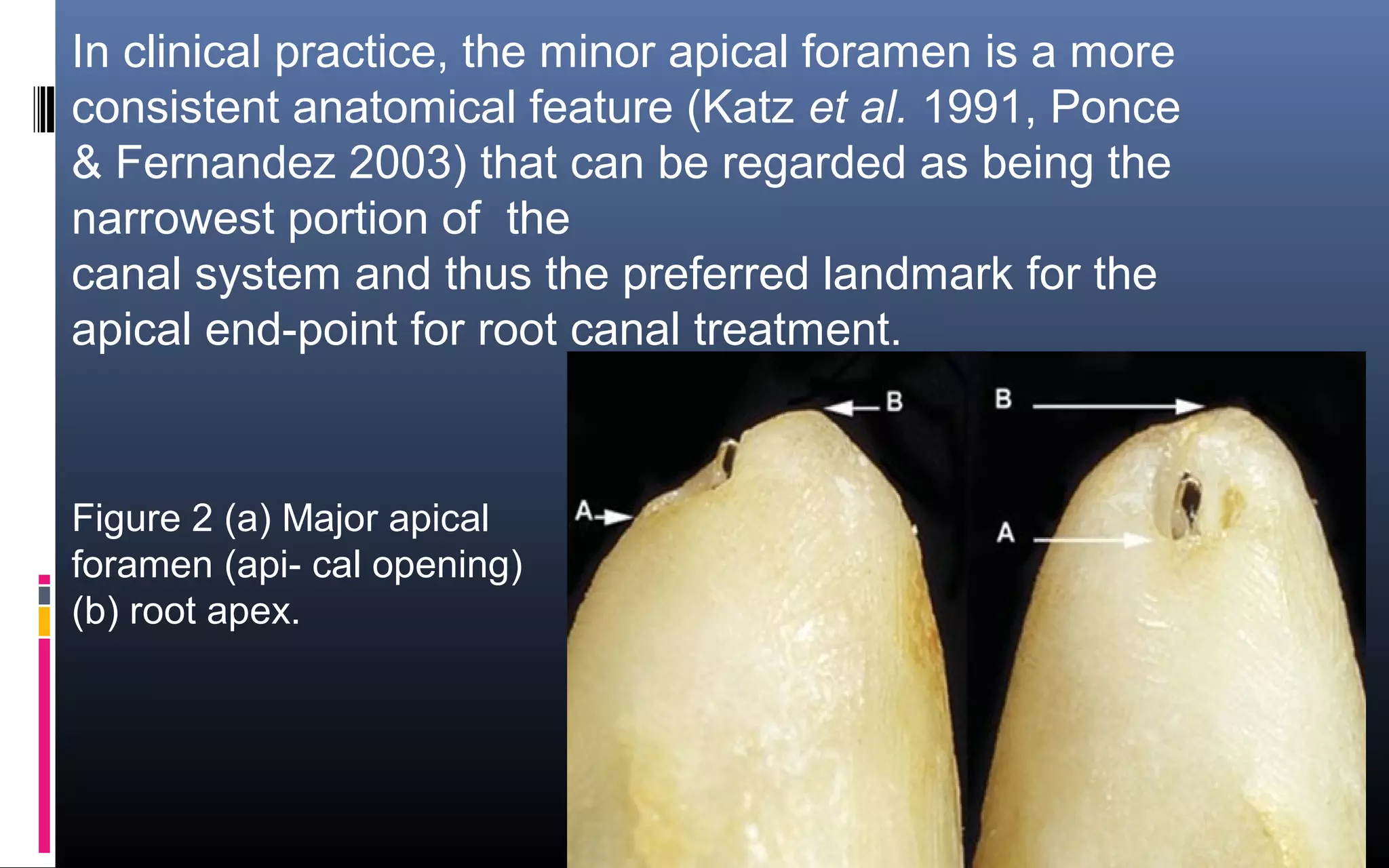
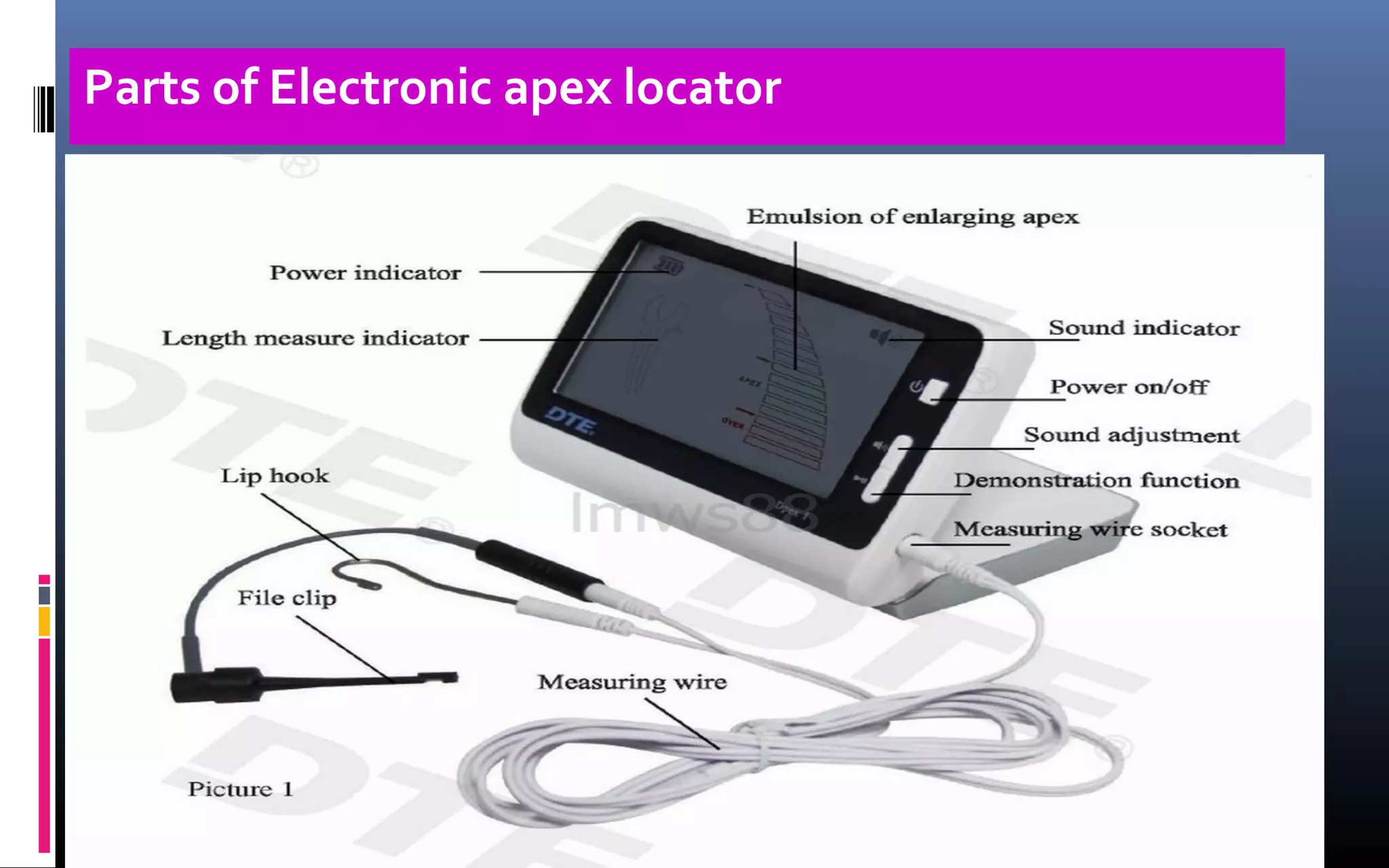
Prepared by – Imran M. Shaikh
Guided by –Dr. Jacob Daniel
National institute of clinical endodontics
Bangalore,India
 Definition
 Other or correct name for device
Anatomy of apical foramen
Limitations of radiographic (traditional)working length
Emergence of electronic apex Locator
Generations /History
Parts of EAL
How to use Electronic Apex Locator?
What Precautions to avoid obtaining false reading?
Advanced models of EAL
Significance of EAL
CONTENTS
An electronic apex locator is 
an electronic device used in endodontics to 
determine the position of the apical foramen 
and its location and thus determine the length 
of the root canal space.
Definition
.Correct name of electronic apex
locator(EAL)
electronic root canal
length measurement
device(ERCLMD)
fandamentals of ERCLMD
international endodontics
Figure 1 Idealized anatomy of apical portion of root (a) 
major apical foramen, (b) minor apical foramen (apical 
constriction) that may be coincident with the cemento-
dentinal junction (CDJ), (c) cementum, (d) dentine and (e) 
root apex
Anatomy of Apical foramen
In clinical practice, the minor apical foramen is a more 
consistent anatomical feature (Katz et al. 1991, Ponce 
& Fernandez 2003) that can be regarded as being the 
narrowest portion of  the
canal system and thus the preferred landmark for the 
apical end-point for root canal treatment.
 
Figure 2 (a) Major apical 
foramen (api- cal opening)  
(b) root apex.
>The  apical  foramen is not always  located  at  the  anatomical 
 apex of the tooth. The foramen of  the  root  canal may be 
located to one side of the anatomical apex,
>sometimes it is upto 2mm short radiographic apex in 50 % of 
roots (Green 1956, Pineda & Kuttler 1972). 
 >The anatomy of the apical foramen changes with age. 
Figure  (a) Position of the apical 
foramen (adapted from Kuttler). (b) 
Anatomy of the root apex .
Radiographic determination of working length has been used fo
many years. The radiographic apex is defined as the anatomical
end of the root as seen on the radiograph,
while the apical foramen is the region where the canal leaves
the root surface next to the periodontal ligament (American
Association of Endodontics1984).
When the apical foramen exits to the side of the root or in a
buccal or lingual direction it becomes difficult to view on the
radiograph
Limitations of traditional working length
assesment
>Dense bone and anatomical structures can make
the visualization of root canal files impossible by
obscuring the apex
>The superimposition of the zygomatic arch has
been shown to interfere radiographically with 20%
of maxillary first molar apices and 42% of second
molar apices (Tamse et al. 1980).
>). The deposition of secondary dentine and
cementum can move the apical constriction further
from accepted limits causing preparation errors
(Stein & Corcoran 1990, Chong & Pitt Ford 1994).
>A radiograph provides a two-dimensional image of a three-
dimensional structure and is technique sensitive in both its
exposure and interpretation.
>Cox et al. found that adjustments were required to working
length radiographicaly. 68% of examiners agreed adjustments up
to 0.5 mm were needed, but there was only 14% agreement when
adjustments greater than 1.0 mm were required.
>radiation exposure is alwayz avoided in pregnant women.
>its difficult to take radiograph in patients have Gag reflex
>also deficulty in taking x ray in patients who have phobia of
radiographic exposure
The preoperative radiograph is essential in endod ontics to
determine the anatomy of the root canal system, the
number and curvature of roots, the presence or absence of
disease, and to act as an initial guide for working length.
The electronic apex locator is an instrument, which used
with appropriate radio- graphs, allows for much greater
accuracy of working length control
Importance of
Radiograph
An electronic method for root length determination was first
investigated by Custer (1918).
The idea was revisited by Suzuki(Japan) in 1942
.Sunada in 1962 .Sunada took these principles and
constructed a simple device
Since then Japan remains the leading country in develping
newer and improved models
EMERGENCE
Generations
First Genration -
1969. It used the resistance method and alternating current
Second Genration-
The change in frequency method of measuring was developed
by Inoue in 1971 as the Sono-Explorer (Hayashi Dental Supply, Tokyo, Japan)
Third Genrations- Third generation apex locators are similar to the second
generation except that they use multiple frequencies to determine the distance
from the end of the canal.
*The Endex/Apit Endex are reported in the literature, which give a combined
accuracy of 81% to within ±0.5 mm of the apical foramen.
(Frank & Torabinejad 1993)
Root ZX 95 -99 % OF ACCURACY
Genrations of EAL
The fourth generation
Bingo 1020/Ray-Pex 4
>Tinaz et al. (2002a) found the Bingo1020
to be as accurate as the Root ZX
in an in vitro study and easier for a beginner to use in
preflared canals.
>This unit has subsequently been marketed by Dentsply as
the Ray-Pex 4.
All modern apex locators are able to detect root perforations
to clinically acceptable limits and are equally able to
distinguish both large and small perforations (Fuss et al. 1996,
Kaufman et al. 1997).
OTHER USES OF APEX LOCATOR
Multiple-function apex locators are becoming more common
and several have vitality testing functions. Combination
electronic apex locators.
Suspected periodontal or pulpal perforation during pinhole
preparation can be confirmed by all apex locators, as a
patent perforation will cause the instrument to complete a
circuit and indicate the instrument is beyond the ‘apex’ (Ingle
et al. 2002).
Parts of Electronic apex locator
HOW to USE???
Clinical use.
1. Analyse the root anatomy for curvature and establish an
estimated working length from the pre operative radiograph.
2. The coronal aspect of the canal should be
opened/prepared to provide straight line access or a “glide
path” to the apical aspect of the root canal.
3. Modern apex locators generally function well in the
presence of fluids and irrigants in the root canal, but prior to
using the apex locator excess irrigating fluids are removed
from the access cavity.
.
4. Once the lip hook and file holder are attached, in most cases a size
15 or 20 file (see troubleshooting) is advanced into the root canal until
the blue scale on the apex locator reaches the “apex and red triangle ”
on the screen of the root ZX. This indicates that the file is now at the
apical foramen .
5. A diagnostic radiograph is taken with the file at this length. If the
radiograph confirms the file to be at the apex this length is effectively the
“canal length”.
6. Since the apical constriction is on average 0.5 mm from the apical
foramen, the working length is calculated by subtracting 0.5 mm from the
canal length. The canal can now be prepared to the working length (see
diagram).
If you are an advocate of patency filing, a size 10 file should be placed to
the canal length to maintain the patency of the root canal

How to Use electronic Apex Locator??
1. The file of the loacator should not contact metal crown
or filling.
2. Ther should not be any fluid contact between the pulp
chamber and the gingiva/periodontal tissues,eigther
through leaky cervical filling or deep or cervical caries
.such an outside contact may cause leakage of
measuring current and inaccurate reading.
3. Genrally the locator should be used in absence of fluid
in the canal but
4. Some newer models ovarcome this limitation
e.g.ROOT ZX ,ENDEX,PROPEX (dentsply)
Precautions taken in order to avoid obtaining false
reading.
5. As much of pulp tissue as possible should be removed
prior to using Locator
6.The largest file that will bind the apex should be used
7.Too loose fitting insrtument should be avoided
8.ALWAYS. Except in unavoidable situation,the reading
should be confirmed in collaberation withradiongraphic
findings
Precautions taken in order to avoid obtaining
false reading.
9.EALs are ineffective in case of teeth with wide open apex
as obtaining correct reading is almost impossible.
10.EALs should be avoided in patients wearing pace
makers.
Precautions taken in order to avoid obtaining
false reading.
Adaptive Apex Locator overcomes as the disadvantages of 
the popular apex locators 4th generation – low accuracy on 
working in wet canals, as well the disadvantages of devices V 
th generation – difficulty on working in dry canals and 
necessarily of compulsory, additional wetting.
 Adaptive Apex Locator  immediately adapts for dry or wet 
canal. On this way is possible to be measured as in dry and in 
additional wetted canals as well, canals with blood or 
exudates, canals with still not-extirpated pulp.
Adaptive Apex Locators
conclusion
Conclusion
No individual technique is truly satisfactory in deter- mining 
endodontic working length. The CDJ is a practical and 
anatomic termination point for the preparation and 
obturation of the root canal and this cannot be determined 
radiographically. Modern elec- tronic apex locators can 
determine this position with accuracies of greater than 90% 
but still have some limitations. Knowledge of apical 
anatomy, prudent use of radiographs and the correct use of 
an electronic apex locator will assist practitioners to achieve 
predictable results.
References:References:
 Books & JournalsBooks & Journals
Ingle,s Endodontics 2013 Special editionIngle,s Endodontics 2013 Special edition
Advanced Endodontics for Clinicinas by Dr.Jacob GAdvanced Endodontics for Clinicinas by Dr.Jacob G
Daniel (Edition1998 )Daniel (Edition1998 )
The fundamental operating principles of electronic root 
canal length measurement devices. International
Endodontic Journal, 39, 595–609, 2006
      >  Electronic   apex locators.
International Endodontic Journal, 37, 425–437, 2004
Electronic apex locator by  dr.imran m.shaikh

Electronic apex locator by dr.imran m.shaikh