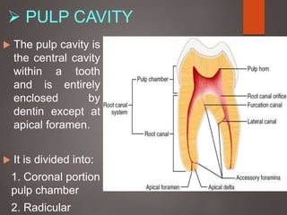
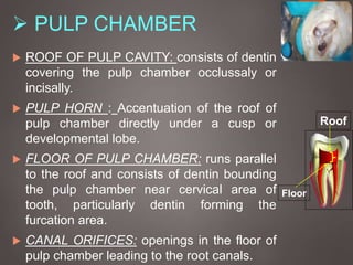
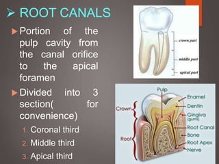
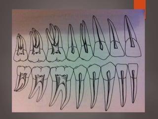
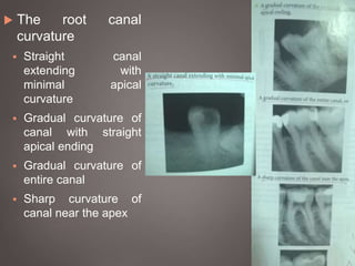
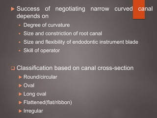
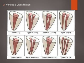
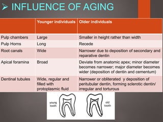
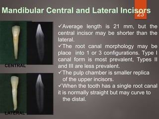
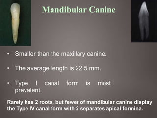
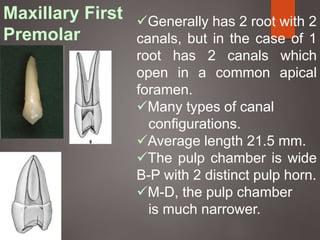
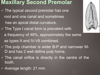
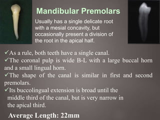
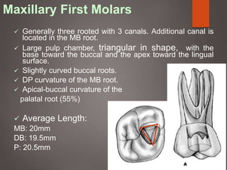
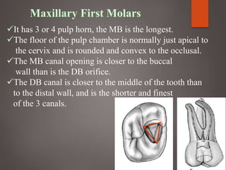
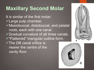
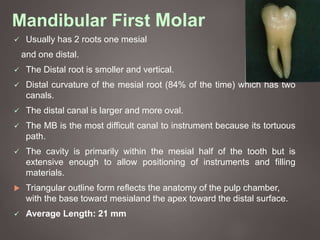
The pulp cavity is the central cavity within a tooth and is entirely enclosed by dentin except at apical foramen.
It is divided into:
1. Coronal portion pulp chamber
2. Radicular portion root canal
PULP CHAMBER
ROOF OF PULP CAVITY: consists of dentin covering the pulp chamber occlussaly or incisally.
PULP HORN : Accentuation of the roof of pulp chamber directly under a cusp or developmental lobe.
FLOOR OF PULP CHAMBER: runs parallel to the roof and consists of dentin bounding the pulp chamber near cervical area of tooth, particularly dentin forming the furcation area.
CANAL ORIFICES: openings in the floor of pulp chamber leading to the root canals.
ROOT CANALS
Portion of the pulp cavity from the canal orifice to the apical foramen
Divided into 3 section( for convenience)
Coronal third
Middle third
Apical third
The root canal curvature
Straight canal extending with minimal apical curvature
Gradual curvature of canal with straight apical ending
Gradual curvature of entire canal
Sharp curvature of canal near the apex
Success of negotiating narrow curved canal depends on
Degree of curvature
Size and constriction of root canal
Size and flexibility of endodontic instrument blade
Skill of operator
Classification based on canal cross-section
Round/circular
Oval
Long oval
Flattened(flat/ribbon)
Irregular
Vertucci’s Classification
Weine’s Classification
ISTHMUS
A narrow passage or anatomic part connecting two larger structures (root canals)
APICAL FORAMEN
In young incompletely developed teeth the apical foramen is funnel shaped with wider portion extending outward
As root develops the apical foramen becomes narrower
Apical foramen is not the most constricted part of root apex\apical foramen is not always located at the centre of the root apex
LATERAL CANALS AND ACCESSARY FORAMINA
Lateral canals frequently occur in apical third of root
May occur in areas of bifurcation and trifurcation of multirooted teeth
With increasing age, number of accessory foramina reduce due to calcification of contained soft tissue
INFLUENCE OF AGING
METHODS OF DETERMINING PULP ANATOMY
CLINICAL METHODS
Anatomy studies
Radiographs
Explorations
High resolution compound tomography
Visualisation endogram
Fiberoptic endoscope
Magnetic resonance imaging
IN VITRO METHODS
sectioning of teeth
use of dyes
Contrasting media
Scanning electron microscope analysis
VARIATIONS IN INTERNAL ANATOMY
Variations in development
Gemination
Fusion
Concrescence
Taurodontism
Talon’s cusp
Dilaceration
Extra root canal
Dens invaginatus
Dens evaginatus
Maxillary Central Incisor
Maxillary Lateral Incisor
Maxillary Canine
Mandibular Central and Lateral Incisors
Mandibular Canine
Maxillary First Premolar
Maxillary Second Premolar
The typical second premolar has one
root and one canal and sometimes
has an apical distal curvature.
The Type I canal form is p
An inlay may cap none, or may cap all but one cusp.
Sturdevant’s 4th ed. page579
Inlays may be used as single-tooth restorations for proximo-occlusal or gingival lesions with minimal to moderate extensions
Shillingburg page 1
An inlay may be defined as a restoration which has been constructed out of mouth from gold, porcelain, or other material & then cemented into the prepared cavity of a tooth.
William McGehee pg410
This presentation specifically deals with the maxillary and mandibular Major connectors used in a cast partial denture. it also mentions the uses, advantages and disadvantages of each,
Bevels and flares are very important components of resin restoration procedure. This presentation focuses on bevels and flares in restorative procedure.
A concise and brief presentation on cleaning and shaping of root canals. Colorful and well pictured. Ideal for UG students and PG students to get a good understanding of BMP techniques.
Coronal and radicular pulp
Apical foramen
Accessory canal
Functions of dental pulp
Components of dental pulp
Functions of pulpal extracellular matrix
Organization of cells in the pulp
The principle cells of the pulp
The pathways of collagen synthesis
Matrix and ground substances
Vasculature and lymphatic supply
Innervation of Dentin- pulp complex
Disorders of the dental pulp
Advances in pulp vitality testing
The Indian Dental Academy is the Leader in continuing dental education , training dentists in all aspects of dentistry and
offering a wide range of dental certified courses in different formats.for more details please visit
www.indiandentalacademy.com
Indian Dental Academy: will be one of the most relevant and exciting training center with best faculty and flexible training programs for dental professionals who wish to advance in their dental practice,Offers certified courses in Dental implants,Orthodontics,Endodontics,Cosmetic Dentistry, Prosthetic Dentistry, Periodontics and General Dentistry.
Palestine last event orientationfvgnh .pptxRaedMohamed3
An EFL lesson about the current events in Palestine. It is intended to be for intermediate students who wish to increase their listening skills through a short lesson in power point.
Embracing GenAI - A Strategic ImperativePeter Windle
Artificial Intelligence (AI) technologies such as Generative AI, Image Generators and Large Language Models have had a dramatic impact on teaching, learning and assessment over the past 18 months. The most immediate threat AI posed was to Academic Integrity with Higher Education Institutes (HEIs) focusing their efforts on combating the use of GenAI in assessment. Guidelines were developed for staff and students, policies put in place too. Innovative educators have forged paths in the use of Generative AI for teaching, learning and assessments leading to pockets of transformation springing up across HEIs, often with little or no top-down guidance, support or direction.
This Gasta posits a strategic approach to integrating AI into HEIs to prepare staff, students and the curriculum for an evolving world and workplace. We will highlight the advantages of working with these technologies beyond the realm of teaching, learning and assessment by considering prompt engineering skills, industry impact, curriculum changes, and the need for staff upskilling. In contrast, not engaging strategically with Generative AI poses risks, including falling behind peers, missed opportunities and failing to ensure our graduates remain employable. The rapid evolution of AI technologies necessitates a proactive and strategic approach if we are to remain relevant.
The French Revolution, which began in 1789, was a period of radical social and political upheaval in France. It marked the decline of absolute monarchies, the rise of secular and democratic republics, and the eventual rise of Napoleon Bonaparte. This revolutionary period is crucial in understanding the transition from feudalism to modernity in Europe.
For more information, visit-www.vavaclasses.com
Welcome to TechSoup New Member Orientation and Q&A (May 2024).pdfTechSoup
In this webinar you will learn how your organization can access TechSoup's wide variety of product discount and donation programs. From hardware to software, we'll give you a tour of the tools available to help your nonprofit with productivity, collaboration, financial management, donor tracking, security, and more.
Unit 8 - Information and Communication Technology (Paper I).pdfThiyagu K
This slides describes the basic concepts of ICT, basics of Email, Emerging Technology and Digital Initiatives in Education. This presentations aligns with the UGC Paper I syllabus.
Synthetic Fiber Construction in lab .pptxPavel ( NSTU)
Synthetic fiber production is a fascinating and complex field that blends chemistry, engineering, and environmental science. By understanding these aspects, students can gain a comprehensive view of synthetic fiber production, its impact on society and the environment, and the potential for future innovations. Synthetic fibers play a crucial role in modern society, impacting various aspects of daily life, industry, and the environment. ynthetic fibers are integral to modern life, offering a range of benefits from cost-effectiveness and versatility to innovative applications and performance characteristics. While they pose environmental challenges, ongoing research and development aim to create more sustainable and eco-friendly alternatives. Understanding the importance of synthetic fibers helps in appreciating their role in the economy, industry, and daily life, while also emphasizing the need for sustainable practices and innovation.
Model Attribute Check Company Auto PropertyCeline George
In Odoo, the multi-company feature allows you to manage multiple companies within a single Odoo database instance. Each company can have its own configurations while still sharing common resources such as products, customers, and suppliers.
June 3, 2024 Anti-Semitism Letter Sent to MIT President Kornbluth and MIT Cor...Levi Shapiro
Letter from the Congress of the United States regarding Anti-Semitism sent June 3rd to MIT President Sally Kornbluth, MIT Corp Chair, Mark Gorenberg
Dear Dr. Kornbluth and Mr. Gorenberg,
The US House of Representatives is deeply concerned by ongoing and pervasive acts of antisemitic
harassment and intimidation at the Massachusetts Institute of Technology (MIT). Failing to act decisively to ensure a safe learning environment for all students would be a grave dereliction of your responsibilities as President of MIT and Chair of the MIT Corporation.
This Congress will not stand idly by and allow an environment hostile to Jewish students to persist. The House believes that your institution is in violation of Title VI of the Civil Rights Act, and the inability or
unwillingness to rectify this violation through action requires accountability.
Postsecondary education is a unique opportunity for students to learn and have their ideas and beliefs challenged. However, universities receiving hundreds of millions of federal funds annually have denied
students that opportunity and have been hijacked to become venues for the promotion of terrorism, antisemitic harassment and intimidation, unlawful encampments, and in some cases, assaults and riots.
The House of Representatives will not countenance the use of federal funds to indoctrinate students into hateful, antisemitic, anti-American supporters of terrorism. Investigations into campus antisemitism by the Committee on Education and the Workforce and the Committee on Ways and Means have been expanded into a Congress-wide probe across all relevant jurisdictions to address this national crisis. The undersigned Committees will conduct oversight into the use of federal funds at MIT and its learning environment under authorities granted to each Committee.
• The Committee on Education and the Workforce has been investigating your institution since December 7, 2023. The Committee has broad jurisdiction over postsecondary education, including its compliance with Title VI of the Civil Rights Act, campus safety concerns over disruptions to the learning environment, and the awarding of federal student aid under the Higher Education Act.
• The Committee on Oversight and Accountability is investigating the sources of funding and other support flowing to groups espousing pro-Hamas propaganda and engaged in antisemitic harassment and intimidation of students. The Committee on Oversight and Accountability is the principal oversight committee of the US House of Representatives and has broad authority to investigate “any matter” at “any time” under House Rule X.
• The Committee on Ways and Means has been investigating several universities since November 15, 2023, when the Committee held a hearing entitled From Ivory Towers to Dark Corners: Investigating the Nexus Between Antisemitism, Tax-Exempt Universities, and Terror Financing. The Committee followed the hearing with letters to those institutions on January 10, 202
Macroeconomics- Movie Location
This will be used as part of your Personal Professional Portfolio once graded.
Objective:
Prepare a presentation or a paper using research, basic comparative analysis, data organization and application of economic information. You will make an informed assessment of an economic climate outside of the United States to accomplish an entertainment industry objective.
3. PULP CAVITY
The pulp cavity is
the central cavity
within a tooth
and is entirely
enclosed by
dentin except at
apical foramen.
It is divided into:
1. Coronal portion
pulp chamber
2. Radicular
4. PULP CHAMBER
ROOF OF PULP CAVITY: consists of dentin
covering the pulp chamber occlussaly or
incisally.
PULP HORN : Accentuation of the roof of
pulp chamber directly under a cusp or
developmental lobe.
FLOOR OF PULP CHAMBER: runs parallel
to the roof and consists of dentin bounding
the pulp chamber near cervical area of
tooth, particularly dentin forming the
furcation area.
CANAL ORIFICES: openings in the floor of
pulp chamber leading to the root canals.
Floor
Roof
5. ROOT CANALS
Portion of the
pulp cavity from
the canal orifice
to the apical
foramen
Divided into 3
section( for
convenience)
1. Coronal third
2. Middle third
3. Apical third
6.
7. The root canal
curvature
Straight canal
extending with
minimal apical
curvature
Gradual curvature of
canal with straight
apical ending
Gradual curvature of
entire canal
Sharp curvature of
canal near the apex
8. Success of negotiating narrow curved canal
depends on
Degree of curvature
Size and constriction of root canal
Size and flexibility of endodontic instrument blade
Skill of operator
Classification based on canal cross-section
Round/circular
Oval
Long oval
Flattened(flat/ribbon)
Irregular
11. ISTHMUS
A narrow passage or anatomic part connecting
two larger structures (root canals)
12. APICAL FORAMEN
In young incompletely developed teeth the apical
foramen is funnel shaped with wider portion extending
outward
As root develops the apical foramen becomes narrower
Apical foramen is not the most constricted part of root
apexapical foramen is not always located at the centre
of the root apex
13. LATERAL CANALS AND
ACCESSARY FORAMINA
Lateral canals frequently occur in apical third of root
May occur in areas of bifurcation and trifurcation of
multirooted teeth
With increasing age, number of accessory foramina
reduce due to calcification of contained soft tissue
14. INFLUENCE OF AGING
Younger individuals Older individuals
Pulp chambers Large Smaller in height rather than width
Pulp Horns Long Recede
Root canals Wide Narrower due to deposition of secondary and
reparative dentin
Apical foramina Broad Deviate from anatomic apex; minor diameter
becomes narrower; major diameter becomes
wider (deposition of dentin and cementum)
Dentinal tubules Wide, regular and
filled with
protoplasmic fluid
Narrower or obliterated y deposition of
peritubular dentin, forming sclerotic dentin/
irregular and torturous
15. METHODS OF DETERMINING PULP
ANATOMY
1. CLINICAL METHODS
I. Anatomy studies
II. Radiographs
III. Explorations
IV. High resolution compound tomography
V. Visualisation endogram
VI. Fiberoptic endoscope
VII. Magnetic resonance imaging
17. 2. IN VITRO METHODS
i. sectioning of teeth
ii. use of dyes
iii. Contrasting media
iv. Scanning electron microscope analysis
18. VARIATIONS IN
INTERNAL ANATOMY
Variations in development
i. Gemination
ii. Fusion
iii. Concrescence
iv. Taurodontism
v. Talon’s cusp
vi. Dilaceration
vii. Extra root canal
viii. Dens invaginatus
ix. Dens evaginatus
19.
20. 20Maxillary Central Incisor
Central incisor are large and on an average of
23 mm.
It has a single canal and a single apical foramen.
The canal form is usually Type I.
The pulp in young patients normally has 3 pulp
horns.
The pulp chambers is noticeably wider in the
faciolingual direction than in the mesiodistal.
All upper anterior teeth
have one root and one
canal.
21. 21
Maxillary Lateral Incisor
It is shorter than central incisor.
Average length of 21- 22 mm.
It has a single canal and a single apical
foramen.
The canal form is usually Type I.
In young patients have two only pulp horn and is
wider in labiopalatal dimension.
The canal is tapered and the apex is often
curved generally in distal direction.
22. 22
Maxillary Canine
It is the longest tooth (26.5 mm)
Seldom has more than one canal
The pulp chamber is quite narrow M- D,
and there is one pulp horn pointed to the
incisal angle.
The pulp space is much wider labiopalatally
and the pulp space follows this outline.
Oval Type I root canal.
The root apex is often tapered and very
thin.
The canal is usually straight but may show
a distal apical curvature.
23. 23
Mandibular Central and Lateral Incisors
CENTRAL
LATERAL
Average length is 21 mm, but the
central incisor may be shorter than the
lateral.
The root canal morphology may be
place into 1 or 3 configurations. Type I
canal form is most prevalent, Types II
and III are less prevalent.
The pulp chamber is smaller replica
of the upper incisors.
When the tooth has a single root canal
it is normally straight but may curve to
the distal.
24. 24
Mandibular Canine
• Smaller than the maxillary canine.
• The average length is 22.5 mm.
• Type I canal form is most
prevalent.
Rarely has 2 roots, but fewer of mandibular canine display
the Type IV canal form with 2 separates apical formina.
25. Maxillary First
Premolar
Generally has 2 root with 2
canals, but in the case of 1
root has 2 canals which
open in a common apical
foramen.
Many types of canal
configurations.
Average length 21.5 mm.
The pulp chamber is wide
B-P with 2 distinct pulp horn.
M-D, the pulp chamber
is much narrower.
26. Maxillary Second Premolar
The typical second premolar has one
root and one canal and sometimes
has an apical distal curvature.
The Type I canal form is prevalent with
a frequency of 48%, approximately the same
as types II and IV-VII combined.
The pulp chamber is wider B-P and narrower M-
D and has 2 well define pulp horns.
The canal orifice is directly in the centre of the
tooth.
Average length: 21 mm.
27. Mandibular Premolars
Usually has a single delicate root
with a mesial concavity, but
occasionally present a division of
the root in the apical half.
As a rule, both teeth have a single canal.
The coronal pulp is wide B-L with a large buccal horn
and a small lingual horn.
The shape of the canal is similar in first and second
premolars.
Its buccolingual extension is broad until the
middle third of the canal, but is very narrow in
the apical third.
Average Length: 22mm
28. Maxillary First Molars
Generally three rooted with 3 canals. Additional canal is
located in the MB root.
Large pulp chamber, triangular in shape, with the
base toward the buccal and the apex toward the lingual
surface.
Slightly curved buccal roots.
DP curvature of the MB root.
Apical-buccal curvature of the
palatal root (55%)
Average Length:
MB: 20mm
DB: 19.5mm
P: 20.5mm
29. It has 3 or 4 pulp horn, the MB is the longest.
The floor of the pulp chamber is normally just apical to
the cervix and is rounded and convex to the occlusal.
The MB canal opening is closer to the buccal
wall than is the DB orifice.
The DB canal is closer to the middle of the tooth than
to the distal wall, and is the shorter and finest
of the 3 canals.
Maxillary First Molars
30. Maxillary Second Molar
It is similar of the first molar:
Large pulp chamber.
Mesiobuccal, distobuccal, and palatal
roots, each with one canal.
Gradual curvature of all three canals.
“Flattened” triangular outline form.
The DB canal orifice is
nearer the centre of the
cavity floor.
31. Mandibular First Molar
Usually has 2 roots one mesial
and one distal.
The Distal root is smoller and vertical.
Distal curvature of the mesial root (84% of the time) which has two
canals.
The distal canal is larger and more oval.
The MB is the most difficult canal to instrument because its tortuous
path.
The cavity is primarily within the mesial half of the tooth but is
extensive enough to allow positioning of instruments and filling
materials.
Triangular outline form reflects the anatomy of the pulp chamber,
with the base toward mesialand the apex toward the distal surface.
Average Length: 21 mm
32. Mandibular Second Molar
It is smaller version of the first molar
with an average length of 20mm.
The mesial root has 2 canals and the
distal one.
The mesial canals tend to fuse in the
apical third to give rise to one main
apical foramen.
Commonly has fused roots.
Editor's Notes
In anterior teeth, the pulp chamber gradually merges into the root canal and the division becomes indistinct.
In multi-rooted tooth, pulp cavity consists of a single pulp chamber and usually 3 or more root canals.
Canal orifices are not separate structures but are continuous with the pulp chamber and root canals
The walls of pulp chamber derive their name from corresponding walls of tooth surface
Angles- corresponding angles like mesiobucccal
Accessary or lateral canals- lateral branching of main root canal generally occurring in apical third or furcation area of root
Apical foramina- aperture at or near the apex f the root thru which the blood vessels and nerves enter or leave the pulp
Accesary foramina
A straight canal extending the entire length of the root is uncommon
Either constriction is present before apex is reached or as is often the case a curvature may be present
Curvature of about 20%-difficult or impossible to negotiate with endodontic instruments
30% if root canal is wide
Type I: 1 canal extends from the pulp chamber to apex. (1)
Type II: 2 separate canals leave the pulp chamber and join short of apex to form 1 canal(2-1)
Type III: 1 canal leaves the pulp chamber and divides into 2 roots; the two then merge to exit as 1 canal. (1-2-1)
Type IV: 2 separate distinct canals extend from the pulp chamber to apex. (2)
Type V: 1 canal exits the pulp chamber and divides short of the apex into 2 separate canals with separate apical foramina. (1-2)
Type VI: 2 separate canals leave the pulp chamber, merge in the body of root and redivide short of apex to exit as 2 distinct canals. (2-1-2)
Type VII: 1 canal leaves the pulp chamber, divides and then rejoins in the body of root and finally redivides into 2 distinct canals short of the apex.
(1-2-1-2)
Type VIII: 3 separate distinct canals extend from pulp chamber to apex (3)
Identified using METHYLENE BLUE DYE
Contains pulp or pulpally derived tissue and acts as store house for bacteria- prepared thoroughly
Type 1- faint communication between two canals
Type 2 complete isthmus with definite connection
Type 3 complete but very short
Type 4 complete or incomplete isthmus between 3/more canals
Type5 2/3 canal openings without visible connections
The periodontal vessels curve around the root apex of a developing tooth n usually =get entrapped in the HERS resulting in formation of lateral canals n accessary foramina
Reparative dentin is devoid of dentinal tubules and moisture content is reduced