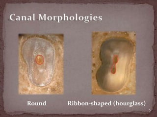
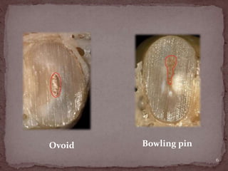
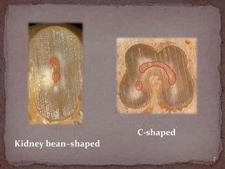
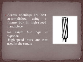
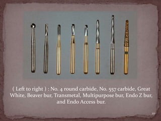
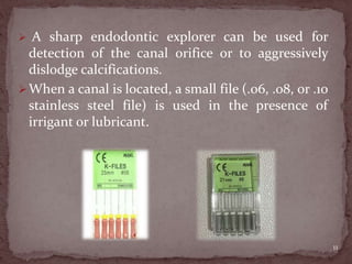
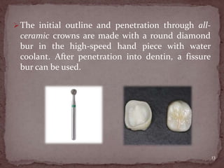
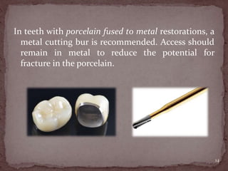
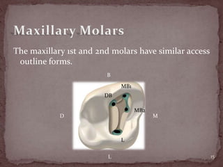
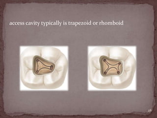
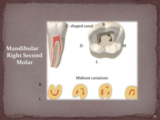
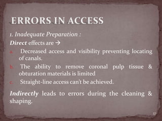
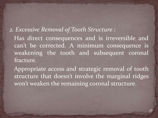
The major objectives of the access openings include :
1. locating all canals
2. unimpeded straight-line access of the instruments
in the canals to the apical one third or first curve
3. removal of the chamber roof and all coronal pulp
tissue
4. conservation of tooth structure.
2
1. Outline form:
is the recommended shape for access of a normal
tooth.
The outline form is a projection of the internal tooth
anatomy onto the external root structure.
2. Convenience form:
allows modification of the ideal outline form to
facilitate unstrained instrument placement and
manipulation.
3
3. Caries removal :
essential for several reasons -->
1st : permits an aseptic environment before entering
the pulp chamber and radicular space.
2nd : allows assessment of restorability.
3rd : provides sound tooth structure for adequate
provisional restoration.
4. Toilet of the cavity :
involves preventing materials and objects from
entering the chamber and canal space.
4
Round Ribbon-shaped (hourglass)
5
Ovoid Bowling pin
6
Kidney bean–shaped
C-shaped
7
In difficult cases the access can be prepared
without the rubber dam in place.
Care must be taken to prevent tooth structure or
restorative materials from entering the radicular
portion.
Before beginning the access the preoperative
radiographs should be assessed to determine the
degree of case difficulty.
8
Access openings are best
accomplished using a
fissure bur in high-speed
hand piece.
9
No single bur type is
superior.
High-speed burs are not
used in the canals.
10
( Left to right ) : No. 4 round carbide, No. 557 carbide, Great
White, Beaver bur, Transmetal, Multipurpose bur, Endo Z bur,
and Endo Access bur.
 A sharp endodontic explorer can be used for
detection of the canal orifice or to aggressively
dislodge calcifications.
When a canal is located, a small file (.06, .08, or .10
stainless steel file) is used in the presence of
irrigant or lubricant.
11
Crowns and fixed partial dentures, may have
changed the coronal landmarks used in canal
location.
Class V restorations may have induced coronal
calcification.
It’s best to remove restorative materials that
interfere with visualization before initiating root
canal treatment.
12
The initial outline and penetration through all-
ceramic crowns are made with a round diamond
bur in the high-speed hand piece with water
coolant. After penetration into dentin, a fissure
bur can be used.
13
In teeth with porcelain fused to metal restorations, a
metal cutting bur is recommended. Access should
remain in metal to reduce the potential for
fracture in the porcelain.
14
The maxillary 1st and 2nd molars have similar access
outline forms.
15
MB1
DB
L
MB2
B
L
MD
The initial movement
of the MB2 canal
from the chamber is
often not toward the
apex but laterally
toward the mesial.
16
Maxillary 1st Molar :
• Erupt: 6-7 Y, calcfic.: 9-10 Y
• Largest tooth volume &
most complex pulp
anatomy
• Chamber wide BL
• Chamber cervical outline
 rhomboid
• P orifice  centered
palatally
• Line connecting 3 main
orifices  Molar Triangle
17
• P root  longest, easiest access
 1, 2 or 3 canals
 curves B at apical 1/3
 type I (100%)
 DB root  conical
 1 or 2 canals
 canal oval then rounded
 type I (100%)
 MB root  1 (oval), 2 or 3 canals ( circular )
 conconavity on D wall .. Thin wall
 type I (45%), type II (37%), type IV (18%) 18
Maxillary 2nd Molar :
• Erupt: 11-13 Y, calcfic.: 14-16 Y
• 3 roots are grouped closer & sometimes fused.
• shorter than roots of 1st molar and not as curved.
• usually has one canal in each root
• 4 canals are less likely to be present
• floor of pulp chamber is convex, which gives the canal
orifices a slight funnel shape.
• When two roots are present, each root may have 1
canal, or B root may have 2 canals that join before
reaching a single foramen.. 2 P roots & 2 P canals occur
in 1.47% of these teeth.
19
• MB canal orifice is located
more to B & M than in 1st
molar;
** type I (71%), type II (17%),
type IV (12%)
• DB orifice approaches
midpoint between MB & P
orifices;
** type I (100%)
• P orifice usually is located at
most palatal aspect of root.
** type I (100%)
** canal orifices in maxillary 2nd
molar are closer mesially
20
Maxillary 3rd Molar :
 Erupt: 17-22 Y, calcific.: 18-25 Y
 root anatomy varies greatly.. can have 1-4 roots & 1-6
canals, C-shaped canals also can occur.
 tooth may be tipped to D, B, or both, which creates
greater access problem.
 Because it typically has 1-3 canals,, the access
preparation can be anything from an oval that is
widest in BL dimension to a rounded triangle.
21
22
D M
3 canals : 40% 4 canals : 60%
Mesial
Midroot
Distal
Single
foramen :
80%
two
foramin
a : 20%
Mesiobuccal
2 canals : 60%
Maxillary
Right First
Molar :
23
3 Roots: 60%
2 mesiobuccal
canals : 38%
Mesial Midroot
Distal
Single foramen : 15%
2 foramina : 10%
Maxillary
Right
Second
Molar
24
2 roots:
25%
L
B
Maxillary
Right Second
Molar
Facial
Facial
Mesial
Midroot
Midroot
L
B
1 root : 10%
Mandibular 1st molar configuration is 2 canals in the mesial
root, although 3 have been reported, & 1 canal in the distal
root.
25
The presence of 2 canals in the
distal root is 30% - 35%. MB DB
ML D
The most common configuration
for the mandibular 2nd molar is 2
canals in the mesial root & 1 canal
in the distal root. The incidence of
4 canals is low.
Mandibular 1st Molar :
• erupt: 6 Y, calcific.: 9-10 Y
• most often requires an endodontic procedure
• It often is extensively restored, & is subjected to heavy
occlusal stress.. Pulp chamber frequently is calcified.
• 2-3 canals in M root ( MB & ML ) & 1, 2 or 3 canals in
D root ( DB, DL, MD ).
• middle mesial (MM) canal sometimes is present in
develpmental groove between other M canals, but it
may only represent a wide anastomosis between 2 M
canals. ( incidence: 1% - 15%)
26
• Orifices to all canals are located in M 2/3 of the crown
• chamber floor is roughly trapezoid or rhomboid.
• M root, the wider.. canals usually are curved, with the
greatest curvature in MB canal
• presence of 2 separate D roots is rare, but if, DL root is
smaller than DB root & usually more curved.
• If 2 canals (DB & DL) are present in D root, they are
more round than oval for their entire length.
• Multiple accessory foramina may be located in
furcation of the mandibular molars.
27
access cavity typically is trapezoid or rhomboid
28
2/3 of 1st mandibular molars found in a Chinese
population had an extra DL root of a type 1 canal
configuration.. occurred in 4% of a Kuwaiti
population.
M  type I (12%), type II (28%), type IV (43%), type V
(8%), type VI (10%), type VIII (1%)
D  type I (70%), type II (15%), type IV (5%), type V
(8%), type VI (2%)
29
Mandibular 2nd Molar :
• Erupt: 11-13 Y, calcific.: 14-15 Y
• identified by the proximity of its roots, the two roots
often sweep distally in a gradual curve.
• pulp chamber and canal orifices not as large as 1st
molar
M  type I (27%), type II (38%), type IV (26%), type V
(9%)
D  type I (92%), type II ( 3%),type IV (4%), type V
(1%)
30
In mandibular 2nd molars with single or fused roots, a
file placed in the MB canal may appear to be in D
canal. This happens because 2 canals sometimes are
connected by a semicircular slit, a variation of the C-
shaped canal.
31
• When 3 canals are present, the access cavity is similar
to mandibular 1st molar.
• The access cavity for a 2 canal 2nd molar is rectangular,
wide MD and narrow BL. The access cavity for a single-
canal mandibular 2nd molar is oval.
32
Mandibular 3rd Molar :
• Erupt: 17-21 Y, calcific.: 18-25 Y
• Fused short, severely curved, or malformed roots
• may have 1-4 roots & 1-6 canals
• C-shaped canals also can occur
• When 3 or more canals are present, a rounded triangle
or rhomboid shaped access is typical.
• When2 canals are present, a rectangular shape is used.
For single-canal molars, an oval shape is customary
33
34
Mandibular
Right First
Molar
M D
Mesial
60%
B
Midroot
Distal
70%
40%
20%
10%
B
L
35
Mandibular
Right Second
Molar
M D
Mesial
40%
B
L
Distal
92%
5%35%
25% 3%
36
Mandibular
Right Second
Molar
MD
B
L
C- shaped canal
B
L
Midroot variations
1. Inadequate Preparation :
Direct effects are 
a. Decreased access and visibility preventing locating
of canals.
b. The ability to remove coronal pulp tissue &
obturation materials is limited
c. Straight-line access can’t be achieved.
37
Indirectly leads to errors during the cleaning &
shaping.
2. Excessive Removal of Tooth Structure :
Has direct consequences and is irreversible and
can’t be corrected. A minimum consequence is
weakening the tooth and subsequent coronal
fracture.
Appropriate access and strategic removal of tooth
structure that doesn’t involve the marginal ridges
won’t weaken the remaining coronal structure.
38
39
40
1. Endodontics - Principles & Practice -
Saunders; 4th Ed.
2. Cohen’s Pathways of the Pulp, 11th Ed.
41
Submit to : Dr. David
Done By : Ola Qatu
42

Molars

  • 2.
    The major objectivesof the access openings include : 1. locating all canals 2. unimpeded straight-line access of the instruments in the canals to the apical one third or first curve 3. removal of the chamber roof and all coronal pulp tissue 4. conservation of tooth structure. 2
  • 3.
    1. Outline form: isthe recommended shape for access of a normal tooth. The outline form is a projection of the internal tooth anatomy onto the external root structure. 2. Convenience form: allows modification of the ideal outline form to facilitate unstrained instrument placement and manipulation. 3
  • 4.
    3. Caries removal: essential for several reasons --> 1st : permits an aseptic environment before entering the pulp chamber and radicular space. 2nd : allows assessment of restorability. 3rd : provides sound tooth structure for adequate provisional restoration. 4. Toilet of the cavity : involves preventing materials and objects from entering the chamber and canal space. 4
  • 5.
  • 6.
  • 7.
  • 8.
    In difficult casesthe access can be prepared without the rubber dam in place. Care must be taken to prevent tooth structure or restorative materials from entering the radicular portion. Before beginning the access the preoperative radiographs should be assessed to determine the degree of case difficulty. 8
  • 9.
    Access openings arebest accomplished using a fissure bur in high-speed hand piece. 9 No single bur type is superior. High-speed burs are not used in the canals.
  • 10.
    10 ( Left toright ) : No. 4 round carbide, No. 557 carbide, Great White, Beaver bur, Transmetal, Multipurpose bur, Endo Z bur, and Endo Access bur.
  • 11.
     A sharpendodontic explorer can be used for detection of the canal orifice or to aggressively dislodge calcifications. When a canal is located, a small file (.06, .08, or .10 stainless steel file) is used in the presence of irrigant or lubricant. 11
  • 12.
    Crowns and fixedpartial dentures, may have changed the coronal landmarks used in canal location. Class V restorations may have induced coronal calcification. It’s best to remove restorative materials that interfere with visualization before initiating root canal treatment. 12
  • 13.
    The initial outlineand penetration through all- ceramic crowns are made with a round diamond bur in the high-speed hand piece with water coolant. After penetration into dentin, a fissure bur can be used. 13
  • 14.
    In teeth withporcelain fused to metal restorations, a metal cutting bur is recommended. Access should remain in metal to reduce the potential for fracture in the porcelain. 14
  • 15.
    The maxillary 1stand 2nd molars have similar access outline forms. 15 MB1 DB L MB2 B L MD
  • 16.
    The initial movement ofthe MB2 canal from the chamber is often not toward the apex but laterally toward the mesial. 16
  • 17.
    Maxillary 1st Molar: • Erupt: 6-7 Y, calcfic.: 9-10 Y • Largest tooth volume & most complex pulp anatomy • Chamber wide BL • Chamber cervical outline  rhomboid • P orifice  centered palatally • Line connecting 3 main orifices  Molar Triangle 17
  • 18.
    • P root longest, easiest access  1, 2 or 3 canals  curves B at apical 1/3  type I (100%)  DB root  conical  1 or 2 canals  canal oval then rounded  type I (100%)  MB root  1 (oval), 2 or 3 canals ( circular )  conconavity on D wall .. Thin wall  type I (45%), type II (37%), type IV (18%) 18
  • 19.
    Maxillary 2nd Molar: • Erupt: 11-13 Y, calcfic.: 14-16 Y • 3 roots are grouped closer & sometimes fused. • shorter than roots of 1st molar and not as curved. • usually has one canal in each root • 4 canals are less likely to be present • floor of pulp chamber is convex, which gives the canal orifices a slight funnel shape. • When two roots are present, each root may have 1 canal, or B root may have 2 canals that join before reaching a single foramen.. 2 P roots & 2 P canals occur in 1.47% of these teeth. 19
  • 20.
    • MB canalorifice is located more to B & M than in 1st molar; ** type I (71%), type II (17%), type IV (12%) • DB orifice approaches midpoint between MB & P orifices; ** type I (100%) • P orifice usually is located at most palatal aspect of root. ** type I (100%) ** canal orifices in maxillary 2nd molar are closer mesially 20
  • 21.
    Maxillary 3rd Molar:  Erupt: 17-22 Y, calcific.: 18-25 Y  root anatomy varies greatly.. can have 1-4 roots & 1-6 canals, C-shaped canals also can occur.  tooth may be tipped to D, B, or both, which creates greater access problem.  Because it typically has 1-3 canals,, the access preparation can be anything from an oval that is widest in BL dimension to a rounded triangle. 21
  • 22.
    22 D M 3 canals: 40% 4 canals : 60% Mesial Midroot Distal Single foramen : 80% two foramin a : 20% Mesiobuccal 2 canals : 60% Maxillary Right First Molar :
  • 23.
    23 3 Roots: 60% 2mesiobuccal canals : 38% Mesial Midroot Distal Single foramen : 15% 2 foramina : 10% Maxillary Right Second Molar
  • 24.
  • 25.
    Mandibular 1st molarconfiguration is 2 canals in the mesial root, although 3 have been reported, & 1 canal in the distal root. 25 The presence of 2 canals in the distal root is 30% - 35%. MB DB ML D The most common configuration for the mandibular 2nd molar is 2 canals in the mesial root & 1 canal in the distal root. The incidence of 4 canals is low.
  • 26.
    Mandibular 1st Molar: • erupt: 6 Y, calcific.: 9-10 Y • most often requires an endodontic procedure • It often is extensively restored, & is subjected to heavy occlusal stress.. Pulp chamber frequently is calcified. • 2-3 canals in M root ( MB & ML ) & 1, 2 or 3 canals in D root ( DB, DL, MD ). • middle mesial (MM) canal sometimes is present in develpmental groove between other M canals, but it may only represent a wide anastomosis between 2 M canals. ( incidence: 1% - 15%) 26
  • 27.
    • Orifices toall canals are located in M 2/3 of the crown • chamber floor is roughly trapezoid or rhomboid. • M root, the wider.. canals usually are curved, with the greatest curvature in MB canal • presence of 2 separate D roots is rare, but if, DL root is smaller than DB root & usually more curved. • If 2 canals (DB & DL) are present in D root, they are more round than oval for their entire length. • Multiple accessory foramina may be located in furcation of the mandibular molars. 27
  • 28.
    access cavity typicallyis trapezoid or rhomboid 28
  • 29.
    2/3 of 1stmandibular molars found in a Chinese population had an extra DL root of a type 1 canal configuration.. occurred in 4% of a Kuwaiti population. M  type I (12%), type II (28%), type IV (43%), type V (8%), type VI (10%), type VIII (1%) D  type I (70%), type II (15%), type IV (5%), type V (8%), type VI (2%) 29
  • 30.
    Mandibular 2nd Molar: • Erupt: 11-13 Y, calcific.: 14-15 Y • identified by the proximity of its roots, the two roots often sweep distally in a gradual curve. • pulp chamber and canal orifices not as large as 1st molar M  type I (27%), type II (38%), type IV (26%), type V (9%) D  type I (92%), type II ( 3%),type IV (4%), type V (1%) 30
  • 31.
    In mandibular 2ndmolars with single or fused roots, a file placed in the MB canal may appear to be in D canal. This happens because 2 canals sometimes are connected by a semicircular slit, a variation of the C- shaped canal. 31
  • 32.
    • When 3canals are present, the access cavity is similar to mandibular 1st molar. • The access cavity for a 2 canal 2nd molar is rectangular, wide MD and narrow BL. The access cavity for a single- canal mandibular 2nd molar is oval. 32
  • 33.
    Mandibular 3rd Molar: • Erupt: 17-21 Y, calcific.: 18-25 Y • Fused short, severely curved, or malformed roots • may have 1-4 roots & 1-6 canals • C-shaped canals also can occur • When 3 or more canals are present, a rounded triangle or rhomboid shaped access is typical. • When2 canals are present, a rectangular shape is used. For single-canal molars, an oval shape is customary 33
  • 34.
  • 35.
  • 36.
  • 37.
    1. Inadequate Preparation: Direct effects are  a. Decreased access and visibility preventing locating of canals. b. The ability to remove coronal pulp tissue & obturation materials is limited c. Straight-line access can’t be achieved. 37 Indirectly leads to errors during the cleaning & shaping.
  • 38.
    2. Excessive Removalof Tooth Structure : Has direct consequences and is irreversible and can’t be corrected. A minimum consequence is weakening the tooth and subsequent coronal fracture. Appropriate access and strategic removal of tooth structure that doesn’t involve the marginal ridges won’t weaken the remaining coronal structure. 38
  • 39.
  • 40.
  • 41.
    1. Endodontics -Principles & Practice - Saunders; 4th Ed. 2. Cohen’s Pathways of the Pulp, 11th Ed. 41
  • 42.
    Submit to :Dr. David Done By : Ola Qatu 42