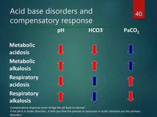
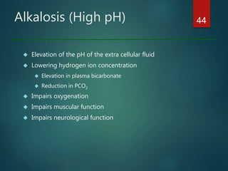
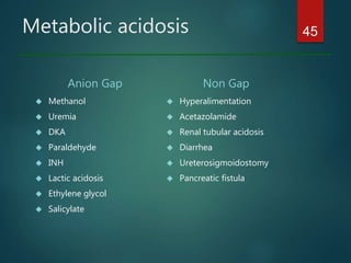
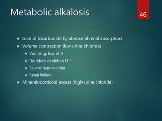
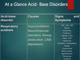
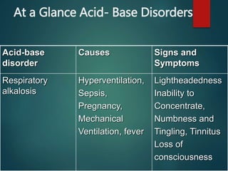
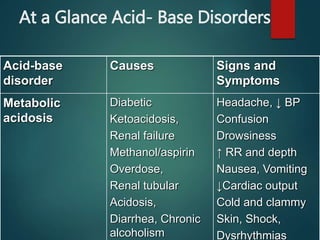
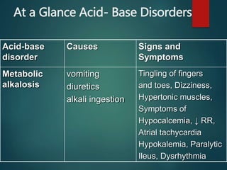
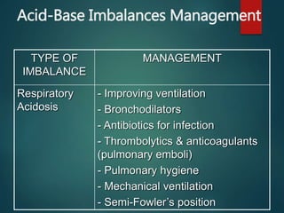
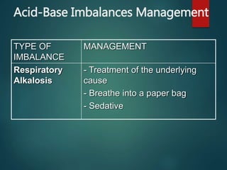
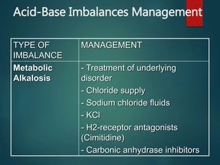
"The body maintains a balance of acids and bases in order to constantly maintain blood pH within a narrow range, despite the continuous generation of metabolic products. In turn, this allows the body to maintain cell enzyme systems in good operation conditions, together with the proper concentration of ionized (active) forms of various electrolytes such as Ca and Mg . This influences the speed of metabolic reactions and trans-membrane transportation systems (pharmacokinetics and pharmacodynamics)." - Luis Núñez Ochoa, Facultad de Medicina Veterinaria y Zootecnia, Unam, Mexico
Acid and base Balance by Dr. Tehmas (Part 1)Tehmas Ahmad
Lecture of Biochemistry about Acid and Base Balance and Imbalance. 1st of 2 Lectures, delivered to students of 2nd professional MBBS students of Bannu Medical College, Bannu
09.12.08(b): An Introduction to Blood Gas Analysis Open.Michigan
Slideshow is from the University of Michigan Medical School’s M2 Respiratory sequence
View additional course materials on Open.Michigan:
openmi.ch/med-M2Resp
Acid and base Balance by Dr. Tehmas (Part 2)Tehmas Ahmad
Lecture of Biochemistry about Acid and Base Balance and Imbalance. 2nd of 2 Lectures, delivered to students of 2nd professional MBBS students of Bannu Medical College, Bannu.
تم تحميل هذا الملف من
منتديات تمريض مستشفى غزة الاوروبي
http://egh-nsg.forumpalestine.com/
لتحميل اجمل واروع المحاضرات فقط قم بزيارتنا وسوف تكون من الاوائل
مع تحيات المدير العام
علاء شعت
An arterial-blood gas test measures the amounts of arterial gases, such as oxygen and carbon dioxide. An ABG test requires that a small volume of blood be drawn from the radial artery with a syringe and a thin needle, but sometimes the femoral artery in the groin or another site is used.
The common indications for ABGs are:
Respiratory compromise, which leads to hypoxia or diminished ventilation.
Peri- or postcardiopulmonary arrest or collapse.
Medical conditions that cause significant metabolic derangement, such as sepsis, diabetic ketoacidosis, renal failure, heart failure, toxic substance ingestion, drug overdose, trauma, or burns.
Evaluating the effectiveness of therapies, monitoring the patient's clinical status, and determining treatment needs. For instance, clinicians often titrate oxygenation therapy, adjust the level of ventilator support, and make decisions about fluid and electrolyte therapy based on ABG results.
During the perioperative phase of major surgeries, which includes the preoperative, intraoperative, and postoperative care of the patient.
The components of an ABG analysis are PaO2, SaO2, hydrogen ion concentration (pH), PaCO2, HCO3-, base excess, and serum levels of hemoglobin, lactate, glucose, and electrolytes (sodium, potassium, calcium, and chloride).
Ozempic: Preoperative Management of Patients on GLP-1 Receptor Agonists Saeid Safari
Preoperative Management of Patients on GLP-1 Receptor Agonists like Ozempic and Semiglutide
ASA GUIDELINE
NYSORA Guideline
2 Case Reports of Gastric Ultrasound
Acute scrotum is a general term referring to an emergency condition affecting the contents or the wall of the scrotum.
There are a number of conditions that present acutely, predominantly with pain and/or swelling
A careful and detailed history and examination, and in some cases, investigations allow differentiation between these diagnoses. A prompt diagnosis is essential as the patient may require urgent surgical intervention
Testicular torsion refers to twisting of the spermatic cord, causing ischaemia of the testicle.
Testicular torsion results from inadequate fixation of the testis to the tunica vaginalis producing ischemia from reduced arterial inflow and venous outflow obstruction.
The prevalence of testicular torsion in adult patients hospitalized with acute scrotal pain is approximately 25 to 50 percent
Tom Selleck Health: A Comprehensive Look at the Iconic Actor’s Wellness Journeygreendigital
Tom Selleck, an enduring figure in Hollywood. has captivated audiences for decades with his rugged charm, iconic moustache. and memorable roles in television and film. From his breakout role as Thomas Magnum in Magnum P.I. to his current portrayal of Frank Reagan in Blue Bloods. Selleck's career has spanned over 50 years. But beyond his professional achievements. fans have often been curious about Tom Selleck Health. especially as he has aged in the public eye.
Follow us on: Pinterest
Introduction
Many have been interested in Tom Selleck health. not only because of his enduring presence on screen but also because of the challenges. and lifestyle choices he has faced and made over the years. This article delves into the various aspects of Tom Selleck health. exploring his fitness regimen, diet, mental health. and the challenges he has encountered as he ages. We'll look at how he maintains his well-being. the health issues he has faced, and his approach to ageing .
Early Life and Career
Childhood and Athletic Beginnings
Tom Selleck was born on January 29, 1945, in Detroit, Michigan, and grew up in Sherman Oaks, California. From an early age, he was involved in sports, particularly basketball. which played a significant role in his physical development. His athletic pursuits continued into college. where he attended the University of Southern California (USC) on a basketball scholarship. This early involvement in sports laid a strong foundation for his physical health and disciplined lifestyle.
Transition to Acting
Selleck's transition from an athlete to an actor came with its physical demands. His first significant role in "Magnum P.I." required him to perform various stunts and maintain a fit appearance. This role, which he played from 1980 to 1988. necessitated a rigorous fitness routine to meet the show's demands. setting the stage for his long-term commitment to health and wellness.
Fitness Regimen
Workout Routine
Tom Selleck health and fitness regimen has evolved. adapting to his changing roles and age. During his "Magnum, P.I." days. Selleck's workouts were intense and focused on building and maintaining muscle mass. His routine included weightlifting, cardiovascular exercises. and specific training for the stunts he performed on the show.
Selleck adjusted his fitness routine as he aged to suit his body's needs. Today, his workouts focus on maintaining flexibility, strength, and cardiovascular health. He incorporates low-impact exercises such as swimming, walking, and light weightlifting. This balanced approach helps him stay fit without putting undue strain on his joints and muscles.
Importance of Flexibility and Mobility
In recent years, Selleck has emphasized the importance of flexibility and mobility in his fitness regimen. Understanding the natural decline in muscle mass and joint flexibility with age. he includes stretching and yoga in his routine. These practices help prevent injuries, improve posture, and maintain mobilit
- Video recording of this lecture in English language: https://youtu.be/lK81BzxMqdo
- Video recording of this lecture in Arabic language: https://youtu.be/Ve4P0COk9OI
- Link to download the book free: https://nephrotube.blogspot.com/p/nephrotube-nephrology-books.html
- Link to NephroTube website: www.NephroTube.com
- Link to NephroTube social media accounts: https://nephrotube.blogspot.com/p/join-nephrotube-on-social-media.html
Explore natural remedies for syphilis treatment in Singapore. Discover alternative therapies, herbal remedies, and lifestyle changes that may complement conventional treatments. Learn about holistic approaches to managing syphilis symptoms and supporting overall health.
ARTIFICIAL INTELLIGENCE IN HEALTHCARE.pdfAnujkumaranit
Artificial intelligence (AI) refers to the simulation of human intelligence processes by machines, especially computer systems. It encompasses tasks such as learning, reasoning, problem-solving, perception, and language understanding. AI technologies are revolutionizing various fields, from healthcare to finance, by enabling machines to perform tasks that typically require human intelligence.
These lecture slides, by Dr Sidra Arshad, offer a quick overview of physiological basis of a normal electrocardiogram.
Learning objectives:
1. Define an electrocardiogram (ECG) and electrocardiography
2. Describe how dipoles generated by the heart produce the waveforms of the ECG
3. Describe the components of a normal electrocardiogram of a typical bipolar leads (limb II)
4. Differentiate between intervals and segments
5. Enlist some common indications for obtaining an ECG
Study Resources:
1. Chapter 11, Guyton and Hall Textbook of Medical Physiology, 14th edition
2. Chapter 9, Human Physiology - From Cells to Systems, Lauralee Sherwood, 9th edition
3. Chapter 29, Ganong’s Review of Medical Physiology, 26th edition
4. Electrocardiogram, StatPearls - https://www.ncbi.nlm.nih.gov/books/NBK549803/
5. ECG in Medical Practice by ABM Abdullah, 4th edition
6. ECG Basics, http://www.nataliescasebook.com/tag/e-c-g-basics
These simplified slides by Dr. Sidra Arshad present an overview of the non-respiratory functions of the respiratory tract.
Learning objectives:
1. Enlist the non-respiratory functions of the respiratory tract
2. Briefly explain how these functions are carried out
3. Discuss the significance of dead space
4. Differentiate between minute ventilation and alveolar ventilation
5. Describe the cough and sneeze reflexes
Study Resources:
1. Chapter 39, Guyton and Hall Textbook of Medical Physiology, 14th edition
2. Chapter 34, Ganong’s Review of Medical Physiology, 26th edition
3. Chapter 17, Human Physiology by Lauralee Sherwood, 9th edition
4. Non-respiratory functions of the lungs https://academic.oup.com/bjaed/article/13/3/98/278874
Lung Cancer: Artificial Intelligence, Synergetics, Complex System Analysis, S...Oleg Kshivets
RESULTS: Overall life span (LS) was 2252.1±1742.5 days and cumulative 5-year survival (5YS) reached 73.2%, 10 years – 64.8%, 20 years – 42.5%. 513 LCP lived more than 5 years (LS=3124.6±1525.6 days), 148 LCP – more than 10 years (LS=5054.4±1504.1 days).199 LCP died because of LC (LS=562.7±374.5 days). 5YS of LCP after bi/lobectomies was significantly superior in comparison with LCP after pneumonectomies (78.1% vs.63.7%, P=0.00001 by log-rank test). AT significantly improved 5YS (66.3% vs. 34.8%) (P=0.00000 by log-rank test) only for LCP with N1-2. Cox modeling displayed that 5YS of LCP significantly depended on: phase transition (PT) early-invasive LC in terms of synergetics, PT N0—N12, cell ratio factors (ratio between cancer cells- CC and blood cells subpopulations), G1-3, histology, glucose, AT, blood cell circuit, prothrombin index, heparin tolerance, recalcification time (P=0.000-0.038). Neural networks, genetic algorithm selection and bootstrap simulation revealed relationships between 5YS and PT early-invasive LC (rank=1), PT N0—N12 (rank=2), thrombocytes/CC (3), erythrocytes/CC (4), eosinophils/CC (5), healthy cells/CC (6), lymphocytes/CC (7), segmented neutrophils/CC (8), stick neutrophils/CC (9), monocytes/CC (10); leucocytes/CC (11). Correct prediction of 5YS was 100% by neural networks computing (area under ROC curve=1.0; error=0.0).
CONCLUSIONS: 5YS of LCP after radical procedures significantly depended on: 1) PT early-invasive cancer; 2) PT N0--N12; 3) cell ratio factors; 4) blood cell circuit; 5) biochemical factors; 6) hemostasis system; 7) AT; 8) LC characteristics; 9) LC cell dynamics; 10) surgery type: lobectomy/pneumonectomy; 11) anthropometric data. Optimal diagnosis and treatment strategies for LC are: 1) screening and early detection of LC; 2) availability of experienced thoracic surgeons because of complexity of radical procedures; 3) aggressive en block surgery and adequate lymph node dissection for completeness; 4) precise prediction; 5) adjuvant chemoimmunoradiotherapy for LCP with unfavorable prognosis.
New Directions in Targeted Therapeutic Approaches for Older Adults With Mantl...i3 Health
i3 Health is pleased to make the speaker slides from this activity available for use as a non-accredited self-study or teaching resource.
This slide deck presented by Dr. Kami Maddocks, Professor-Clinical in the Division of Hematology and
Associate Division Director for Ambulatory Operations
The Ohio State University Comprehensive Cancer Center, will provide insight into new directions in targeted therapeutic approaches for older adults with mantle cell lymphoma.
STATEMENT OF NEED
Mantle cell lymphoma (MCL) is a rare, aggressive B-cell non-Hodgkin lymphoma (NHL) accounting for 5% to 7% of all lymphomas. Its prognosis ranges from indolent disease that does not require treatment for years to very aggressive disease, which is associated with poor survival (Silkenstedt et al, 2021). Typically, MCL is diagnosed at advanced stage and in older patients who cannot tolerate intensive therapy (NCCN, 2022). Although recent advances have slightly increased remission rates, recurrence and relapse remain very common, leading to a median overall survival between 3 and 6 years (LLS, 2021). Though there are several effective options, progress is still needed towards establishing an accepted frontline approach for MCL (Castellino et al, 2022). Treatment selection and management of MCL are complicated by the heterogeneity of prognosis, advanced age and comorbidities of patients, and lack of an established standard approach for treatment, making it vital that clinicians be familiar with the latest research and advances in this area. In this activity chaired by Michael Wang, MD, Professor in the Department of Lymphoma & Myeloma at MD Anderson Cancer Center, expert faculty will discuss prognostic factors informing treatment, the promising results of recent trials in new therapeutic approaches, and the implications of treatment resistance in therapeutic selection for MCL.
Target Audience
Hematology/oncology fellows, attending faculty, and other health care professionals involved in the treatment of patients with mantle cell lymphoma (MCL).
Learning Objectives
1.) Identify clinical and biological prognostic factors that can guide treatment decision making for older adults with MCL
2.) Evaluate emerging data on targeted therapeutic approaches for treatment-naive and relapsed/refractory MCL and their applicability to older adults
3.) Assess mechanisms of resistance to targeted therapies for MCL and their implications for treatment selection
Couples presenting to the infertility clinic- Do they really have infertility...Sujoy Dasgupta
Dr Sujoy Dasgupta presented the study on "Couples presenting to the infertility clinic- Do they really have infertility? – The unexplored stories of non-consummation" in the 13th Congress of the Asia Pacific Initiative on Reproduction (ASPIRE 2024) at Manila on 24 May, 2024.
Triangles of Neck and Clinical Correlation by Dr. RIG.pptx
ABG Seminar for PNA Cebu
1. Acid-base disorders and its
implications: Therapy in a Nursing
Perspective
(July 23, 2014)
EMILIANO IAN B. SUSON II, ED.D, USRN, MAN, RN
“SIR EYE”
2. Objectives
Describe physiology involved in acid base balance of
the body
Review causes and treatments of acid base disorders
Identify normal arterial blood gas values
Interpret results of ABG samples
Interpret oxygenation state of a patient using reported
ABG values
2
3. Interpretation of ABG
» Very important for health care providers
» Usefulness of this tool depends on being able to
interpret correctly the results
» Critically ill patients
3
4. Acid base disorders
12000 to 15000 mEq of volatile acids are
produced daily by body and excreted as CO2
by lungs
1 mEq / kg / day of non-volatile acids
(sulfuric and phosphoric acids) are produced
daily by body and excreted by the kidneys
The most important buffers in the body are,
hemoglobin, plasma proteins and
bicarbonate
4
5. Acid base disorders
SIMPLE ACID BASE DISORDER:
when there is only one primary
disorder
MIXED ACID BASE DISORDER: when
there are two or more primary
disorders present at the same time
5
6. Normal values
pH 7.35-7.45 7.40
PaCO2 35-45 mmHg 40
PaO2 70-100 mmHg
HCO3- 24±2 24
Met-Hb <2%
CO-Hb <3%
BE -2 to 2 mEq/L
CaO2 16-22 ml/dL
A gap 10±2 12
6
14. 14Protein Buffers
Includes hemoglobin, work in blood and ISF
Carboxyl group gives up H+
Amino Group accepts H+
Side chains that can buffer H+ are present on 27 amino acids.
The 10 essential amino acids are:(PVTMATHILL)
Phenylalanine, Valine, Tryptophan, Methionine, Arginine,
Threonine, Histidine, Isoleucine, Leucine, Lysine
10 Amino acids the body produces:
alanine, asparagine, aspartic acid, cysteine, glutamic acid,
glutamine, glycine, proline, serine and tyrosine
15. 152. Respiratory mechanisms
Exhalation of carbon dioxide
Powerful, but only works with volatile acids
Doesn’t affect fixed acids like lactic acid
CO2 + H20 ↔ H2CO3 ↔ H+ + HCO3
-
Body pH can be adjusted by changing rate and depth
of breathing
16. 163. Kidney excretion
Can eliminate large amounts of acid
Can also excrete base
Can conserve and produce bicarb ions
Most effective regulator of pH
If kidneys fail, pH balance fails
17. 17Rates of correction
Buffers function almost instantaneously
Respiratory mechanisms take several
minutes to hours
Renal mechanisms may take several
hours to days
21. 21Acidosis
Principal effect of acidosis is depression of the CNS through ↓ in synaptic
transmission.
Generalized weakness
Deranged CNS function the greatest threat
Severe acidosis causes
Disorientation, Coma, Death
22. 22
Alkalosis
Alkalosis causes over excitability of the central
and peripheral nervous systems.
Numbness
Lightheadedness
It can cause :
Nervousness
muscle spasms or tetany
Convulsions
Loss of consciousness
Death
25. 25Signs and Symptoms of
Respiratory Acidosis
Breathlessness
Restlessness
Lethargy and disorientation
Tremors, convulsions, coma
Respiratory rate rapid, then gradually depressed
Skin warm and flushed due to vasodilation caused by
excess CO2
29. 29Treatment of Respiratory
Alkalosis
Treat underlying cause
Breathe into a paper bag
IV Chloride containing solution – Cl- ions
replace lost bicarbonate ions
30. 30Metabolic Acidosis
Bicarbonate deficit - blood concentrations of bicarb drop below 22mEq/L
Causes:
Loss of bicarbonate through diarrhea or renal dysfunction
Accumulation of acids (lactic acid or ketones)
Failure of kidneys to excrete H+
32. 32Compensation for Metabolic
Acidosis
Increased ventilation
Renal excretion of hydrogen ions if
possible
K+ exchanges with excess H+ in ECF
( H+ into cells, K+ out of cells)
34. 34Metabolic Alkalosis
Bicarbonate excess - concentration in blood is greater than
26 mEq/L
Causes:
Excess vomiting = loss of stomach acid
Excessive use of alkaline drugs
Certain diuretics
Endocrine disorders
Heavy ingestion of antacids
Severe dehydration
35. 35Symptoms of Metabolic
Alkalosis
Respiration slow and shallow
Hyperactive reflexes ; tetany
Often related to depletion of electrolytes
Atrial tachycardia
Dysrhythmias
37. 37Example
A patient is in intensive care because he suffered a
severe myocardial infarction 3 days ago. The lab
reports the following values from an arterial blood
sample:
pH 7.3
HCO3- = 20 mEq / L ( 22 - 26)
pCO2 = 32 mm Hg (35 - 45)
39. Acid base disorders
METABOLIC ACIDOSIS: HCO3 <24 OR Anion Gap
>12
METABOLIC ALKALOSIS: HCO3 >24
RESPIRATORY ALKALOSIS: PCO2 <40 or PCO2 less
than expected for primary metabolic abnormality
RESPIRATORY ACIDOSIS: PCO2 >40 or PCO2 higher
than expected for primary metabolic abnormality
HIGH ANION GAP (>12-20) always indicates primary
metabolic acidosis
39
40. Acid base disorders and
compensatory response
pH HCO3- PaCO2
Metabolic
acidosis
Metabolic
alkalosis
Respiratory
acidosis
Respiratory
alkalosis
Compensatory response never brings the pH back to normal
if the pH is in acidic direction, it tells you that the process or processes in acidic direction are the primary
disorders
40
41. Anion Gap
Na-(Cl+HCO3)= 12±2
Estimates unmeasured anions
Normal is 12
Hypoalbuminemia:
Correct anion gap: 2.5 per gram of albumin below 4
Calculate osmolal gap if anion gap is elevated
OSM gap = measured OSM-2(Na)-glu/18-BUN/2.8 =
<10
41
42. Urinary anion gap
Useful in differential diagnosis of non gap
acidosis
U anion gap= Na + K – Cl
A negative U. Anion Gap ie Cl >> Na + K
suggests appropriate urinary NH4
excretion and G.I. loss of HCO3
A positive U. Anion Gap ie. Cl << Na + K
suggests RTA with distal acidification
defect and inadequate NH4 excretion in
urine
42
43. Acidosis (Low pH)
Lowering extracellular pH by rising concentration of
hydrogen ions
Fall in bicarbonate concentration
Elevation in PCO2
Decreases force of cardiac contractions
Decreases vascular response to catecholamines
Decreases response of certain medications
43
44. Alkalosis (High pH)
Elevation of the pH of the extra cellular fluid
Lowering hydrogen ion concentration
Elevation in plasma bicarbonate
Reduction in PCO2
Impairs oxygenation
Impairs muscular function
Impairs neurological function
44
51. Acid-Base Imbalances Management
TYPE OF
IMBALANCE
MANAGEMENT
Respiratory
Acidosis
- Improving ventilation
- Bronchodilators
- Antibiotics for infection
- Thrombolytics & anticoagulants
(pulmonary emboli)
- Pulmonary hygiene
- Mechanical ventilation
- Semi-Fowler’s position
52. Acid-Base Imbalances Management
TYPE OF
IMBALANCE
MANAGEMENT
Respiratory
Alkalosis
- Treatment of the underlying
cause
- Breathe into a paper bag
- Sedative
53. Acid-Base Imbalances Management
TYPE OF
IMBALANCE
MANAGEMENT
Metabolic
Acidosis
- Treatment is correcting the
underlying defect.
- Eliminating source of chloride
- Bicarbonate
- Alkalizing agents
- Hemodialysis
- Peritoneal dialysis
58. 58
Hemoglobin – this is the part of your red blood cell which carries
oxygen all over the body. If your hemoglobin levels are decreased it
could pertain to anemia, bleeding, patients with chronic kidney
disease, or a possible blood dyscrasia. If your hemoglobin levels
are elevated we could consider polycythemia vera and dehydration.
Red Blood Cell (RBC) – this part of the CBC tells you the number
of cells that could carry oxygen in the body. Same with hemoglobin,
decrease levels of RBC could also be secondary to anemia or
bleeding and increase levels could also be secondary to
polycythemia or dehydration.
Hematocrit – this measures the amount of space in the blood
being occupied by your red blood cells. Causes for the increased
and decreased of hematocrit are the same with your hemoglobin
and red blood cells.
59. 59
White Blood Cell (WBC) – this are the cells in the body that fight
off invaders like infections. An increase or decrease in WBC count
could represent an ongoing infection or a malignancy like your
leukemia. Also included in the CBC is the 5 differential count for
your WBC, namely:
Neutrophils or segmenters – this type of WBC are the primary
cells that respond to a bacterial infection. High levels of your
neutrophils usually represent and ongoing infection, an
inflammation, malignancy, cause by some drugs, etc. Low levels of
your neutrophils could be seen in patients with viral infection,
autoimmune diseases, some medications and malignancy.
Lymphocytes – this type of WBC represent 20-40% of your
circulating WBC in the blood. An increased in lymphocyte count
usually represents an acute infection especially viral infections,
leukemia, smoking, etc. Low lymphocyte count is usually not
significant.
60. 60
Monocytes – this comprises 3-8% of all white blood cells in the
body. An increase in monocyte could signify a chronic infection like
your tuberculosis or a chronic inflammation condition like your
inflammatory bowel disease and malignancy. Low levels of
monocytes are usually none significant if other cells are normal.
Basophils – this comprises only 0.01-0.3% of all white blood cells
in the blood. This type could produce histamine. Increased
numbers could represent a myeloproliferative disorder.
Eosinophils – comprises 1 – 6% of all white blood cells in the
blood stream. They are usually increase in cases of allergy, asthma
and in parasitic infections. Low levels are usually not significant.
Platelet Count – the normal platelet count ranges from 150,000 –
400,000 /L and this cell is involved in the clotting cascade of the
body. Low levels of this cell could cause easy bruising and
bleeding. Causes of low platelet count include infections (ex:
dengue fever), autoimmune disease, liver disease, idiopathic
thrombocytopenic purpura, etc.
61. 61
Red Cell Indices – this are investigated when considering
diseases like your thalassemia or sickle cell anemia.
MCV (mean corpuscle volume) – telling you the average size of
the red blood cell (80-100)
MCH (mean corpuscle hemoglobin) – shows the average
amount of hemoglobin in each red blood cell (26-34)
MCHC (mean corpuscle hemoglobin concentration) – average
amount of hemoglobin in the red blood cell compared to their
average size. (31-37)
RDW – (11.5 -14.5)
62. ABG SHORTCUT
Laboratory studies
ABG: pH/PaCO2/PaO2/HCO3/O2sat
ABG: 7.38-7.44/35-45/80-100/22-26/95-100
What is the acid base disturbance and what is the cause
Na
135-145
62
K
3.5-5.1
AG
7-16
CO2
22-30
BUN
5-25
Crea
0.6-1.2
Glu
70-110
Ca
8.5-10.1
Cl
98-107
66. Indications
To assess.
Respiratory Status
Assess oxygenation and ventilation
Acid Base Balance
Phlebotomy. Used if venous route is unavailable or
inaccessible due to trauma or burns. Usually a
femoral puncture, uncommon variation.
67. Contraindications
Overlying infection or burn at insertion site.
Absent collateral circulation.
Arteriovenous shunt. Often radial or brachial.
Severe atherosclerosis
Raynauds disease.
Coagulopathy.
68. Sites
Preferred radial or femoral arteries.
Less common. Dorsalis pedis and posterior tibial.
Avoid. Branches without collateral supply. Example is
the brachial artery.
69. Complications
Bleeding causing hematoma.
Arterial occlusion causing thrombus or
dissection.
Infection causing arteritis or cellulitis.
Embolization
Last 3 uncommon.
70. Normal Values
pH, 7.36 to 7.44. For acid base status of blood.
pCO2, 38 to 44 mmHg. Reflects ventillation.
pO2, 85 to 95 mmHg. Reflects oxygenation.
HCO3, 21 to 27 meq per litre. Key blood buffer.
Base excess, plus or minus 2 meq per litre
ABG quiz. http://www.vectors.cx/med/apps/abg.cgi
72. Initial Preparation
Wash hands
Gloves
Protective eye wear
Iodine swab. Povidone-iodine, betadine. Followed by
alcohol swab
Arterial blood gas sampling kit
2 x 2 cm gauze
Bag of ice. To store sample
73. Allens Test
Indicates collateral circulation to hand.
Radial artery on non dominant hand.
Palpate radial artery.
Simultaneouslys palpate ulnar artery, or as
close to that area as possible.
Patient makes a fist. Palpate both arteries
for10 seconds.
Release ulnar artery and witness blood flow
and pinking of the hand via collateral radial
artery
Radial artery is now a candidate for testing.
74. Set Up
Patient seated on stretcher
Rolled up towel under wrist. That hyperextends wrist,
bringing artery closer to surface.
Clean area in a cicular motion with iodine. Allow to dry.
Wipe away iodine with alcohol. While drying, open
sampling kit.
75. Sampling Kit
3 pieces
1. Orange air ball or cube. Used to expel excess air
from the syringe.
2. Black cap for syringe, used for transport.
3. 3 cc, cubic centimetres heparinised syringe. With
needle attached.
76. Sampling Kit Use
Pull back slightly on plunger, so once needle is in
artery, natural pulsations will fill the syringe.
Remove clear needle cap. Locate the bevel. Bevel is a
slanted opening on one side of the needle tip. We
want bevel facing upward, so you can see it.
77. Syringe Use
45 degrees, sharper angle.
Hold like a dart or pen.
Feeling pulse under non syringe finger is the only
landmark for orientation.
Before piercing skin, roll finger back slightly from
artery, so you dont stab yourself in the finger.
Flash of blood into hub of needle. Artery has been
accessed.
Blood will pulse into syringe. 1.5 to 2.0 cc
required.
Cover needle with gauze. Quickly remove needle.
78. After Care
Physician applies pressure to gauze for 5 minutes. 10
minutes if patient is on anticoaggulant therapy.
Optional to ask patient to do this instead.
79. Blood Care
Insert needle into orange air cube or ball. Want
bevel covered, dont want needle to go through
cube.
Push down on plunger to expell excess air. So it
doesnt affect results. Key point because we are
measuring air component levels in blood.
Remove cube and needle as one.
Attach black cap to syringe.
Roll test tube between hands, to ensure blood
heparinisation.
Place in iced bag. Send to lab.
Needle and cube to sharps container.
81. References
Bishop, M., Fody, E., & Schoeff, l. (2010). Clinical Chemistry:
Techniques, principles, Correlations. Baltimore: Wolters
Kluwer Lippincott Williams & Wilkins.
Carreiro-Lewandowski, E. (2008). Blood Gas Analysis and
Interpretation. Denver, Colorado: Colorado Association for
Continuing Medical Laboratory Education, Inc.
Sunheimer, R., & Graves, L. (2010). Clinical Laboratory
Chemistry. Upper Saddle River: Pearson .
81
82. Resources to learn more
www.acid-base.com
www.acidbasedisorders.com
Haber RJ. A practical approach to acid-base
disorders. West J Med 1991; 155: 146
www.postgradmed.com/issues/2000/03_00/fall.ht
m
http://medicine.ucsf.edu/housestaff/handbook/H
ospH2002_C5.htm