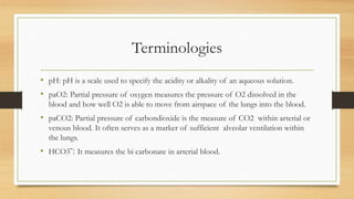
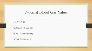
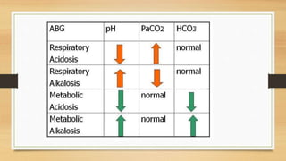
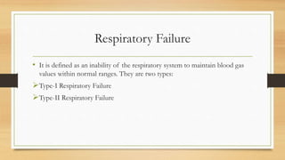
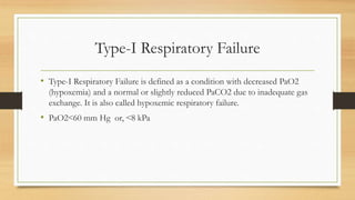
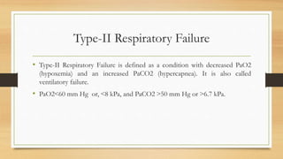
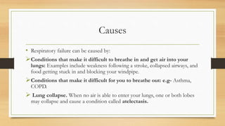
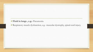
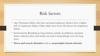
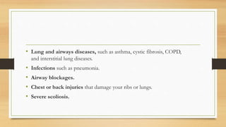
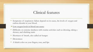
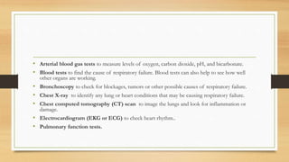
💚😋Kolkata Escort Service Call Girls, ₹5000 To 25K With AC💚😋
Abg and Respiratory failure
1. Arterial Blood Gas Analysis
&
Respiratory Failure
Dr. Shilpasree Saha (PT)
MPT- Cardio-Thoracic Disorders)
Assistant Professor, JVWU
2. What do it mean by ABG analysis?
• Arterial blood gas (ABG) analysis is an essential part of
diagnosing and managing a patient’s oxygenation status and acid–
base balance.
• Blood gas analysis is a commonly used diagnostic tool to evaluate
the partial pressures of gas in blood as well as acid-base content.
• Disorders of acid–base balance can create complications in many
disease states, and occasionally the abnormality may be so severe
so as to become a life-threatening risk factor.
•
3. Terminologies
• pH: pH is a scale used to specify the acidity or alkality of an aqueous solution.
• paO2: Partial pressure of oxygen measures the pressure of O2 dissolved in the
blood and how well O2 is able to move from airspace of the lungs into the blood.
• paCO2: Partial pressure of carbondioxide is the measure of CO2 within arterial or
venous blood. It often serves as a marker of sufficient alveolar ventilation within
the lungs.
• HCO3ˉ: It measures the bi carbonate in arterial blood.
4. Normal Blood Gas Value
• pH: 7.35-7.45
• PaCO2: 35-45 mm Hg
• PaO2: 75-100 mm Hg
• HCO3: 22-26 meq/L
5. Sample For ABG Analysis
• Site of choice:
The first choice is the radial artery.
Alternative sites for access are:
Brachial and Femoral artery.
6. Assessing acid-base disorders
• A low pH, ˂7.35 indicates a tendency towards acidosis, a high pH˃7.45
indicates a tendency towards alkalosis.
• Increase in paCO2 indicates acidosis, decrease in paCO2 indicates alkalosis.
• Increase HCO3ˉ leads to alkalosis, decrease HCO3ˉ leads to acidosis.
7. Assessment
• Establish whether the patient’s pH is acidotic, alkalotic or normal.
• If the pH is acidotic & PaCO2 increased, then it is RESPIRATORY ACIDOSIS.
• If the pH is acidotic & HCO3ˉ decreased, then it is METABOLIC ACIDOSIS.
• If the pH is alkalotic & PaCO2 decreased, then it is RESPIRATORY ALKALOSIS.
• If the pH is alkalotic & HCO3ˉ increased, then it is METABOLIC ALKALOSIS.
8.
9. Respiratory Failure
• It is defined as an inability of the respiratory system to maintain blood gas
values within normal ranges. They are two types:
Type-I Respiratory Failure
Type-II Respiratory Failure
10. Type-I Respiratory Failure
• Type-I Respiratory Failure is defined as a condition with decreased PaO2
(hypoxemia) and a normal or slightly reduced PaCO2 due to inadequate gas
exchange. It is also called hypoxemic respiratory failure.
• PaO2˂60 mm Hg or, ˂8 kPa
11. Type-II Respiratory Failure
• Type-II Respiratory Failure is defined as a condition with decreased PaO2
(hypoxemia) and an increased PaCO2 (hypercapnea). It is also called
ventilatory failure.
• PaO2˂60 mm Hg or, ˂8 kPa, and PaCO2 ˃50 mm Hg or ˃6.7 kPa.
12. Causes
• Respiratory failure can be caused by:
Conditions that make it difficult to breathe in and get air into your
lungs: Examples include weakness following a stroke, collapsed airways, and
food getting stuck in and blocking your windpipe.
Conditions that make it difficult for you to breathe out: e.g- Asthma,
COPD.
Lung collapse. When no air is able to enter your lungs, one or both lobes
may collapse and cause a condition called atelectasis.
14. Risk factors
• Age: Premature babies who have neonatal respiratory distress have a higher
risk of respiratory failure. Older adults have more risk factors for respiratory
failure.
• Environment: Breathing in lung irritants, include air pollution, chemical
fumes, asbestos, dust, and smoke can lead to lung damage over the long
term.
• Nerve and muscle disorders such as amyotrophic lateral sclerosis.
15. • Lung and airways diseases, such as asthma, cystic fibrosis, COPD,
and interstitial lung diseases.
• Infections such as pneumonia.
• Airway blockages.
• Chest or back injuries that damage your ribs or lungs.
• Severe scoliosis.
16. Clinical features
• Symptoms of respiratory failure depend on its cause, the levels of oxygen and
carbon dioxide in your blood.
Low oxygen levels in blood can cause:
• Difficulty or extreme tiredness with routine activities such as dressing, taking a
shower, and climbing stairs
• Shortness of breath, also called air hunger.
• Drowsiness
• A bluish color on your fingers, toes, and lips
17. High carbon dioxide levels in blood can cause:
• Blurred vision
• Confusion
• Headaches
• Rapid breathing
Symptoms of respiratory failure in newborns include rapid breathing, grunting,
widening of the nostrils with each breath, a bluish tone to baby's skin and lips, and a
pulling inward of the muscles between the ribs between the ribs while breathing.
18. Complications
Respiratory failure can cause serious or life-threatening problems such as:
• Arrhythmias (irregular heartbeats)
• Brain injury
• Kidney failure
• Lung damage
19. Diagnosis
1. Medical history: Look for the risk factors & the symptoms of respiratory
failure such as shortness of breath, rapid breathing, and confusion.
2. Physical examination:
• Check for cyanosis.
• Auscultation to check heart beat & lung sound.
• Blood oxygen level with pulse oximetry.
• Blood pressure measurement.
20. • Arterial blood gas tests to measure levels of oxygen, carbon dioxide, pH, and bicarbonate.
• Blood tests to find the cause of respiratory failure. Blood tests can also help to see how well
other organs are working.
• Bronchoscopy to check for blockages, tumors or other possible causes of respiratory failure.
• Chest X-ray to identify any lung or heart conditions that may be causing respiratory failure.
• Chest computed tomography (CT) scan to image the lungs and look for inflammation or
damage.
• Electrocardiogram (EKG or ECG) to check heart rhythm..
• Pulmonary function tests.
21. Management
• Antibiotics to treat bacterial lung infections such as pneumonia.
• Bronchodilators to open your airways or treat an asthma attack.
• Corticosteroids to shrink swollen airways and treat any inflammation.
22. • Fluids may be given to improve blood flow throughout body. Fluids are usually given
through an intravenous (IV) line.
• Nutritional support.
• Physical therapy :
• Helps to maintain & improve muscle strength and prevent sores from forming. Movement
may also help shorten the time of ventilatory support and improve recovery.
• Positioning your body. For severe respiratory failure, prone position is preferred which
helps the lungs to get more oxygen.
• Pulmonary rehabilitation program to educate and teach exercises & breathing techniques
that can improve your oxygen levels.