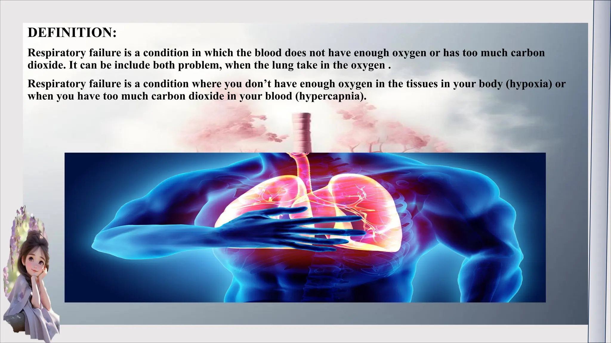
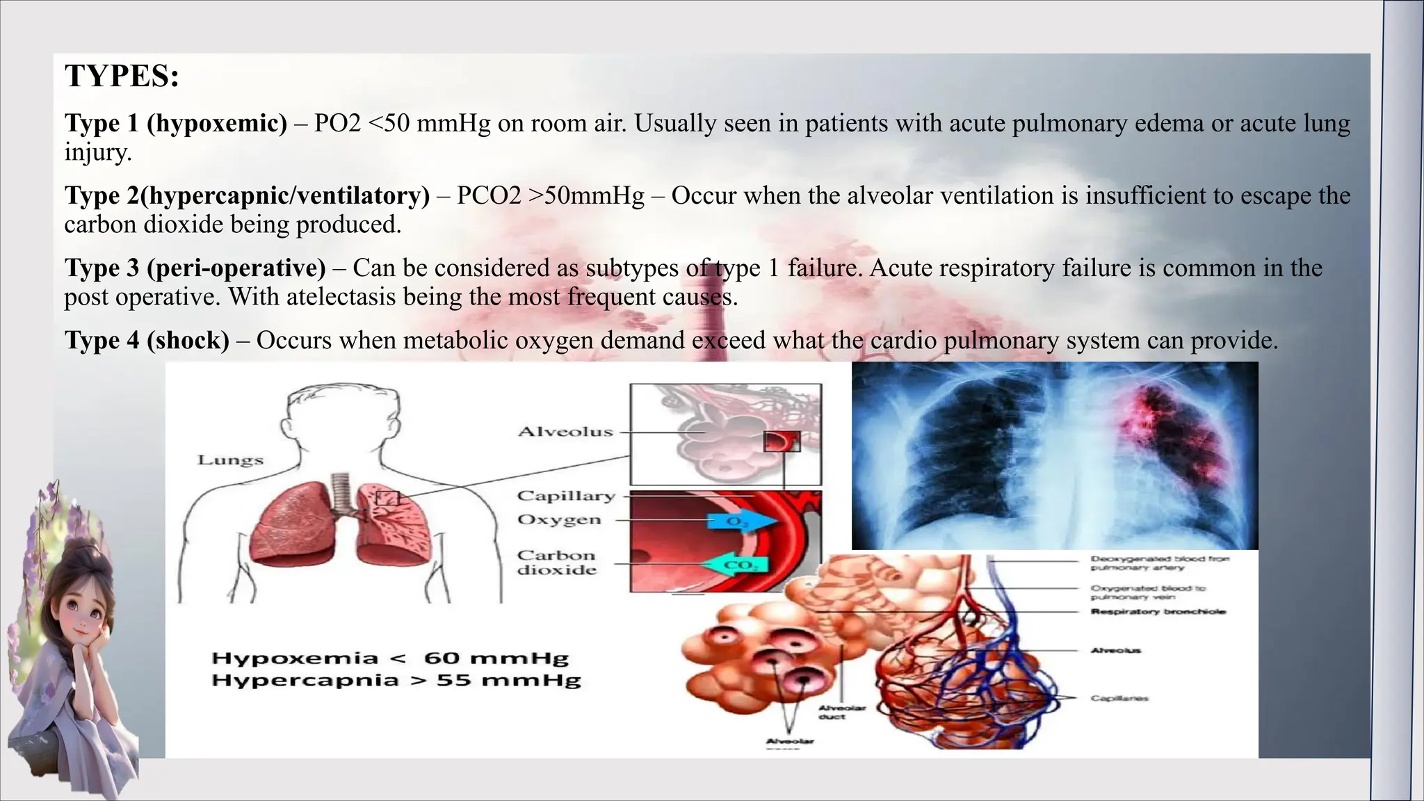
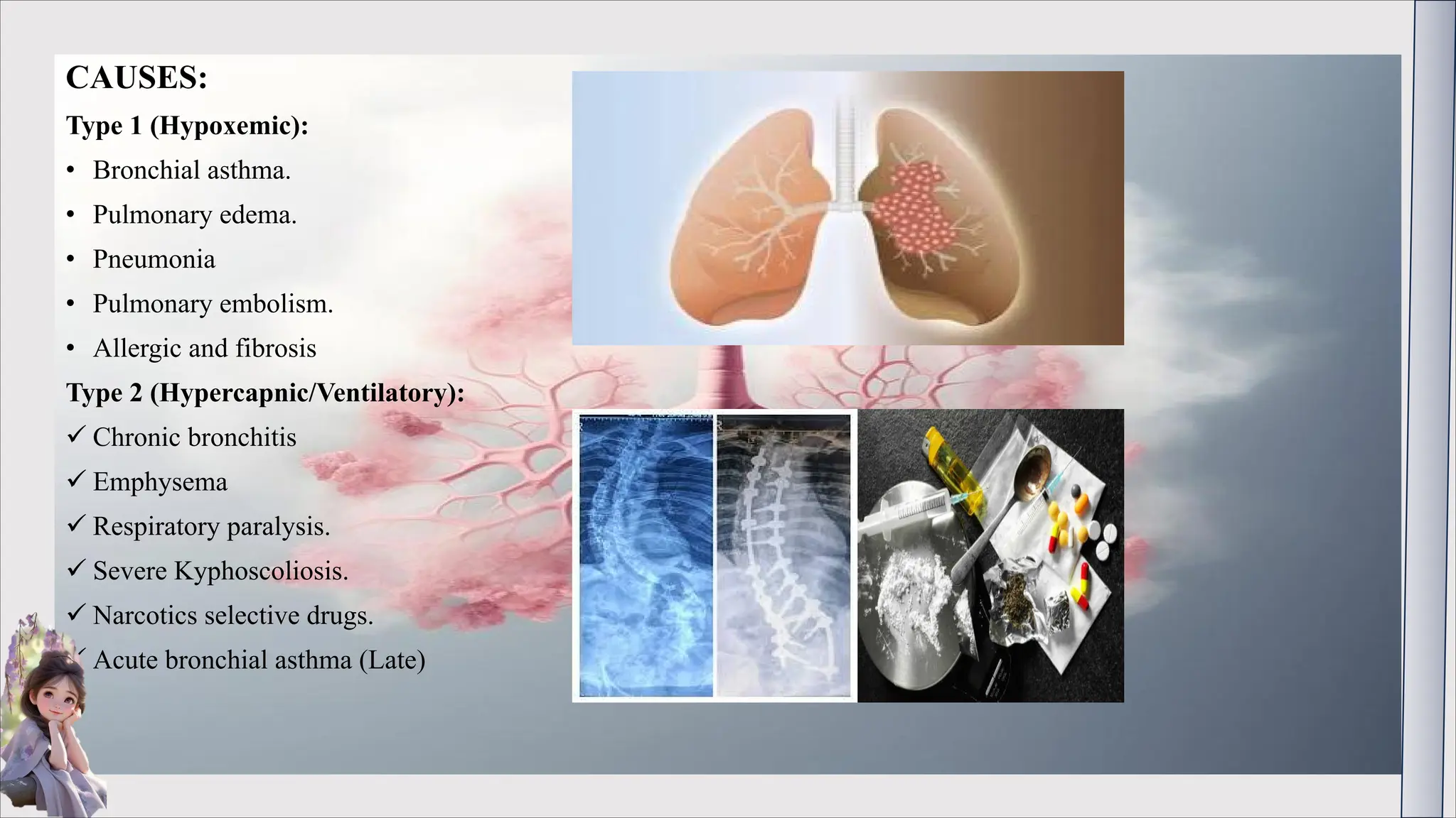
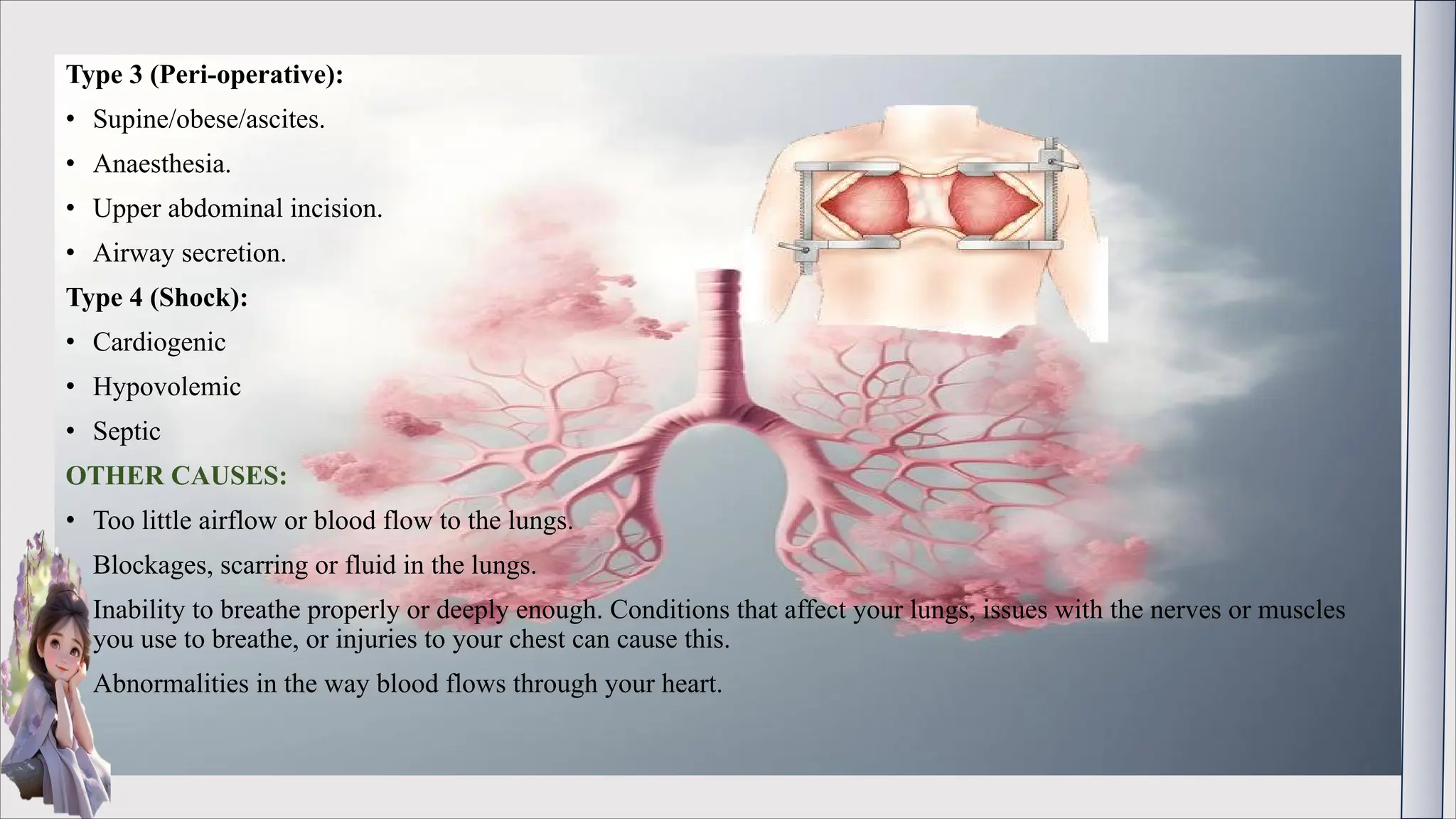
.
RESPIRATORY FAILURE
PREPARED BY DOLISHA WARBI
DEFINITION:
Respiratory failure is a condition in which the blood does not have enough oxygen or has too much carbon
dioxide. It can be include both problem, when the lung take in the oxygen .
Respiratory failure is a condition where you don’t have enough oxygen in the tissues in your body (hypoxia) or
when you have too much carbon dioxide in your blood (hypercapnia).
TYPES:
Type 1 (hypoxemic) – PO2 <50 mmHg on room air. Usually seen in patients with acute pulmonary edema or acute lung
injury.
Type 2(hypercapnic/ventilatory) – PCO2 >50mmHg – Occur when the alveolar ventilation is insufficient to escape the
carbon dioxide being produced.
Type 3 (peri-operative) – Can be considered as subtypes of type 1 failure. Acute respiratory failure is common in the
post operative. With atelectasis being the most frequent causes.
Type 4 (shock) – Occurs when metabolic oxygen demand exceed what the cardio pulmonary system can provide.
CAUSES:
Type 1 (Hypoxemic):
• Bronchial asthma.
• Pulmonary edema.
• Pneumonia
• Pulmonary embolism.
• Allergic and fibrosis
Type 2 (Hypercapnic/Ventilatory):
ü Chronic bronchitis
ü Emphysema
ü Respiratory paralysis.
ü Severe Kyphoscoliosis.
ü Narcotics selective drugs.
ü Acute bronchial asthma (Late)
Type 3 (Peri-operative):
• Supine/obese/ascites.
• Anaesthesia.
• Upper abdominal incision.
• Airway secretion.
Type 4 (Shock):
• Cardiogenic
• Hypovolemic
• Septic
OTHER CAUSES:
• Too little airflow or blood flow to the lungs.
• Blockages, scarring or fluid in the lungs.
• Inability to breathe properly or deeply enough. Conditions that affect your lungs, issues with the nerves or muscles
you use to breathe, or injuries to your chest can cause this.
• Abnormalities in the way blood flows through your heart.
SYMPTOMS:
• Shortness of breath or feeling like you can’t get enough air (dyspnea).
• Rapid breathing (tachypnea).
• Extreme tiredness (fatigue).
• Fast heart rate (feeling like your heart’s racing) or heart palpitations.
• Spitting or coughing blood or bloody mucus (hemoptysis).
• Excessive sweating.
• Restlessness.
• Pale skin.
• Bluish skin, lips or nails (cyanosis).
• Headaches.
• Blurred vision.
• Agitation, confusion or being unable to think straight.
• Behavioral changes, not acting like yourself.
DIAGNOSIS:
• Pulse oximetry
• Arterial blood gas (ABG) test
• Imaging - X-rays and CT scans
• Electrocardiogram (EKG)
• Blood test
TREATMENT:
• Mechanical ventilation
• Extracorporeal membrane oxygenation (ECMO). Providers use a bypass machine to add oxygen to the blood and
remove carbon dioxide.
• Oxygen therapy.
• Fluids.
• Managing underlying conditions
• Lung surgery or lung resection related to the particular conditions
NURSING DIAGNOSIS:
• Impair gas exchange related to increase alveolar capillary permeability interstitial edema and decrease long-
complaince.
• Ineffective breathing pattern.
• Ineffective airway clearance.
• Activity intolerance.
• Risk for aspiration.
NURSING MANAGEMENT:
• Manage nutrition.
• Treating the underlying causes or injury.
• Improve oxygenation with mechanical ventilation.
• Suction oral cavity.
• Give antibiotic.
• Deep venous thrombosis prophylaxis
• Stress ulcer prophylaxis
• Monitor fluid and electrolyte level.
THANKS YOU

RESPIRATORY FAILURE AND ITS TYPES....pdf

  • 1.
  • 2.
    DEFINITION: Respiratory failure isa condition in which the blood does not have enough oxygen or has too much carbon dioxide. It can be include both problem, when the lung take in the oxygen . Respiratory failure is a condition where you don’t have enough oxygen in the tissues in your body (hypoxia) or when you have too much carbon dioxide in your blood (hypercapnia).
  • 3.
    TYPES: Type 1 (hypoxemic)– PO2 <50 mmHg on room air. Usually seen in patients with acute pulmonary edema or acute lung injury. Type 2(hypercapnic/ventilatory) – PCO2 >50mmHg – Occur when the alveolar ventilation is insufficient to escape the carbon dioxide being produced. Type 3 (peri-operative) – Can be considered as subtypes of type 1 failure. Acute respiratory failure is common in the post operative. With atelectasis being the most frequent causes. Type 4 (shock) – Occurs when metabolic oxygen demand exceed what the cardio pulmonary system can provide.
  • 4.
    CAUSES: Type 1 (Hypoxemic): •Bronchial asthma. • Pulmonary edema. • Pneumonia • Pulmonary embolism. • Allergic and fibrosis Type 2 (Hypercapnic/Ventilatory): ü Chronic bronchitis ü Emphysema ü Respiratory paralysis. ü Severe Kyphoscoliosis. ü Narcotics selective drugs. ü Acute bronchial asthma (Late)
  • 5.
    Type 3 (Peri-operative): •Supine/obese/ascites. • Anaesthesia. • Upper abdominal incision. • Airway secretion. Type 4 (Shock): • Cardiogenic • Hypovolemic • Septic OTHER CAUSES: • Too little airflow or blood flow to the lungs. • Blockages, scarring or fluid in the lungs. • Inability to breathe properly or deeply enough. Conditions that affect your lungs, issues with the nerves or muscles you use to breathe, or injuries to your chest can cause this. • Abnormalities in the way blood flows through your heart.
  • 6.
    SYMPTOMS: • Shortness ofbreath or feeling like you can’t get enough air (dyspnea). • Rapid breathing (tachypnea). • Extreme tiredness (fatigue). • Fast heart rate (feeling like your heart’s racing) or heart palpitations. • Spitting or coughing blood or bloody mucus (hemoptysis). • Excessive sweating. • Restlessness. • Pale skin. • Bluish skin, lips or nails (cyanosis). • Headaches. • Blurred vision. • Agitation, confusion or being unable to think straight. • Behavioral changes, not acting like yourself.
  • 7.
    DIAGNOSIS: • Pulse oximetry •Arterial blood gas (ABG) test • Imaging - X-rays and CT scans • Electrocardiogram (EKG) • Blood test TREATMENT: • Mechanical ventilation • Extracorporeal membrane oxygenation (ECMO). Providers use a bypass machine to add oxygen to the blood and remove carbon dioxide. • Oxygen therapy. • Fluids. • Managing underlying conditions • Lung surgery or lung resection related to the particular conditions
  • 8.
    NURSING DIAGNOSIS: • Impairgas exchange related to increase alveolar capillary permeability interstitial edema and decrease long- complaince. • Ineffective breathing pattern. • Ineffective airway clearance. • Activity intolerance. • Risk for aspiration. NURSING MANAGEMENT: • Manage nutrition. • Treating the underlying causes or injury. • Improve oxygenation with mechanical ventilation. • Suction oral cavity. • Give antibiotic. • Deep venous thrombosis prophylaxis • Stress ulcer prophylaxis • Monitor fluid and electrolyte level.
  • 9.