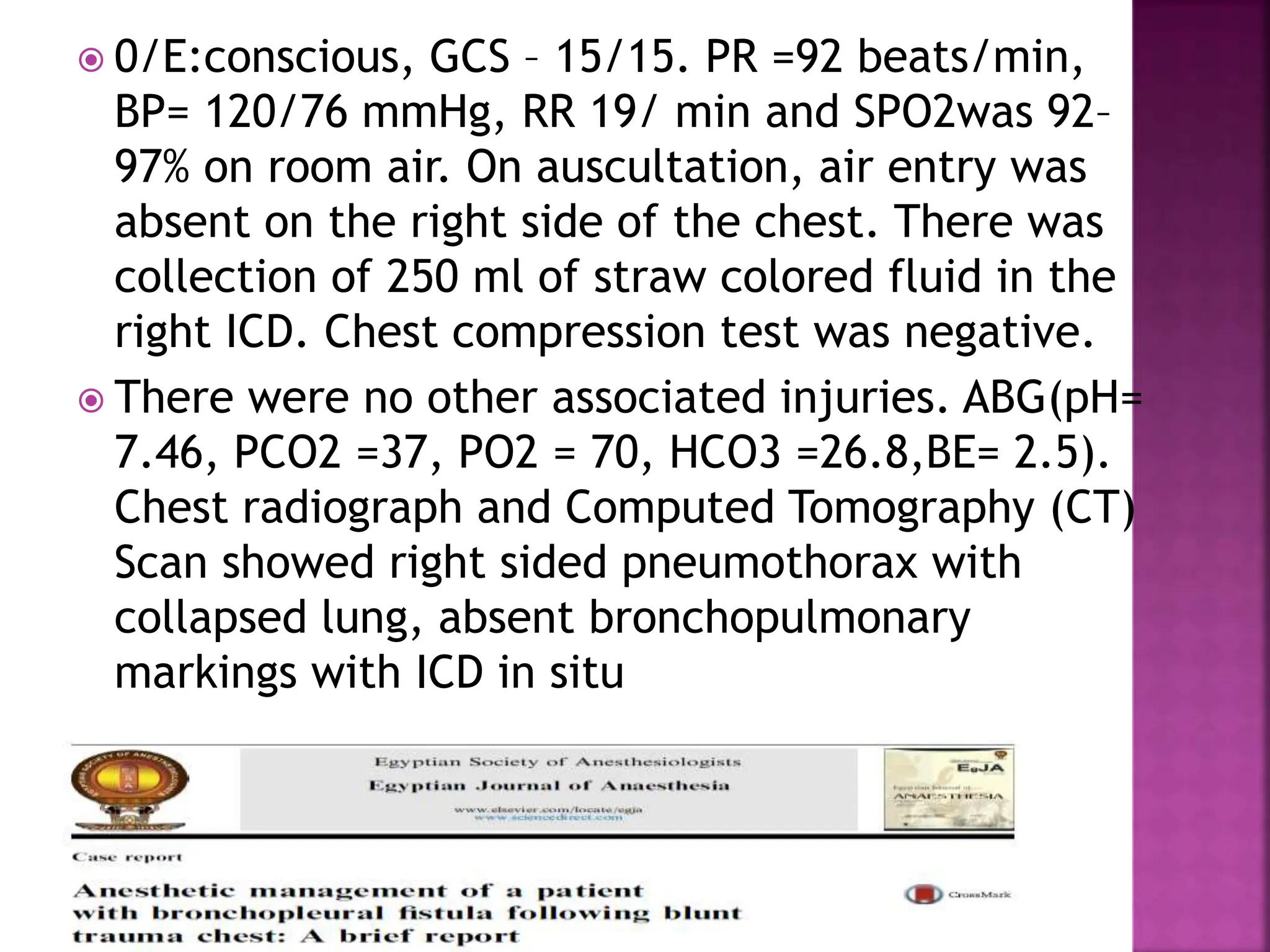
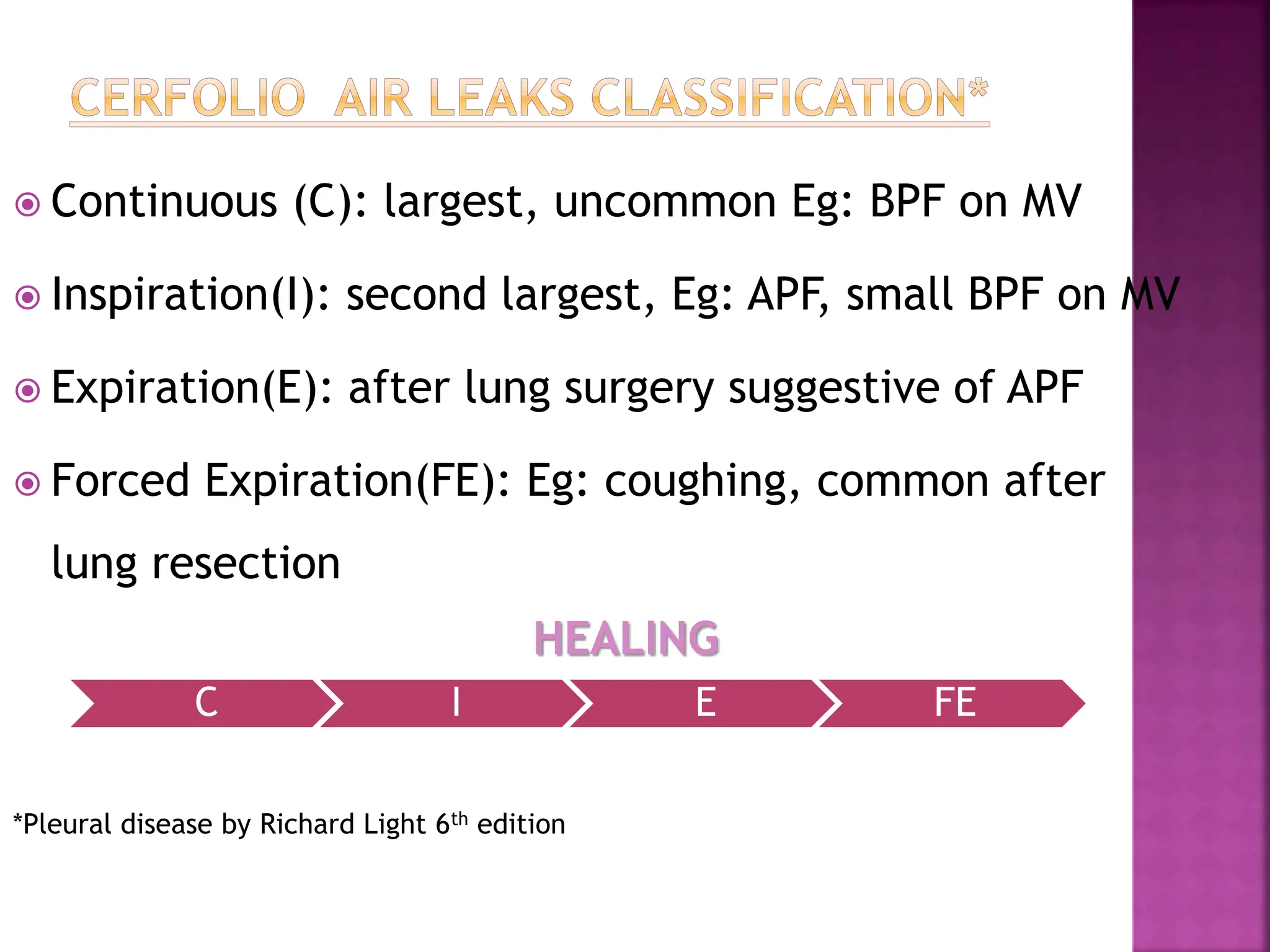
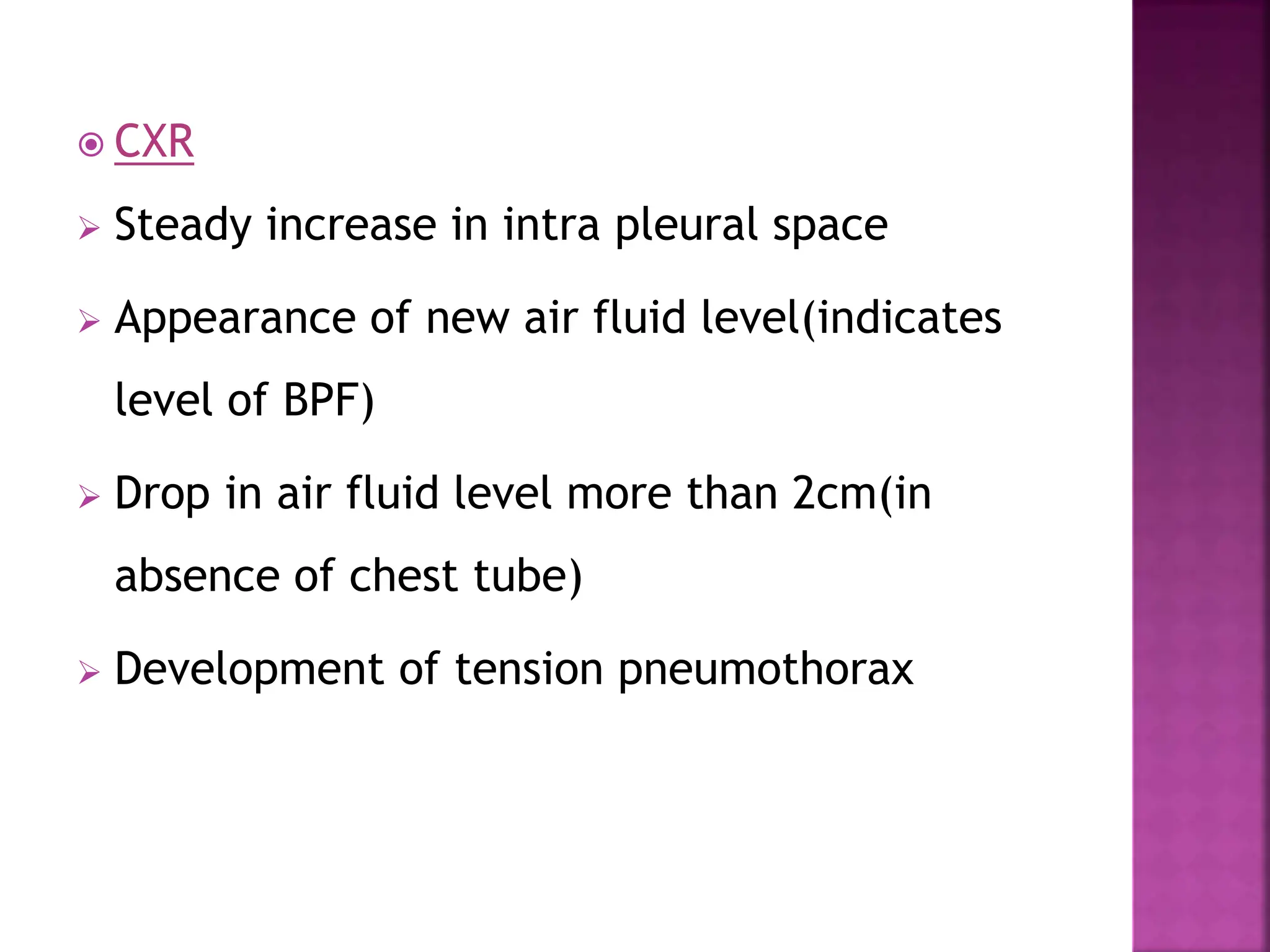
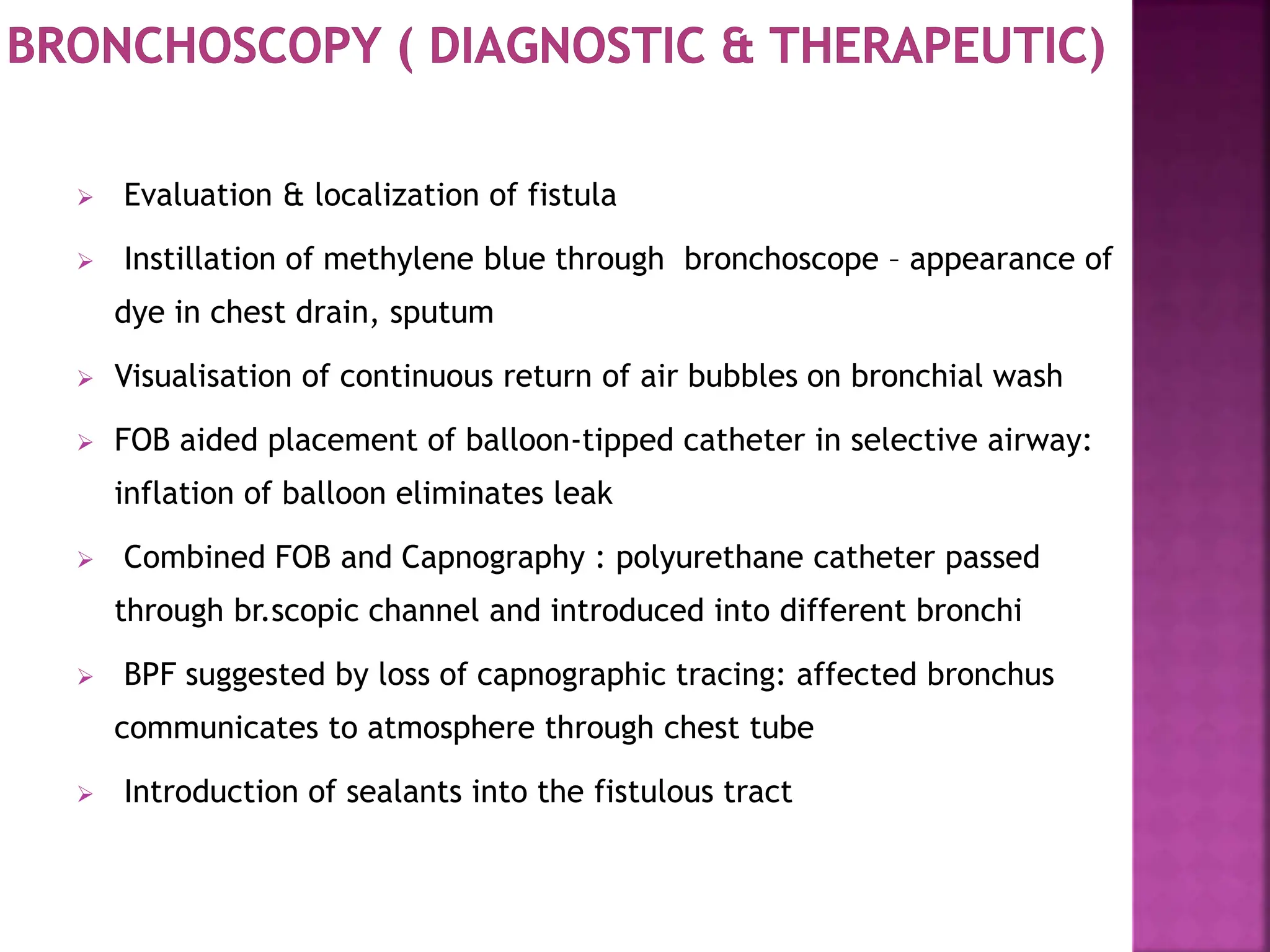
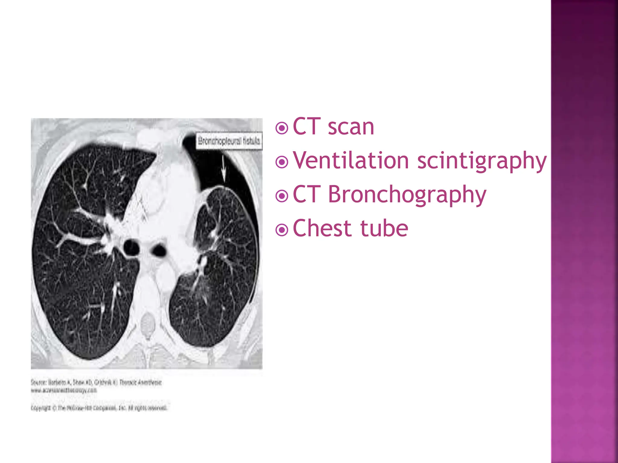
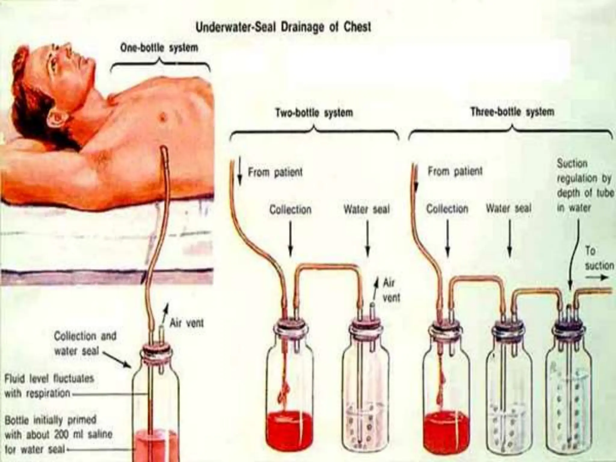
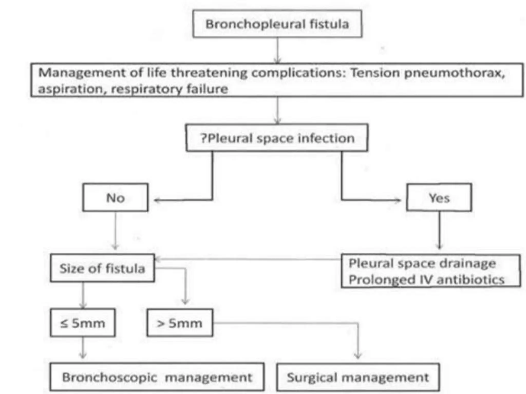
2. A 15-year-old male, weighing 49 kg was
brought to the emergency Department 19 days
after an injury to the right side of his chest by
the steering wheel of a tractor.
Patient had complaints of pain over the right
side of chest and breathlessness soon after the
injury. He had no upper airway obstruction and
there was no other associated injury.
He was initially treated at a peripheral
hospital, where intercostal drainage (ICD) was
placed & on fiberoptic bronchoscopy near
complete transaction of right main bronchus
with bronchopleural fistula.
3. 0/E:conscious, GCS – 15/15. PR =92 beats/min,
BP= 120/76 mmHg, RR 19/ min and SPO2was 92–
97% on room air. On auscultation, air entry was
absent on the right side of the chest. There was
collection of 250 ml of straw colored fluid in the
right ICD. Chest compression test was negative.
There were no other associated injuries. ABG(pH=
7.46, PCO2 =37, PO2 = 70, HCO3 =26.8,BE= 2.5).
Chest radiograph and Computed Tomography (CT)
Scan showed right sided pneumothorax with
collapsed lung, absent bronchopulmonary
markings with ICD in situ
4. A Broncho pleural fistula (BPF) is a communication
between the pleural space and the bronchial
tree
5.
6. ALVEOLOPLEURAL FISTULA(APF): Pulmonary pleural
communication distal to segmental bronchus and
pleural space.
Common after lung resection except pneumonectomy
BRONCHOPLEURAL FISTULA: Communication between
mainstem, lobar or segmental bronchus or the pleura
lined cavity
*Pleural disease by Richard Light 6th edition
Anesthesia for thoracic surgery – Slinger P
7. Continuous (C): largest, uncommon Eg: BPF on MV
Inspiration(I): second largest, Eg: APF, small BPF on MV
Expiration(E): after lung surgery suggestive of APF
Forced Expiration(FE): Eg: coughing, common after
lung resection
HEALING
*Pleural disease by Richard Light 6th edition
C I E FE
8. POSTOPERATIVE
Incidence 1.5 to 28% for pnemonectomies (2% -
11%) and for lobectomies (0.5%)
Mortality: 5% to 70%
Risk of dehiscence* :
1.8% : manual suture
5.0% : stapling device
1.9% : reinforcement suture at distal side
1.0% : reinforcement suture at proximal side
*Sonobe et al. Eur J Cardiothorac Surg 2000; 18:519–523
9. Advanced case ( main bronchus)
Previous ipsilateral thorocotomy
H/O chemotherapy and radiotherapy
H/O fever, steroid, diabetes
Anemia, leukocytosis, ESR, hypoalbuminemia
Residual tumor on stump
Excess peribronchial, paratracheal dissection
Intrathoracic chemotherapy
Tightness of sutures
Continous Mechanical ventilation > 24hrs
*Sato et al. Nippon Kyobu Geka Gekkai Zasshi 1989;37:498–503
10. Infection: bacterial , fungal , parasitic
Spontaneous rupture of bullae, cyst
After procedures
Persistent spontaneous pneumothorax.
Thoracic trauma
Erosion of bronchial wall : by malignancy,
foreign body, chronic inflammation
GERD
11. Varoli et al* classified according to time of onset
Early- 1 to 7 days
Immediate – 8 to 30 days
Late- more than 30 days
Usually presents after 7-15 days following lung
resections
Early indicators- reappearance of fever, increased
cough with purulent or serosanguinous sputum
*Varoli F et al. Endoscopic treatment of bronchopleural fistulas. Ann Thorac Surg 1998;65:807-9.
12. Life-threatening , due to tension pneumothorax or
asphyxiation secondary to massive pulmonary flooding,
Clinical presentation
sudden onset of dyspnoea,
hypotension,
subcutaneous emphysema, cough with expectoration,
There may be antecedent tracheal shift
Acute form is typically related to stump dehiscence &
requires immediate re-exploration.
13.
14. Ass infection, I/C
state
Generalised wasting
Malaise
Fever
minimally productive
cough
It may be associated with
an infectious process and
there may be coexistant
fibrosis of the pleural
space and mediastinum
**
15. CLINICAL
After pneumonectomy- development of sudden
onset dysponea, subcutaneous emphysema
After lobectomies - persistent air leak, purulent
drainage and expectoration of purulent material
After ICD removal – fever, purulent sputum,
new air fluid on CXR
16. CXR
Steady increase in intra pleural space
Appearance of new air fluid level(indicates
level of BPF)
Drop in air fluid level more than 2cm(in
absence of chest tube)
Development of tension pneumothorax
17.
18. Evaluation & localization of fistula
Instillation of methylene blue through bronchoscope – appearance of
dye in chest drain, sputum
Visualisation of continuous return of air bubbles on bronchial wash
FOB aided placement of balloon-tipped catheter in selective airway:
inflation of balloon eliminates leak
Combined FOB and Capnography : polyurethane catheter passed
through br.scopic channel and introduced into different bronchi
BPF suggested by loss of capnographic tracing: affected bronchus
communicates to atmosphere through chest tube
Introduction of sealants into the fistulous tract
23. Indications
High flow BPF
Drainage of empyema
Pneumothorax
Advantages
Add positive intrapleural pressure during expiration to reduce
air leak and maintain PEEP
Intermittent occlusion during inspiratory phase to decrease
BPF flow
Useful in ARDS
Chest tube can be used for pleurodesis
24. Disadvantages
Can function as foreign body and delay healing
Predispose to infection at insertion site and pleural space
Loss of tidal volume
Abnormal gas exchange
Inappropiate ventilator cycling
• FANNING EQUATION
• Larger diameter for drainage of air
Size of tube
25.
26. 1977 - Ratliff et al pioneered endoscopic
management of BPF
INDICATIONS
Suitable for small fistulas(<5mm).
Proximally located fistulas- main stem, lobar,
or segmental bronchi are more suitable.
Useful for patients who are not proper
candidate for surgery.
27. Injecting absolute ethanol
directly into submucosal
layer of a fistula is first-line
therapy for patients with a
postoperative central BPF
with an orifice < 3 mm
Lead Shots
Polyethylene Glycol
Cyanoacrylate Glue , Fibrin
Glue , Blood Clot
Antibiotics
Albumin-Glutaraldehyde
Tissue Adhesive
Gel Foam
Coils
Intrabronchial stents, valves
Calf Bone
Balloon Catheter Occlusion
Cellulose, silver nitrate
*Kinoshita et al. Chest 2000; 17:790–795
*Potaris et al Med Sci Monit 2003; 9:P179–P183
*Chest 2008; 133(6): 1481-4
28.
29.
30. Revision of bronchial stump (CLAGETT procedure)
Decortication of lung
Closure of fistula with muscle flap from intercostal space
Thoracoplasty with pedicle muscle flap to cover bronchial stump.
Resection of diseased chronically infected segments
First stage: chest cavity drainage consisting of muscle flap operation
Second stage: After aggressive nutritional & physical rehabilitation,
second procedure for chest cavity obliterationwith an omental flap is
performed
*Turk et al. Plastic Surg 2000; 45:560–564
31. HISTORY
EXAMINATION
Estimation of volume loss through fistula
1. Check bubble flow through chest drain- continous (large) or
intermittent (small)
2. Quantification of size of BPF- Difference of inhaled vs exhaled tidal
volumes
Intubated patients: direct attatchment of spirometer to ETT tube
Non intubated patients: tight fitting mask and fast responding
spirometer
32. Routine – CBC, RBS, SE, KFT, LFT, Coag profile
urine r/m, CXR , ECG, ECHO
Specific – ABG, Bronchoscopy, CT, VS
33. Optimize respiratory function(smoking, infection)
Optimize associated diseases
Predict difficult endobronchial intubation
Predict desaturation during OLV
If chest tube present- check for patency
Mild sedation
A
B
C
35. PATIENT
• Diseased lung( air leak, pneumothorax, soilage, V/Q
mismatch)
• Elderly , comorbidities
SURGERY
• Open chest,
• Bleeding
ANESTHESIA
• Position
• Mechanical ventilation
• OLV & HPV
• Blood loss
• Diseased lung
• Chest tube
• Choice of anesthetic & CO
•Temperature
• Fluid
• Emergence
• Post op icu stay
•Pain control
•Mortality
36. ASA I
ASA II – ECG , SpO2, NIBP,
EtCO2, Temperature
urine output.
Thoracic surgery:
(Paw)
1) Arterial line - IBP
2) central line
3) side stream spirometry (NR)
4) TEE
5) EDU
*Hemodynamic monitoring in thoracic surgery – Jacob Raphael 2017
38. Position for induction- Head up with 30 degree lateral tilt keeping
diseased lung down
Options to secure airway include
1)Awake fibreoptic intubation with DLT,- Induction of GA once lung
isolation achieved
Safest but technically difficult
Requires patient cooperation and thorough airway topicalization
2) Induction of GA maintaining maintaining spontanoeus ventilation,
DLT insertion when deep , aviod IPPV
3) If airway thought to be easy, Modified RSI can be done avoiding IPPV
until lung isolation achieved
Thoracic epidural + IV sedation
(SLT safe to use: if fistula small, chronic, uninfected)
* Anesthesia for thoracic surgery – Slinger P
39.
40. Air leak
Loss of TV, PEEP
V/Q mismatch
Incomplete lung expansion
CO2 retension
Auto trigger
Paw < Critical opening
Pressure
Pleural decompression for
lung expansion GOALS
CHALLENGES
41. To keep reduced airway pressures
Low tidal volume
Limiting PEEP
Short inspiratory time ( high PIFR)
Reducing respiratory rate
Reducing minute ventilation
Pleural decompression
Chest drain: lowest possible suction pressure or
no suction as increased suction pressure increases
the flow.
For large fistula: Independent lung ventilation or
HFJV can be considered
*Pierson DJ. Management of bronchopleural fistula in patients on mechanical ventilation.
42. Chest tube manipulation
Intermittent inspiratory chest tube occlusion
Application of intrapleural pressure at expiration
Independent lung ventilation
• CPAP
• High frequency ventilation
Extracorporeal oxygenation
• HFPPV
• HFJV
• HFOV
• HFFI
• HFPV
HFV
46. Inhalational agents and narcotics or epidural
High FiO2
IPPV to healthy lung
Ventilate healthy lung with normal tidal volume.
Ventilate affected lung with smaller volume
Titrate ventilation to normal PaCO2
Blood loss
Fluid management
EMERGENCE : Avoid high Paw ( SIMV, Hand ventilation)
Examine airway – suction , extubation
*Thoracic surgery -James Mitchell (December 24, 2003)
48. Assesed EVLWT, PaO2/FiO2,blood lactate,ScvO2,BE,CVP,CI,GEDI
Carbohydrate
drink
BSS 3-4ml/kg/hr
vasopressor
Meta analysis
have shown
that GDFT is
better than
conventional
49. Thoracotomy – 80%
a. Atelectasis
b. Pneumonia
c. RF
d. DVT
Types
1) Acute
2) chronic
Thoracotomy T4 – T6
CHEST DRAIN T7 – T8
MEDIASTINAL PLEURA X
CENTRAL DIAPHRAGMATIC
PLEURA
C3 – C5
I/L SHOULDER PAIN BRACHIAL
PLEXUS
Thoracic compression test
Assess if symptoms are due to vertebra compression.pain disc proplapse or nerve compression
Flow varies with 5th power of tube radius in
clinical situations due to turbulent flow of
moist air( Fanning equation )
Loss of air so loss of tv and loss of lung expansion hence vq mismatch
Short inspiratort so air leak is less
Flow through an air leak is proportional to:
Cross-sectional area of the leak
Time held at high airway pressure
∴High frequency ventilation may reduce fistula leak