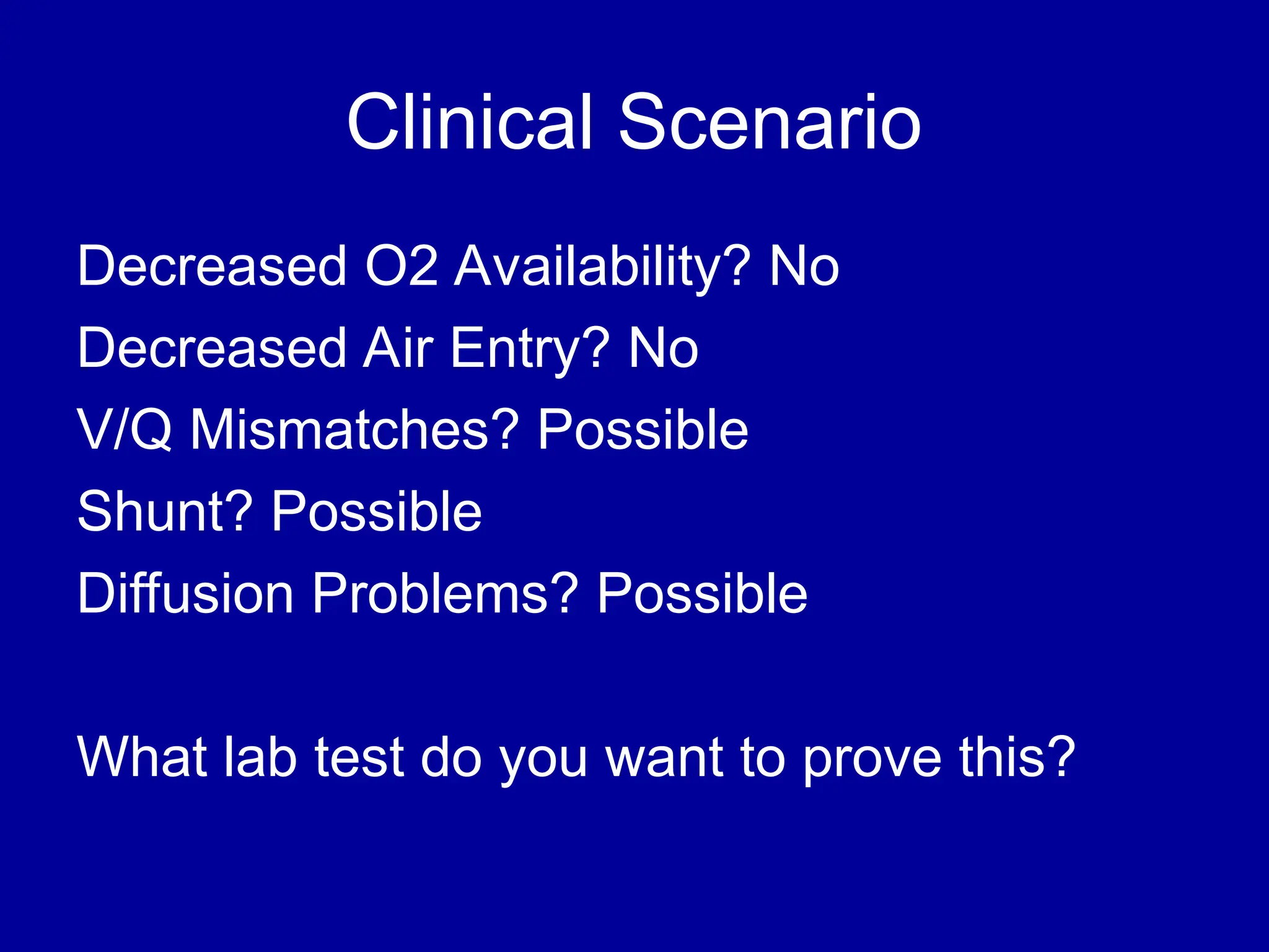
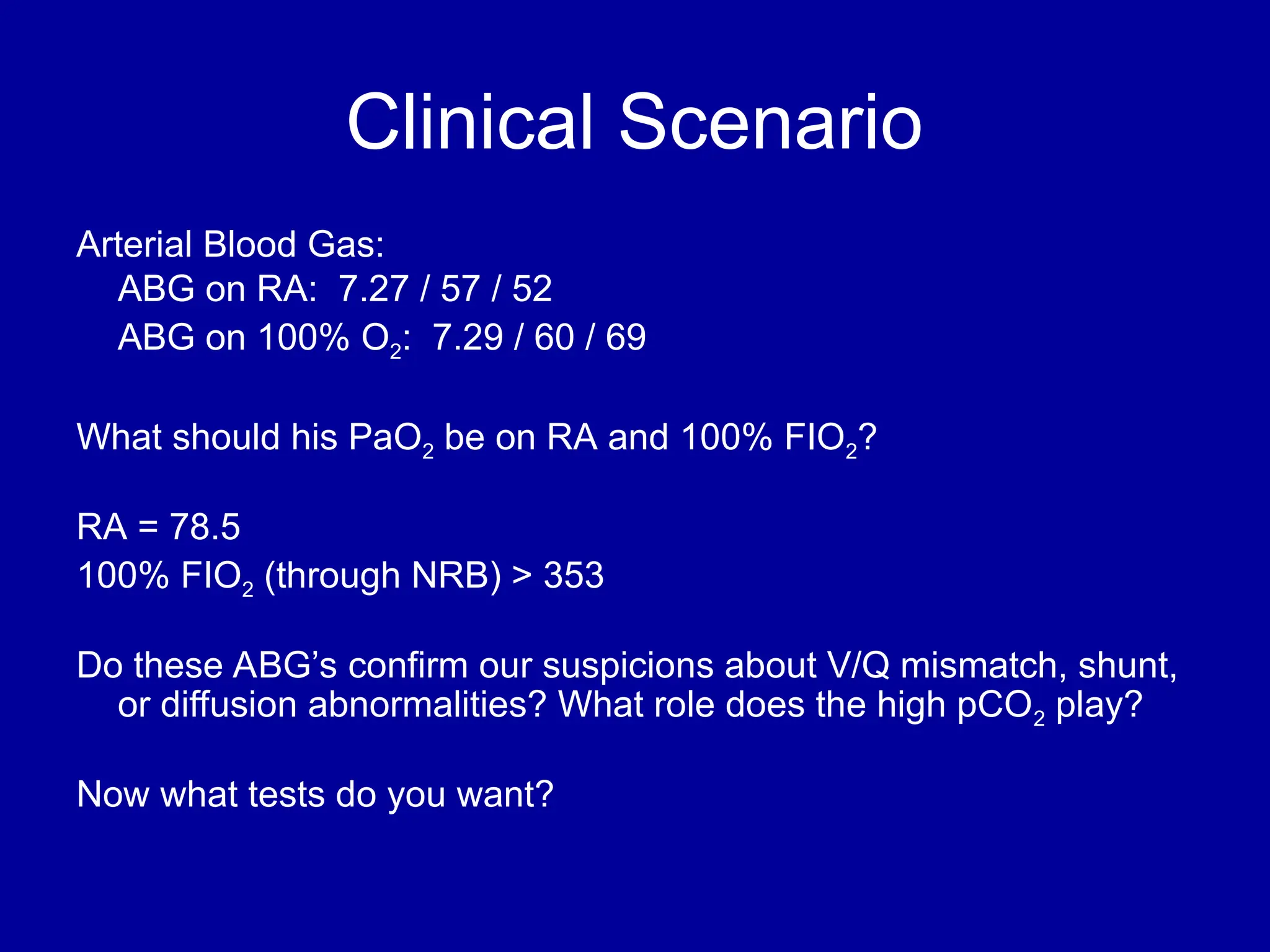
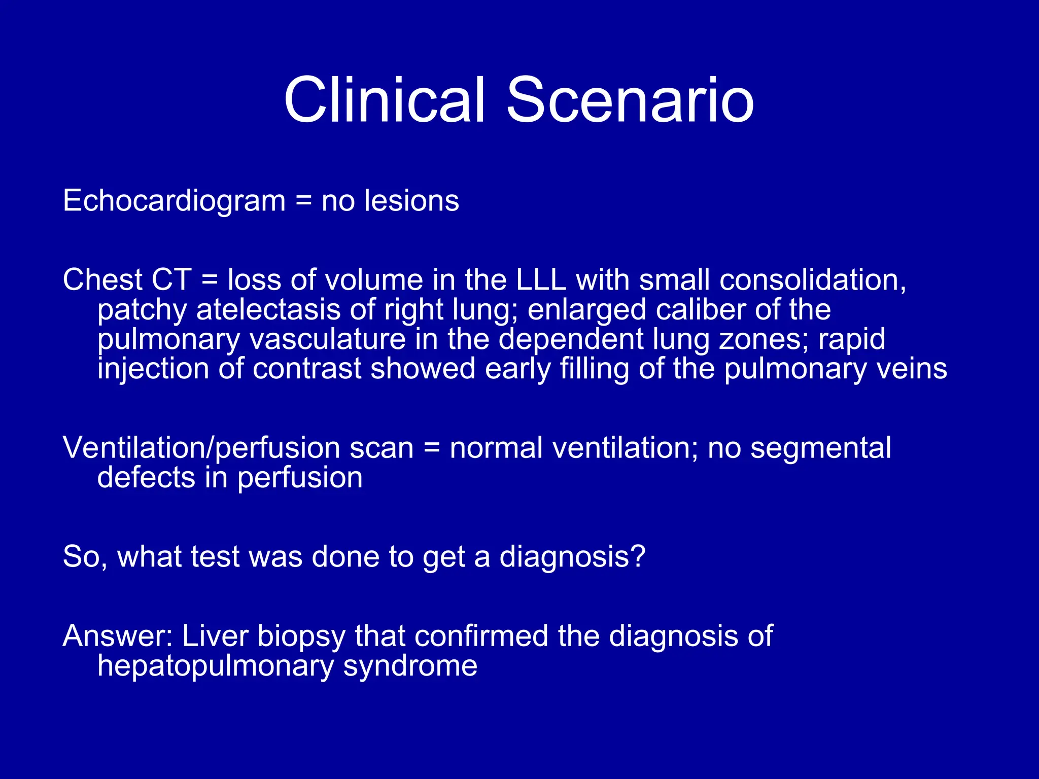
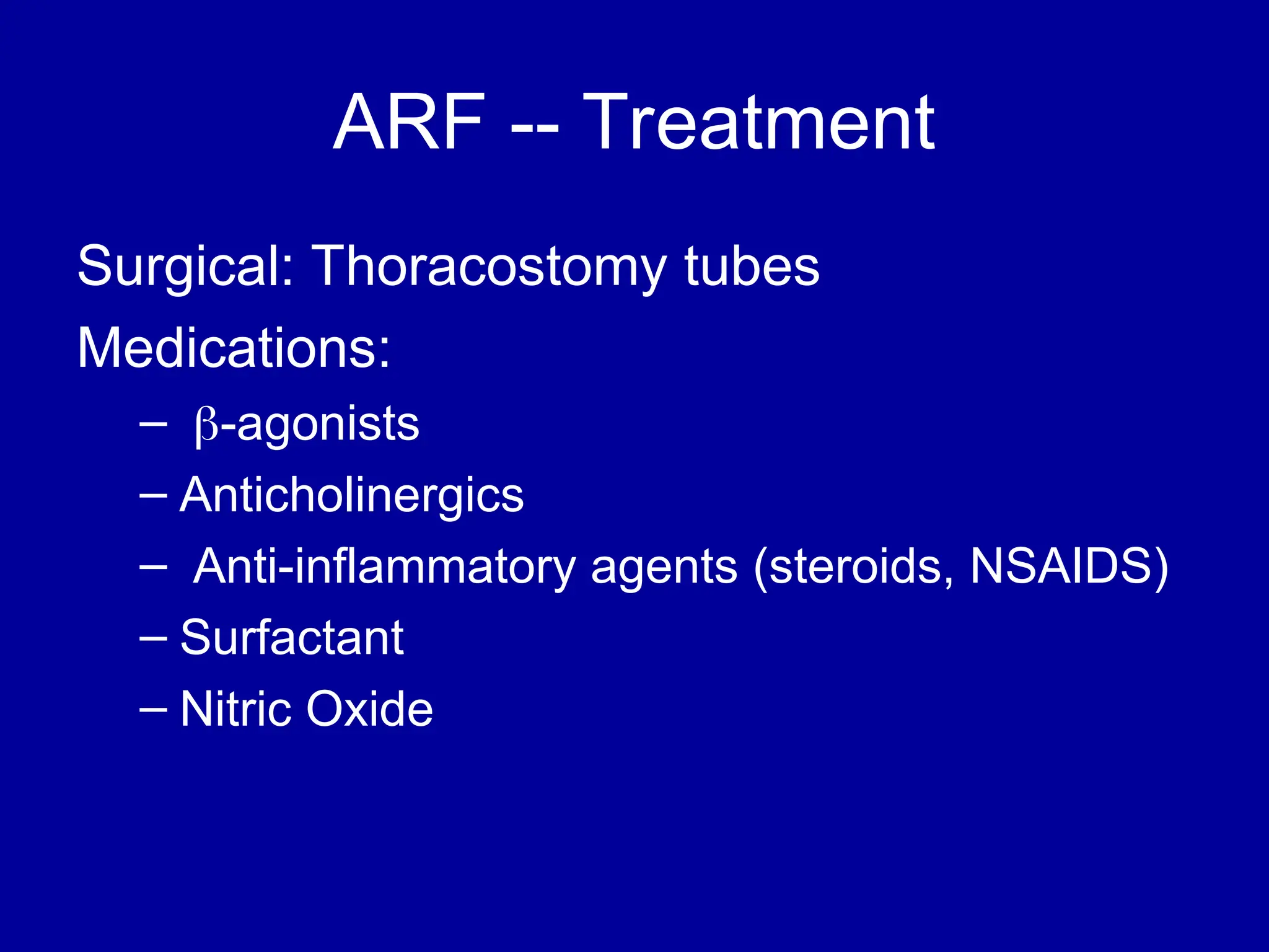
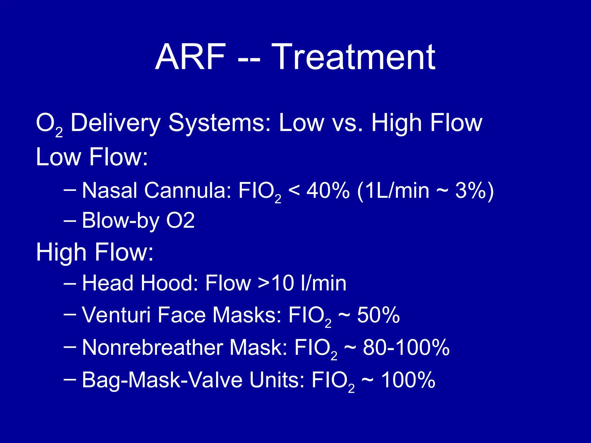
Respiratory Failure in Children
Daniel Sloniewsky, MD
Associate Professor
Division of Critical Care
Department of Pediatrics
Stony Brook University Hospital
Definitions: Acute Respiratory
Failure
ARF is the inability of the respiratory system to
deliver O2 and remove CO2 at a sufficient rate to
meet the body’s metabolic demands
Can be hypoxic or hypercarbic
ABG abnormalities
– PaCO2 > 55 mm Hg (with low pH)
– PaO2 < 60 mm Hg
– SaO2 < 90% (in absence of cyanotic CHD)
Problems with Oxygenation
Hypoxemia: Decrease in tissue O2 delivery
– Hypoxic Hypoxemia -- Lung Disease
– Ischemic Hypoxemia -- Decreased Blood Flow
-- DO2 = O2 content x Cardiac Output
– Anemic Hypoxemia -- Decreased O2-Carrying
Capacity
-- O2 Content = (1.39 x HgB x SaO2) + (0.003)
(PaO2)
Hypoxic Hypoxemia: Where is the
problem?
FIO2 (Ex. Altitude)
Decreased Air Entry
-- Sedation (Ex. Opioid Overdose)
-- Respiratory Muscle Weakness (Ex. SMA)
-- Airway Obstruction (Ex. Croup, Asthma, etc)
V/Q Mismatches (Ex. Atelectasis, ARDS)
Shunting (Ex. CHD, pulmonary hypertension)
Diffusion Abnormalities (Ex. Pulmonary Fibrosis,
ARDS)
Acute Respiratory Failure:
Problems with Ventilation
Hypercapnea (increased CO2) can be seen with:
1. Increased Dead Space (areas not involved in gas
exchange) (Ex. Asthma, Pulmonary HTN)
2. Decreased Alveolar Ventilation
- Decreased Tidal Volume or Respiratory Rate (Ex.
Coma, Overdose, MG, ARDS, etc)
3. Obstructed Airways (Ex. Asthma)
4. Increased CO2 production (Ex.
Burns,Overfeeding)
CO2 diffuses easily across alveolar-capillary membrane so
diffusion problems usually do not cause hypercapnea
Acute Respiratory Failure: How to
Diagnose
Clinical Exam -- MOST IMPORTANT
1. Changes in RR and Breathing Effort:
Question - Why do infants “tire out?”
2. Changes in lung exam (stridor, wheeze, etc.)
3. Cyanosis – definition?
Flushing, tachycardia, HA, confusion – seen with
acute hypercarbia
Mental Status
Chronic
Hypoxemia/Hypercarbia
Clinical signs/lab evidence of chronic
hypoxia:
-- Clubbing – seen with chronic hypoxemia
-- What might you see on abdominal
exam?
-- Labs: Polycythemia
Lab evidence of hypercarbia
Hypoxic Hypoxemia: How to
Diagnose
Pulse oximetry – How does it work? What
are its limitations?
Arterial Blood Gas – What information does
this provide that pulse-ox does not?
Pulse-oximetry: How does it work?
Current pulse-ox machines measure 2 types
of light: red (wavelength=660 nm) and
infrared (wavelength= 940 nm)
Red light is better absorbed by deoxy-Hgb,
infrared light is better absorbed by oxy-
Hgb, so the ratio gives you the SaO2
Pulse-oximetry Problems
1. Doesn’t work well with poor perfusion
2. Other light sources (Ex. phototherapy
light) can interfere with the results
3. Abnormal Hgb can lead to overestimation
of a true SaO2 (specifically carboxy-, and
metHgb)
What laboratory test do you order in
someone with met- or carboxy-hgb
Arterial Blood Gas and
Oxygenation
ABG – can give you carboxy and metHgb
levels (if you ask); will give you co-
oximetry values (multiple wavelengths
measured)
-- will also give you a PaO2 which can
help you diagnose a patient with
hypoxemic hypoxia using an equation
called ………..
Alveolar Gas Equation
PAO2 = FIO2(PB-PH2O) – PCO2/R
– PAO2 = Alveolar oxygen partial pressure
– FIO2 = fraction of inspired oxygen
– PB = barometric pressure (760 mm Hg at sea
level)
– PH2O = partial pressure of water vapor (47 mm
Hg)
– PCO2 = partial pressure of CO2
– R = respiratory quotient (usually ~ 0.8)
Alveolar Gas Equation
So at room air at sea level, your PAO2
should be 100
On 100% FIO2 at sea level, your PAO2
should be 663
The difference between Alveolar (PAO2) and
arterial (PaO2) oxygen is called the A-a
gradient and should be less than 20; this
will help you figure out if this is hypoxic
hypoxemia and the degree of hypoxia
Given a PaO2, what is the SaO2
(and vice-versa)
Curve moves to right with
 Lower pH
Lower pH
 Higher Temperature
Higher Temperature
 Increased 2,3 DPG
Increased 2,3 DPG
Hypercarbic Respiratory Failure:
Who cares about CO2 and pH?
What makes you breathe?
Primarily pH (can be PaO2 in some patients)
What happens to you if you are too alkalotic or acidotic?
Acidosis: AMS, impaired cardiac contractility, pulmonary
vasoconstriction, metabolic abnormalities, etc.
Alkalosis: tetany (sec. to decreased iCa), arrhythmias, etc.
Does it matter what the source of the altered pH is?
Yes. For example: metabolic acidosis is worse for you than
respiratory acidosis.
Arterial Blood Gas, pH, and
Ventilation
pH < 7.35 with high CO2: respiratory acidosis
pH > 7.35 with high CO2: compensated
respiratory acidosis
pH < 7.35 with low bicarb: metabolic acidosis
pH > 7.35 with low bicarb: compensated
metabolic acidosis
What is a base deficit?
Base Deficit
Given a normal pCO2, how much base would
be needed to correct the pH.
Ex. 7.35/28/80/-15
Arterial Blood Gas, pH, and
Ventilation
pH > 7.45 with low CO2: respiratory alkalosis
pH < 7.45 with low CO2: compensated
respiratory alkalosis
pH > 7.45 with high bicarb: metabolic
alkalosis
pH < 7.45 with high bicarb: compensated
metabolic alkalosis
How to Approach an ABG
First – Is this an arterial or venous blood gas?
Second - does the patient have an acidosis or an
alkalosis
– Look at the pH
Third, what is the primary problem – metabolic or
respiratory
– Look at the pCO2
– If the pCO2 change is in the opposite direction of the
pH change, the primary problem is respiratory
– You never overcompensate
How to Approach an ABG
Next, don’t forget to look at the
effectiveness of oxygenation, (and look at
the patient)
– your patient may have a significantly
increased work of breathing in order to
maintain a “normal” blood gas
– metabolic acidosis with a concomitant
respiratory acidosis is concerning
Case 1
Sameer got into some of Dad’s barbiturates.
He suffers a significant depression of
mental status and respiration. You see
him in the ER 3 hours after ingestion with
a respiratory rate of 12. A blood gas is
obtained. It shows pH = 7.16, pCO2 = 70,
HCO3 = 22
Case 1
What is the acid/base abnormality?
1. Uncompensated metabolic acidosis
2. Compensated respiratory acidosis
3. Uncompensated respiratory acidosis
4. Compensated metabolic alkalosis
Case 2
You are evaluating a 15 year old female in the
ER who was brought in by EMS from school
because of abdominal pain and vomiting.
Review of system is negative except for a 10 lb.
weight loss over the past 2 months and polyuria
for the past 2 weeks. She has no other medical
problems and denies any sexual activity or drug
use. On exam, she is alert and oriented,
afebrile, HR 115, RR 26 and regular, BP 114/75,
pulse ox 95% on RA.
Case 2
Exam is unremarkable except for mild abdominal
tenderness on palpation in the midepigastric
region and capillary refill time of 3 seconds. The
nurse has already seen the patient and has sent
off “routine” blood work. She hands you the
result of the blood gas. pH = 7.21 pCO2= 24
pO2 = 45 HCO3 = 10 BE = -10 saturation =
72%
Case 2
What is the blood gas interpretation?
1. Uncompensated respiratory acidosis with
severe hypoxia
2. Uncompensated metabolic alkalosis
3. Combined metabolic acidosis and respiratory
acidosis with severe hypoxia
4. Metabolic acidosis with respiratory
compensation
Case 3
10 year old with history of ALL and
neutropenia presents with tachypnea. He
has no O2 requirement but is breathing 30
– 40 times/minute. Lung exam (other than
the tachypnea) is normal. CXR shows no
infiltrate. An ABG is done: 7.45/30/90/22
on room air. Does this patient need a
bronchoscopy to diagnose his respiratory
compromise? Why or why not?
Case 3
Answer: No; This is a trick question
because he doesn’t have respiratory
compromise
The patient is tachypneic for some other
reason than acidosis, hypercarbia, or
hypoxia (i.e. increased intracranial
pressure, burgeoning sepsis, etc)
Other Laboratory Findings in ARF
CXR Abnormalities
Complete Blood Count (look at WBC and
Hgb, which may suggest chronic hypoxia)
Electrolyte Abnormalities (look at
bicarbonate)
Foreign Body Aspiration
Right Lung
Atelectasis
Left Lung
Pneumonia
with Effusion
Pneumothorax
ARDS
(Bilateral
Infiltrates)
Flail Chest
Flail Chest
A Case of Hypoxia
4 yo presents to the ER with fever and cough. On examination, the patient has
the following vital signs: T 39.9, P 130, RR 32, O2 sats 87-90% on RA, Nl BP’s
PE: Lungs – tachypneic with good breathing effort, clear lung sounds,
Cardiac -- 2/6 SEM at LSB, good pulses in all extremities
Extremities -- mild clubbing of fingers and toes
Labs: WBC ct = 9.6, H/H = 14/41, platelets = 192k
Elytes normal except bicarbonate = 20; LFT’s Nl except AP = 358
CXR showed normal sized heart, possible infiltrate in hilar area
Patient is admitted to the hospital with a diagnosis of pneumonia and is
started on appropriate abx and supplemental oxygen. He defervesces after 24
hours but still has O2 saturations in the low 90’s that increase to mid-90’s on
4L NC. Physical exam remains the same and the repeat CXR is negative.
What is the cause of his hypoxia?
Clinical Scenario
Decreased O2 Availability? No
Decreased Air Entry? No
V/Q Mismatches? Possible
Shunt? Possible
Diffusion Problems? Possible
What lab test do you want to prove this?
Clinical Scenario
Arterial Blood Gas:
ABG on RA: 7.27 / 57 / 52
ABG on 100% O2: 7.29 / 60 / 69
What should his PaO2 be on RA and 100% FIO2?
RA = 78.5
100% FIO2 (through NRB) > 353
Do these ABG’s confirm our suspicions about V/Q mismatch, shunt,
or diffusion abnormalities? What role does the high pCO2 play?
Now what tests do you want?
Clinical Scenario
Echocardiogram = no lesions
Chest CT = loss of volume in the LLL with small consolidation,
patchy atelectasis of right lung; enlarged caliber of the
pulmonary vasculature in the dependent lung zones; rapid
injection of contrast showed early filling of the pulmonary veins
Ventilation/perfusion scan = normal ventilation; no segmental
defects in perfusion
So, what test was done to get a diagnosis?
Answer: Liver biopsy that confirmed the diagnosis of
hepatopulmonary syndrome
ARF -- Treatment
Monitoring
– invasive (ABG, PAC) or noninvasive (pulse-
oximeter)
Prevention
– Encourage coughing, frequent position
changes, reflux precautions, decompress
abdomen, etc.
ARF -- Treatment
Surgical: Thoracostomy tubes
Medications:
– -agonists
– Anticholinergics
– Anti-inflammatory agents (steroids, NSAIDS)
– Surfactant
– Nitric Oxide
ARF -- Treatment
O2 Delivery Systems: Low vs. High Flow
Low Flow:
– Nasal Cannula: FIO2 < 40% (1L/min ~ 3%)
– Blow-by O2
High Flow:
– Head Hood: Flow >10 l/min
– Venturi Face Masks: FIO2 ~ 50%
– Nonrebreather Mask: FIO2 ~ 80-100%
– Bag-Mask-Valve Units: FIO2 ~ 100%
ARF -- Treatment
Continuous or Bilevel Positive Pressure
(CPAP or BIPAP) -- applied through a
tight-fitting mask
Best applied in an awake, cooperative
patient who is expected to improve in 48-
72 hours.
ARF – Treatment with Mechanical
Ventilation
Always Remember: Bag-mask ventilation is an
effective method to oxygenate patients: use
BMV if conditions are not ideal for intubation
Mechanical Ventilation: Indications
– Airway Protection
– Respiratory Failure
– Shock
– Treatment of Intracranial Hypertension
– Other (for procedures, pulmonary toilet, etc.)
Mechanical Ventilation: Modes
AC – assist control
– no spontaneous breaths
– delivers full breath with any ventilator and patient
initiated effort
SIMV -- synchronized intermittent mandatory
ventilation
– ventilator breaths synchronized with patient’s own
breaths
– weaning mode
Mechanical Ventilation: Modes
Pressure Ventilation (Control)
– the size of the breath is determined by the pressure
that is set
– the tidal volume then depends on the lung compliance
(it is the dependent variable)
Volume Ventilation (Control)
– the size of the breath is determined by the volume
that is set
– the pressure is the dependent variable
Mechanical Ventilation: Goals
Your goals for gas exchange when a patient is on
a ventilator must not be so rigid as to cause
further injury to the lung when trying to obtain
them.
Oxygenation is more important than ventilation
You should try to keep the FIO2 < 60%, the PIP <
35-40 cm H20, and the TV ~6-8 cc/kg
Mechanical Ventilation:
Terminology
Mean Airway Pressure (MAP): the average
pressure over a respiratory cycle, measured at
the proximal airway
Peak Inspiratory Pressure (PIP): the maximum
amount of pressure needed to deliver a breath
Positive End Expiratory Pressure (PEEP):
pressure applied at the end of exhalation
I time: total time spent during inspiration
Mechanical Ventilation:
Terminology
Tidal Volume: the size (volume) of the breath
– goal ~ 6-10 cc/kg
Minute Ventilation: total volume of air inspired in
one minute
Compliance: relationship of volume to pressure
– C = V / P
Mechanical Ventilation
Oxygenation
– determined by FIO2 and Mean Airway
Pressure
– MAP is determined by PEEP, PIP, and
inspiratory time (I time)
Ventilation
– determined by rate and TV
– rate x TV = MV
Mechanical Ventilation --
Supportive Care
Fluids and electrolytes: permissive
hypercapnia
Nutrition
HOB up at 30o
Suctioning/Chest PT
Mechanical Ventilation --
Monitoring
ABG/VBG
Pulse-oximetry
EtCO2 -- depends on reason for respiratory
failure (not good with obstructive diseases
like asthma or if there is a big air leak
around the ETT)
Mechanical Ventilation --
Weaning
Is the problem solved?
Is the patient awake?
Is the patient NPO
Is there an airleak around the ETT
What are the mode of ventilation and settings?
Mechanical Ventilation --
Complications
Oxygen Toxicity: keep O2 < 60%
Barotrauma/Volutrauma: Keep PIP < 35-40 and TV < or =
to 6-8 cc/kg
Atelectasis
Ventilator Associated Pneumonia: keep HOB up at 30o
Fluid Retention
Airway Trauma: uncuffed tubes when <6 yo
ARF -- Alternate Therapies
HFOV -- High Frequency Oscillatory Ventilation:
used for problems with oxygenation, not very
good for ventilation
ECMO -- Extra-corporeal membrane oxygenation
Liquid Ventilation – oxygen dissolves well in
perfluorocarbons which can be used for gas
exchange: only used in the lab
Summary
When a patient is in respiratory failure, you
must decide the primary gas exchange
problem.
While the clinical exam is the most important
method of diagnosing someone with ARF,
an ABG can help you with the diagnosis
and can tell you the degree of hypoxia or
hypercarbia
Summary
If hypoxia is the problem, going through the
algorithm may help you decide which tests and
therapies you need
If hypercarbia is the problem, going through that
algorithm may help you manage the patient
A thoughtful, goal-directed approach to therapies
must also be used in treating respiratory failure.

Resident Resp Failure Lecture........ppt

  • 1.
    Respiratory Failure inChildren Daniel Sloniewsky, MD Associate Professor Division of Critical Care Department of Pediatrics Stony Brook University Hospital
  • 2.
    Definitions: Acute Respiratory Failure ARFis the inability of the respiratory system to deliver O2 and remove CO2 at a sufficient rate to meet the body’s metabolic demands Can be hypoxic or hypercarbic ABG abnormalities – PaCO2 > 55 mm Hg (with low pH) – PaO2 < 60 mm Hg – SaO2 < 90% (in absence of cyanotic CHD)
  • 3.
    Problems with Oxygenation Hypoxemia:Decrease in tissue O2 delivery – Hypoxic Hypoxemia -- Lung Disease – Ischemic Hypoxemia -- Decreased Blood Flow -- DO2 = O2 content x Cardiac Output – Anemic Hypoxemia -- Decreased O2-Carrying Capacity -- O2 Content = (1.39 x HgB x SaO2) + (0.003) (PaO2)
  • 4.
    Hypoxic Hypoxemia: Whereis the problem? FIO2 (Ex. Altitude) Decreased Air Entry -- Sedation (Ex. Opioid Overdose) -- Respiratory Muscle Weakness (Ex. SMA) -- Airway Obstruction (Ex. Croup, Asthma, etc) V/Q Mismatches (Ex. Atelectasis, ARDS) Shunting (Ex. CHD, pulmonary hypertension) Diffusion Abnormalities (Ex. Pulmonary Fibrosis, ARDS)
  • 5.
    Acute Respiratory Failure: Problemswith Ventilation Hypercapnea (increased CO2) can be seen with: 1. Increased Dead Space (areas not involved in gas exchange) (Ex. Asthma, Pulmonary HTN) 2. Decreased Alveolar Ventilation - Decreased Tidal Volume or Respiratory Rate (Ex. Coma, Overdose, MG, ARDS, etc) 3. Obstructed Airways (Ex. Asthma) 4. Increased CO2 production (Ex. Burns,Overfeeding) CO2 diffuses easily across alveolar-capillary membrane so diffusion problems usually do not cause hypercapnea
  • 6.
    Acute Respiratory Failure:How to Diagnose Clinical Exam -- MOST IMPORTANT 1. Changes in RR and Breathing Effort: Question - Why do infants “tire out?” 2. Changes in lung exam (stridor, wheeze, etc.) 3. Cyanosis – definition? Flushing, tachycardia, HA, confusion – seen with acute hypercarbia Mental Status
  • 7.
    Chronic Hypoxemia/Hypercarbia Clinical signs/lab evidenceof chronic hypoxia: -- Clubbing – seen with chronic hypoxemia -- What might you see on abdominal exam? -- Labs: Polycythemia Lab evidence of hypercarbia
  • 8.
    Hypoxic Hypoxemia: Howto Diagnose Pulse oximetry – How does it work? What are its limitations? Arterial Blood Gas – What information does this provide that pulse-ox does not?
  • 9.
    Pulse-oximetry: How doesit work? Current pulse-ox machines measure 2 types of light: red (wavelength=660 nm) and infrared (wavelength= 940 nm) Red light is better absorbed by deoxy-Hgb, infrared light is better absorbed by oxy- Hgb, so the ratio gives you the SaO2
  • 10.
    Pulse-oximetry Problems 1. Doesn’twork well with poor perfusion 2. Other light sources (Ex. phototherapy light) can interfere with the results 3. Abnormal Hgb can lead to overestimation of a true SaO2 (specifically carboxy-, and metHgb) What laboratory test do you order in someone with met- or carboxy-hgb
  • 11.
    Arterial Blood Gasand Oxygenation ABG – can give you carboxy and metHgb levels (if you ask); will give you co- oximetry values (multiple wavelengths measured) -- will also give you a PaO2 which can help you diagnose a patient with hypoxemic hypoxia using an equation called ………..
  • 12.
    Alveolar Gas Equation PAO2= FIO2(PB-PH2O) – PCO2/R – PAO2 = Alveolar oxygen partial pressure – FIO2 = fraction of inspired oxygen – PB = barometric pressure (760 mm Hg at sea level) – PH2O = partial pressure of water vapor (47 mm Hg) – PCO2 = partial pressure of CO2 – R = respiratory quotient (usually ~ 0.8)
  • 13.
    Alveolar Gas Equation Soat room air at sea level, your PAO2 should be 100 On 100% FIO2 at sea level, your PAO2 should be 663 The difference between Alveolar (PAO2) and arterial (PaO2) oxygen is called the A-a gradient and should be less than 20; this will help you figure out if this is hypoxic hypoxemia and the degree of hypoxia
  • 14.
    Given a PaO2,what is the SaO2 (and vice-versa) Curve moves to right with Curve moves to right with  Lower pH Lower pH  Higher Temperature Higher Temperature  Increased 2,3 DPG Increased 2,3 DPG
  • 15.
    Hypercarbic Respiratory Failure: Whocares about CO2 and pH? What makes you breathe? Primarily pH (can be PaO2 in some patients) What happens to you if you are too alkalotic or acidotic? Acidosis: AMS, impaired cardiac contractility, pulmonary vasoconstriction, metabolic abnormalities, etc. Alkalosis: tetany (sec. to decreased iCa), arrhythmias, etc. Does it matter what the source of the altered pH is? Yes. For example: metabolic acidosis is worse for you than respiratory acidosis.
  • 16.
    Arterial Blood Gas,pH, and Ventilation pH < 7.35 with high CO2: respiratory acidosis pH > 7.35 with high CO2: compensated respiratory acidosis pH < 7.35 with low bicarb: metabolic acidosis pH > 7.35 with low bicarb: compensated metabolic acidosis What is a base deficit?
  • 17.
    Base Deficit Given anormal pCO2, how much base would be needed to correct the pH. Ex. 7.35/28/80/-15
  • 18.
    Arterial Blood Gas,pH, and Ventilation pH > 7.45 with low CO2: respiratory alkalosis pH < 7.45 with low CO2: compensated respiratory alkalosis pH > 7.45 with high bicarb: metabolic alkalosis pH < 7.45 with high bicarb: compensated metabolic alkalosis
  • 19.
    How to Approachan ABG First – Is this an arterial or venous blood gas? Second - does the patient have an acidosis or an alkalosis – Look at the pH Third, what is the primary problem – metabolic or respiratory – Look at the pCO2 – If the pCO2 change is in the opposite direction of the pH change, the primary problem is respiratory – You never overcompensate
  • 20.
    How to Approachan ABG Next, don’t forget to look at the effectiveness of oxygenation, (and look at the patient) – your patient may have a significantly increased work of breathing in order to maintain a “normal” blood gas – metabolic acidosis with a concomitant respiratory acidosis is concerning
  • 21.
    Case 1 Sameer gotinto some of Dad’s barbiturates. He suffers a significant depression of mental status and respiration. You see him in the ER 3 hours after ingestion with a respiratory rate of 12. A blood gas is obtained. It shows pH = 7.16, pCO2 = 70, HCO3 = 22
  • 22.
    Case 1 What isthe acid/base abnormality? 1. Uncompensated metabolic acidosis 2. Compensated respiratory acidosis 3. Uncompensated respiratory acidosis 4. Compensated metabolic alkalosis
  • 23.
    Case 2 You areevaluating a 15 year old female in the ER who was brought in by EMS from school because of abdominal pain and vomiting. Review of system is negative except for a 10 lb. weight loss over the past 2 months and polyuria for the past 2 weeks. She has no other medical problems and denies any sexual activity or drug use. On exam, she is alert and oriented, afebrile, HR 115, RR 26 and regular, BP 114/75, pulse ox 95% on RA.
  • 24.
    Case 2 Exam isunremarkable except for mild abdominal tenderness on palpation in the midepigastric region and capillary refill time of 3 seconds. The nurse has already seen the patient and has sent off “routine” blood work. She hands you the result of the blood gas. pH = 7.21 pCO2= 24 pO2 = 45 HCO3 = 10 BE = -10 saturation = 72%
  • 25.
    Case 2 What isthe blood gas interpretation? 1. Uncompensated respiratory acidosis with severe hypoxia 2. Uncompensated metabolic alkalosis 3. Combined metabolic acidosis and respiratory acidosis with severe hypoxia 4. Metabolic acidosis with respiratory compensation
  • 26.
    Case 3 10 yearold with history of ALL and neutropenia presents with tachypnea. He has no O2 requirement but is breathing 30 – 40 times/minute. Lung exam (other than the tachypnea) is normal. CXR shows no infiltrate. An ABG is done: 7.45/30/90/22 on room air. Does this patient need a bronchoscopy to diagnose his respiratory compromise? Why or why not?
  • 27.
    Case 3 Answer: No;This is a trick question because he doesn’t have respiratory compromise The patient is tachypneic for some other reason than acidosis, hypercarbia, or hypoxia (i.e. increased intracranial pressure, burgeoning sepsis, etc)
  • 28.
    Other Laboratory Findingsin ARF CXR Abnormalities Complete Blood Count (look at WBC and Hgb, which may suggest chronic hypoxia) Electrolyte Abnormalities (look at bicarbonate)
  • 29.
  • 30.
  • 31.
  • 32.
  • 33.
  • 35.
  • 36.
    A Case ofHypoxia 4 yo presents to the ER with fever and cough. On examination, the patient has the following vital signs: T 39.9, P 130, RR 32, O2 sats 87-90% on RA, Nl BP’s PE: Lungs – tachypneic with good breathing effort, clear lung sounds, Cardiac -- 2/6 SEM at LSB, good pulses in all extremities Extremities -- mild clubbing of fingers and toes Labs: WBC ct = 9.6, H/H = 14/41, platelets = 192k Elytes normal except bicarbonate = 20; LFT’s Nl except AP = 358 CXR showed normal sized heart, possible infiltrate in hilar area Patient is admitted to the hospital with a diagnosis of pneumonia and is started on appropriate abx and supplemental oxygen. He defervesces after 24 hours but still has O2 saturations in the low 90’s that increase to mid-90’s on 4L NC. Physical exam remains the same and the repeat CXR is negative. What is the cause of his hypoxia?
  • 37.
    Clinical Scenario Decreased O2Availability? No Decreased Air Entry? No V/Q Mismatches? Possible Shunt? Possible Diffusion Problems? Possible What lab test do you want to prove this?
  • 38.
    Clinical Scenario Arterial BloodGas: ABG on RA: 7.27 / 57 / 52 ABG on 100% O2: 7.29 / 60 / 69 What should his PaO2 be on RA and 100% FIO2? RA = 78.5 100% FIO2 (through NRB) > 353 Do these ABG’s confirm our suspicions about V/Q mismatch, shunt, or diffusion abnormalities? What role does the high pCO2 play? Now what tests do you want?
  • 39.
    Clinical Scenario Echocardiogram =no lesions Chest CT = loss of volume in the LLL with small consolidation, patchy atelectasis of right lung; enlarged caliber of the pulmonary vasculature in the dependent lung zones; rapid injection of contrast showed early filling of the pulmonary veins Ventilation/perfusion scan = normal ventilation; no segmental defects in perfusion So, what test was done to get a diagnosis? Answer: Liver biopsy that confirmed the diagnosis of hepatopulmonary syndrome
  • 40.
    ARF -- Treatment Monitoring –invasive (ABG, PAC) or noninvasive (pulse- oximeter) Prevention – Encourage coughing, frequent position changes, reflux precautions, decompress abdomen, etc.
  • 41.
    ARF -- Treatment Surgical:Thoracostomy tubes Medications: – -agonists – Anticholinergics – Anti-inflammatory agents (steroids, NSAIDS) – Surfactant – Nitric Oxide
  • 42.
    ARF -- Treatment O2Delivery Systems: Low vs. High Flow Low Flow: – Nasal Cannula: FIO2 < 40% (1L/min ~ 3%) – Blow-by O2 High Flow: – Head Hood: Flow >10 l/min – Venturi Face Masks: FIO2 ~ 50% – Nonrebreather Mask: FIO2 ~ 80-100% – Bag-Mask-Valve Units: FIO2 ~ 100%
  • 43.
    ARF -- Treatment Continuousor Bilevel Positive Pressure (CPAP or BIPAP) -- applied through a tight-fitting mask Best applied in an awake, cooperative patient who is expected to improve in 48- 72 hours.
  • 44.
    ARF – Treatmentwith Mechanical Ventilation Always Remember: Bag-mask ventilation is an effective method to oxygenate patients: use BMV if conditions are not ideal for intubation Mechanical Ventilation: Indications – Airway Protection – Respiratory Failure – Shock – Treatment of Intracranial Hypertension – Other (for procedures, pulmonary toilet, etc.)
  • 45.
    Mechanical Ventilation: Modes AC– assist control – no spontaneous breaths – delivers full breath with any ventilator and patient initiated effort SIMV -- synchronized intermittent mandatory ventilation – ventilator breaths synchronized with patient’s own breaths – weaning mode
  • 46.
    Mechanical Ventilation: Modes PressureVentilation (Control) – the size of the breath is determined by the pressure that is set – the tidal volume then depends on the lung compliance (it is the dependent variable) Volume Ventilation (Control) – the size of the breath is determined by the volume that is set – the pressure is the dependent variable
  • 47.
    Mechanical Ventilation: Goals Yourgoals for gas exchange when a patient is on a ventilator must not be so rigid as to cause further injury to the lung when trying to obtain them. Oxygenation is more important than ventilation You should try to keep the FIO2 < 60%, the PIP < 35-40 cm H20, and the TV ~6-8 cc/kg
  • 48.
    Mechanical Ventilation: Terminology Mean AirwayPressure (MAP): the average pressure over a respiratory cycle, measured at the proximal airway Peak Inspiratory Pressure (PIP): the maximum amount of pressure needed to deliver a breath Positive End Expiratory Pressure (PEEP): pressure applied at the end of exhalation I time: total time spent during inspiration
  • 49.
    Mechanical Ventilation: Terminology Tidal Volume:the size (volume) of the breath – goal ~ 6-10 cc/kg Minute Ventilation: total volume of air inspired in one minute Compliance: relationship of volume to pressure – C = V / P
  • 50.
    Mechanical Ventilation Oxygenation – determinedby FIO2 and Mean Airway Pressure – MAP is determined by PEEP, PIP, and inspiratory time (I time) Ventilation – determined by rate and TV – rate x TV = MV
  • 51.
    Mechanical Ventilation -- SupportiveCare Fluids and electrolytes: permissive hypercapnia Nutrition HOB up at 30o Suctioning/Chest PT
  • 52.
    Mechanical Ventilation -- Monitoring ABG/VBG Pulse-oximetry EtCO2-- depends on reason for respiratory failure (not good with obstructive diseases like asthma or if there is a big air leak around the ETT)
  • 53.
    Mechanical Ventilation -- Weaning Isthe problem solved? Is the patient awake? Is the patient NPO Is there an airleak around the ETT What are the mode of ventilation and settings?
  • 54.
    Mechanical Ventilation -- Complications OxygenToxicity: keep O2 < 60% Barotrauma/Volutrauma: Keep PIP < 35-40 and TV < or = to 6-8 cc/kg Atelectasis Ventilator Associated Pneumonia: keep HOB up at 30o Fluid Retention Airway Trauma: uncuffed tubes when <6 yo
  • 55.
    ARF -- AlternateTherapies HFOV -- High Frequency Oscillatory Ventilation: used for problems with oxygenation, not very good for ventilation ECMO -- Extra-corporeal membrane oxygenation Liquid Ventilation – oxygen dissolves well in perfluorocarbons which can be used for gas exchange: only used in the lab
  • 59.
    Summary When a patientis in respiratory failure, you must decide the primary gas exchange problem. While the clinical exam is the most important method of diagnosing someone with ARF, an ABG can help you with the diagnosis and can tell you the degree of hypoxia or hypercarbia
  • 60.
    Summary If hypoxia isthe problem, going through the algorithm may help you decide which tests and therapies you need If hypercarbia is the problem, going through that algorithm may help you manage the patient A thoughtful, goal-directed approach to therapies must also be used in treating respiratory failure.