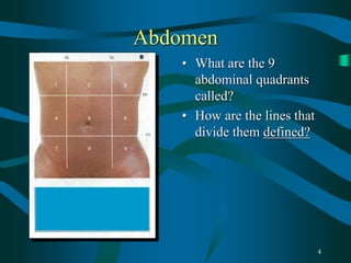
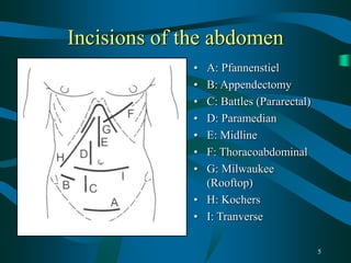
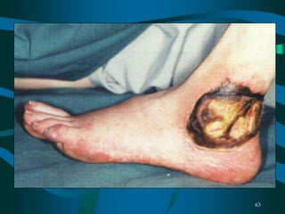
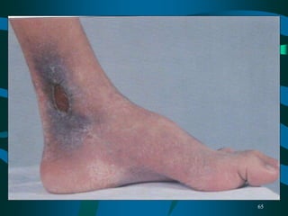
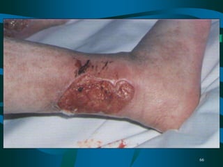
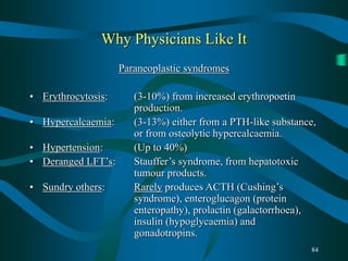
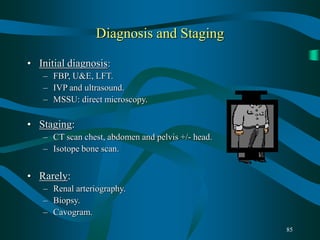
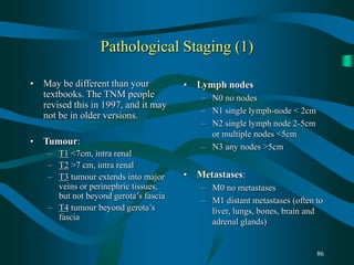
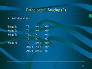
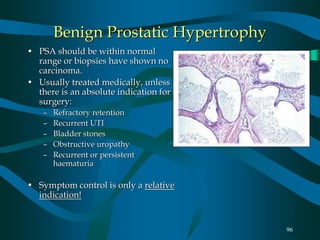
SlideShare a Scribd company logo
1
Final Year Students
Surgery and Urology
Mr Little
2
Objectives
• Standard history taking
• Physical examination
• Diagnosis and investigation
• Frequently asked questions
3
FIRST SENTENCE OF
ANSWER
• “I will take a full
history and perform a
full physical
examination.”
• Examiners expect this
• Remember to say that
you will do a PR, if
appropriate.
4
Abdomen
• What are the 9
abdominal quadrants
called?
• How are the lines that
divide them defined?
5
Incisions of the abdomen
• A: Pfannenstiel
• B: Appendectomy
• C: Battles (Pararectal)
• D: Paramedian
• E: Midline
• F: Thoracoabdominal
• G: Milwaukee
(Rooftop)
• H: Kochers
• I: Tranverse
6
7
Hernias
• One of the few parts of final MB where surface
anatomy is crucial! Inguinal surface anatomy is used
to distinguish inguinal hernias into direct and indirect,
and to demonstrate femoral hernias.
• Most likely to get inguinal hernias, incisional hernias,
femoral hernias, para-umbilical hernias and epigastric
hernias.
8
Inguinal surface anatomy
• A: Inferior
epigastric artery
• B: Femoral nerve
• C: Femoral artery
• D: Femoral vein
• E is the most
important …
• THE PUBIC
TUBERCLE
9
Examination of the groin
• Usually means either a hernia, a testicular swelling or
lymph nodes.
• Groin Hernias:
• Either an inguinal lump or a scrotal lump you can’t get above.
If not clinically obvious, as the patient to stand and/or cough.
• Femoral hernias do not usually enter the scrotum.
• Try to gently reduce it.
• Surface anatomy: If it is below and lateral to the pubic
tubercle it’s a femoral hernia. If it’s superior or medial to
the pubic tubercle it’s an inguinal hernia. Inguinal is
commoner.
• Occlusion at the mid-inguinal point (i.e. halfway between the
pubic tubercle and the anterior superior iliac spine, i.e. the deep
ring) prevents an INDIRECT inguinal hernia from popping
out. In the middle-aged male, direct is commoner.
10
Other hernias
• Incisional: 10% of hernias. Either reduceable or not.
• Para-umbilical: True umbilical hernias are rare after childhood. Para-
umbilical hernias are found in fat middle-aged women. Repaired
using Mayo repair
• Epigastric hernias: Fit young men
• Frequent questions:
• What is a hernia?
• What are the three commonest hernias?
• Who gets most inguinal hernias? Why?
• What is the commonest female hernia?
• What are the complications of hernias?
• What hernias occur ouside the abdomen?
• What is a Richter’s hernia?
11
Scrotal swellings (i)
• CAN I GET ABOVE IT? If you can then it’s coming from the
scrotal structures.
• Common: Hydrocoeles, Epididymal cysts, spematocoeles.
• Uncommon: Tumours
• Examine as for any lump or bump:
• The 3 ‘S’s: Site, Size, Shape
• The 3 ‘C’s: Colour, Contour, Consistency
• The 3 ‘T’s: Tenderness, Tethering, Transillumination
• The ‘F’er: Fluctulence.
12
Scrotal swellings (ii)
• Hydrocoeles: Surround the testis and make it difficult to feel.
Transilluminate!!!
• Spermatocoeles and epididymal cysts: Smaller, and arise from
adnexal structures. Do not obscure testis usually.
• Tumours: Stony hard. Arise from testis.
• Frequent questions:
• What investigation is appropriate for a newly diagnosed hydrocoele?
• What is a hydrocoele?
• What are the risk factors for testicular cancer?
• What blood tests would you consider for testicle carcinoma?
• What are the common types of testicle cancer and how are they
treated?
13
Stomas (i)
• Most likely to be either an iliostomy or an end-colostomy.
• Less frequently, loop colostomy or ileal conduit(urinary
diversion)
• An iliostomy is in the RIF, and protrudes from the abdominal
wall. Most commonly fashioned following pan-proctocolectomy
in ulcerative colitis, or less frequently for caecal obstruction,
polyposis coli or severe Crohns colitis.
• An end colostomy is most frequently formed in the LIF, and
sits flush with the abdominal wall. Most commonly formed
after a Hartmann’s procedure or some rectal excisions.
14
Stomas (ii)
• Loop colostomies are uncommon, and tend to be formed in the
epigastrium from the transverse colon, or the LIF from the sigmoid
colon. It is usually a palliative procedure for carcinomatous
obstruction.
• Ileal conduit urinary diversions look just like ileostomies, but the
bag will contain urine, even if only a little. They are usually formed
when the patient has had a cystectomy, often for carcinoma of the
bladder.
• Frequent questions:
• What is this?
• Why is it there?
15
Stomas (iii)
• Complications:
• ILIOSTOMY:
• Metabolic: High output, B12 and Folate deficieny, Stones,
Anaemia.
• Anatomical: Stoma Prolapse and retraction, less frequently
stomal stenosis or parastomal herniation
• COLOSTOMY:
• Mostly Anatomical:
– EARLY: Stomal necrosis, ischaemia
– LATE: Stomal retraction, prolapse, stenosis, parastomal
herniation etc
16
Small bowel obstruction
• Intestinal obstruction is characterised by vomiting,
abdominal distension, constipation and pain.
• In high obstruction, the vomiting tends to occur earlier then the
constipation. The reverse is the case for lower GI obstruction. In
high obstruction, the distension is minimal.
• The commonest causes are: Adhesions (60-80% of cases) and
Hernias (10-15%).
• Others include:
– Extrinsic: Volvulus, Non-GI neoplastic or inflammatory masses
– In the wall: Crohn’s disease, Intussussception, Strictures, Atresias.
– In the Lumen: Meconium ileus, Gallstones, Foreign body,
Faecolith.
17
Small bowel obstruction
• Treatment:
• Assess for strangulation (Localised tenderness & pain)
• If simple (ie no strangulation):
– IV fluids, NBM, Analgesia
– FBP, U&E
– Monitor urine output
– NG aspiration for vomiting, if required.
• Remember, the rule is that 80% of simple small bowel
obstruction will settle with conservative treatment.
18
Large Bowel obstruction
• Causes:
• Colonic Carcinoma (65%)
• Diverticular disease (10%)
• Volvulus (5%)
• Crohn’s disease
• Hernia
• Strictures: Ischaemic,
anastamotic, inflammatory etc
19
Abdominal Masses
• Non-pathological: Gravid Uterus, Faeces in colon, Riedels lobe
of liver, kidneys in a thin person.
• Standard questiion is to distinguish a kidney from a spleen
• RIF:
– Sore: Appendix mass/abscess, Crohns mass
– Not Sore: Cancer (Caecal, Ovarian, Renal), Fibroids and ovarian
cysts, transplanted kidney.
• Epigastric
– Ca stomach, colon, liver, pancreas
• RUQ
– Liver, Colon, Right kidney, GB, Adrenal
20
Colorectal Disorders
• Colorectal Carcinoma
• Important point is that the history is different depending on the
side of the colon the carcinoma is:
– RIGHT sided carcinomas Present insidiously because the colonic
contents are liquid and the colon relatively spacious at this point.
Presentation is with the development of anaemia : Fatigue,
malaise, weight loss, dark blood in stools.
– LEFT sided carcinomas present with the symptoms of narrowing
the lumen to more solid matter, Altered bowel habit, alternating
constipation and diarrhoea, brighter red PR bleeding.
– RECTAL carcinomas also have the symptoms of tenesmus and
passage of mucus PR
• Colonic carcinomas can also perforate and fistulate
21
Colorectal Disorders
• Risk Factors:
– Longstanding Ulcerative colitis
– Polyposis Coli
– Family History
• Differential diagnosis:
– DIVERTICULAR DISEASE
• Any cause of large bowel obstruction
• PR Bleeding: Colorectal CA, Diverticular disease, Angiodysplasia,
Haemorrhoids, Anal fissure
• Investigations of choice: Barium enema and sigmoidoscopy,
with CT if carcinoma is confirmed.
22
Colorectal Disorders
• TREATMENT:
• Surgery
• Chemotherapy with 5-Fluorouracil for tumours of stage Dukes
C or greater. Some would also give it for Dukes B
• Colorectal Carcinoma Complications: Bleeding, Perforation and
Fistulation into adjacent organs and obstruction…
23
24
Colorectal Disorders
• DIVERTICULAR DISEASE:
• The great pretender. Can mimic colorectal carcinoma
remarkably. Both cause PR bleeding, can cause intestinal
obstruction, can perforate and can fistulate.
• Related to low-fibre diet
• Chronic symptoms investigated with Ba enema and
Sigmoidoscopy. (Aren’t they all)
• Often presents acutely with LIF pain and tenderness and
rebound. Settles with conservative Rx and Antibiotics usually.
• Majority do not require surgery, and are controlled with dietary
advice
25
Colorectal Disorders
• Inflammatory Bowel disease: Summary table:
26
Colorectal Disorders
• Ulcerative colitis
• Palpable masses rare, never
fistulates
• Anorectal fissures and
infection occur less
frequently
• Medical Rx successful in
80%
• Surgery can be curative
• Crohn’s Disease
• Occasionally forms
inflammatory masses or
fistulae
• Anorectal sepsis common
• Medical Rx inadequate in
80%
• Surgery for complications
only
27
Colorectal Disorders
• Medical treatment of ulcerative colitis:
• 5-ASA derivatives, such as Sulphasalazine or Mesalazine can be used
to reduce relapse rate. Delivered either orally, Enemas or suppository
form
• Prednisolone can be used topically as above, or can be given orally for
relapses, up to 60mg per day. It must be weaned off, rather than
stopped suddenly.
• Anti-diarrhoeals: Codeine, Loperamide.
• Endoscopy is used for surveillance
• Surgery is used for failure of medical Rx, For Carcinoma, Failure of
steroids to induce response in few days, and Toxicity: >8 bloody stools
per day, pulse >100, Temp > 38.5 C, Transverse colon dilated beyond
5cm and hypoalbuminaemia.
• Often a panproctocolectomy and iliostomy
28
Colorectal Disorders
• Crohns disease
• Usually not accessable by topical preparations, so generally
treated with systemic 5-ASA derivatives or steroids where flare
up occurs.
• Surgery is often used for the complications: Abscess drainage,
resection of fistulas, and removal of strictured segments.
• Cannot be cured by surgery. Colonic crohns is a contraindication
for continent pouch procedures.
29
30
31
32
Oesophagus and Stomach
• More likely to be used in
the long case setting, as
signs are uncommon, but
history is usually detailed.
• Haematemesis and
melaena.
• Use of NSAIDS eg
Aspirin, Smoking,
Alcohol, Steroids,
WARFARIN
• Remember to say PR
33
Stomach and Oes. History (i)
• This is actually the history of upper GI bleeding, Upper abdominal
pain, and upper GI obstruction, with history.
• Upper GI Bleeding:
– Was it haematemesis or melaena
– Did one start before the other
– When did it start
– How many times has it happened
– When did it happen last
– Was it precipitated by anything
– How much blood was there
– Coffee-grounds vs fresh blood
– Did it make them collapse
– Have they felt tired / easily fatigued / easily short of breath… ANAEMIA!
34
Stomach and Oes. History (ii)
• Upper GI obstruction: Oesophagus or Gastric?
– At the level of the Oesopagus: Dysphagia, regurgitation of food
– At the level of the Stomach: Early satiety, Forceful vomiting, Sucussion
splash if you’re lucky!
– Both give marked weight loss
• History
– Drug history: NSAIDS + Aspirin, Warfarin, Steroids.
– Smoking: Amount per day, and duration. “Have you ever smoked?”
– Alcohol: As for smoking.
– Family history
• Upper GI pain and differential later
35
Stomach and Oesophagus signs
• Signs: THINK: Bleeding, Baccy and Booze.
• Bleeding: Pale conjunctivae, pallor. Melaena. (ie more chronic signs)
Unlikely to see tachycardia, hypotension etc, as this tends to be quite
acute. Remember NSAIDS and Warfarin. Trap for the unwary:
Guinness or Iron tablets give you black stools.
• Smoking: The most obvious is often nicotine stained fingers! Also
remember the signs of COAD: It implies a smoking history. Smoking
also significant in the context of cancer: Look for wasting, an LIF
mass, Hepatomegaly etc.
• Alcohol: This essentially means the signs of chronic liver disease.
36
Stomach and Oesophagus
• Oral Questions relating to haematemesis / melaena:
• What risk factors did this patient have for Upper GI bleeding?
• Why are they on Aspirin / Warfarin / Steroids ?
• What investigations will you do AND WHY?:
• FBP, Coag, U&E, LFT, G&Csm 4 units, OGD.
• If the OGD is negative what investigation will you do (Melaena)?
• BARIUM ENEMA
• What are the commonest causes of upper GI bleeding:
• Gastritis, Duodenitis
37
Stomach and Oesophagus
• What are the causes of upper GI bleeding:
• Oesophagus: Oesophagitis, Oesophageal erosions, Mallory-Weiss tears,
Oesphageal varices, Oesophageal carcinoma (rarely)
• Stomach: Gastritis, Gastric erosions, Gastric ulcers, Gastric carcinoma.
• Duodenum: Duodenitis, Duodenal Ulcers, Aorto-Enteric fistulas.
• How do you treat upper GI bleeding?
• Most settle with conservative Rx: IV fluids, NBM, PPI. Can use endoscopy
therapeutically for both ulcers and varices. Open resection of ulcer etc rare.
• What are oesophageal varices?
• What causes them?
• What are the other sites of porto-systemic anastamosis?
38
Stomach and Oesophagus
• Difficult
Question
• How would
you see this?
• What is it?
39
Stomach and Oesophagus
• This is a
barium
swallow.
• What is the
likely
diagnosis?
• Difficult
question.
40
Stomach and Oesophagus
• How would you investigate the patient with a tumour of the
stomach or oesophagus on OGD ?
• Stage them, using a CT scan of Chest, Abdomen and Pelvis. If
resectable then treat surgeically if possible. Radiotherapy is second-
line option for oesophagus. Many Gastric and oesophageal tumours are
irresectable, so remember to consider palliative care options such as
bypass and stenting, laser resection of oesophagus.
• What is Barrett’s Oesophagus?
• What is it’s significance?
• How do you manage it?
• What might cause dysphagia?
• What drugs are associated with upper GI bleeding?
41
• What kind of X-ray is this?
• What does it show?
42
The Liver, biliary system and pancreas
• History features:
• Jaundice, Upper abdominal pain.
• Jaundice: The A to I varies from book to book, but I use:
– Alcohol
– Blood tranfusion
– Contact
– Drugs (eg Paracetamol)
– Extrahepatic
– Family History, Foreign Travel
– Gallstones
– Homosexuality
– Infections ( HBV, EBV, Leptospirosis)
43
The Liver, biliary system and pancreas
• Upper Abdominal pain:
• Epigastric:
– Upper GI: Gastritis, Oesophageal reflux, Peptic Ulcer, Perforation of
gastric or duodenal ulcer.
– Hepatobiliary: Biliary Colic, Pancreatitis
– REMEMBER MYOCARDIAL INFARCTION
• Right Hypochondrium
– Hepatobiliary: Biliary Colic, Cholecystitis, Cholangitis, Hepatitis,
Pancreatitis, Fitz-Hugh-Curtis syndrome.
– Upper GI: Peptic Ulcer, Perforation of Ulcer
– Other Abdominal: Subphrenic abscess, Appendixitis, Renal Colic
– Extra-Abdominal: Right lower lobe pneumonia, Pulmonary embolus.
44
The Liver, biliary system and pancreas
• Physical examination often involves looking for the signs of
chronic liver disease in the case of jaundice or pancreatitis.
Cholecystitis is most frequently abdominal signs only, if un-
complicated.
• Surgeons most frequently look after Cholecystitis and
Pancreatitis.
• Cholecystitis: Remember the 5 F’s! It gives the game away.
– Right Upper quadrant pain, aching/colicky, may radiate around to
back, often onset at night or in response to fatty foods, may be
associated with vomiting. Gallstones in the gallbladder do not
cause jaundice unless they move into the common bile duct.
– Complications: Jaundice, Pancreatitis.
45
The Liver, biliary system and pancreas
• Physical examination usually only shows subcostal tenderness
and murphy’s sign. Rarely palpable. 5 F’s
• The investigation of cholecystitis is essentially the investigation
of upper abdominal pain.
• FBP, U&E, AMYLASE, LFT’s, Erect Chest X-Ray, Abdominal
film.
• Initial Rx: IV Fluids, Nil by mouth.
– Cefuroxime for infection. Most add Metronidazole as well.
Cholangitis is Jaundice, Fever and Rigors. This is known as
Charcot’s triad.
• Investigation: Ultrasound of abdomen. OGD if it’s normal.
• Treatment: Laparoscopic cholecystectomy or open
cholecystectomy. (Don’t use laparoscopic surgery if there are
adhesions, or a common bile duct stone)
46
The Liver, biliary system and pancreas
• Stones in the common bile duct may
produce jaundice and pancreatitis.
They are suspected when ultrasound
detects a dilated common bile duct, or
visualised duct stones.
• An ERCP can the be carried out to
prove the fact, and the sphincter of
oddi divided to allow the stones to
pass harmlessly into the duodenum.
• They are not removed by a
laparoscopic cholecystectomy. At the
time of open surgery, an intraoperative
cholangiogram is performed to ensure
that none are present. If there are, then
they are removed with specialised
forceps.
47
The Liver, biliary system and pancreas
• Pancreatitis:
• Presentation is similar to Cholecystitis, but pain is more centralised
and is more severe, possibly radiating to the back. Classically the
patient lies still with their knees bent. They are generally more unwell.
May be shocked, jaundiced.
• Physical signs are often absent or very scanty! Consider the signs of
chronic liver disease, Cullens sign and Gray-Turner’s sign. Abdominal
tenderness to palpation can be poorly localised and hard to define.
Pyrexia, Tachycardia.
• Initial investigations are the same as for cholecystitis: FBP, U&E, LFT,
Amylase, CXR, AXR. Amylase is considered diagnostic for
pancreatitis if it is greater than 1000 u/ml. Abdo Xray may show a
sentinel loop or loss of psoas shadow.
48
The Liver, biliary system and pancreas
• Causes: Gallstones and Alcohol most common. Also GET
SMASH’D: Trauma and Surgery, Steroids, Mumps, AI, Scorpions,
Hyperlipidaemia and drugs.
• Treatment:
• Once diagnosis is established do an arterial blood gas and a serum calcium &
glucose. Treat any hypocalcaemia.
• IV fliuds, nil by mouth. Aim to keep urine output above 30 mls per hour.
Consider central line in elderly or CCF patients.
• Opiate analgesia
• NG tube
• Monitor
– Hourly urometer, Pulse and BP
– 12-hourly arterial bolld gases
– Daily FBP, U&E, Calcium and amylase
49
The Liver, biliary system and pancreas
You must learn the Ranson criteria, as it is a measure of
severity. 0-2: 2% motality, rising to 100% for 7+
• AT PRESENTATION
• Age >55
• WCC>16
• Glucose>11
• LDH>350
• AST>60
• DURING 1st 48 HOURS
• Haematocrit falls >10%
• Urea rises >10
• Serum Ca2+ <2
• Base excess <-4
• PaO2 <8 kPa
• Fluid sequestered> 6 litres
50
Jaundice
• Most surgeons look after post-hepatic jaundice. This type of
jaundice is charcterised by dark urine and pale stools. This is
because bilirubin products are not allowed to pass into the GI
tract via the biliary tree, but conjugated bilirubin is water
soluble and so darkens the urine.
• Jaundice is divided into pre-hepatic, hepatic and post-hepatic
aetiologies.
• Pre-Hepatic: Haemolysis, Heriditary eg Gilbert’s
• Hepatic: Cirrhosis, Carcinoma infiltration, Viral, Autoimmune,
Paracetamol and other drugs. Halothane is no longer used.
• Post-Hepatic (Obstructive): CBD gallstones, Pancreatic
carcinoma, Nodes in the porta-Hepatis, Cholangiocarcinoma.
Complication of lap chole?
51
Jaundice
• The physical signs of chronic liver disease is a gob-standard
exam question. Remember to start at the hands and work up!
• Hands: Finger clubbing, Leukonechia, Palmar erythema,
Dupuytren’s, Asterixis
• Skin: Bruising, Spider Naevi, hair loss
• Face & Head: Jaundiced sclera. Constructional apraxia, Foetor
hepaticus.
• Chest: Gynaecomastia
• Abdomen: Enlarged liver or spleen, Ascites, Caput Medusae.
Remember to ascertain the nature of the liver edge, consistency,
tenderness. Bruits etc astronomically rare.
• Legs: Peripheral pitting oedema from hypoalbuminaemia.
52
Jaundice
• Investigation of the jaundiced patient:
• FBP, U&E, LFT, Coagulation, Hepatitis
• Remember Macrocytosis as a sign of alcoholism
• In obstructive jaundice, the Alkaline Phosphatase will rise out
of proportion to the transaminases (These may be slightly up),
and the bilirubin is conjugated.
• Hepatocellular jaundice is characterised by marked
transaminase rise, and a mix of conjugated and unconjugated
bilirubin.
• Prehepatic Jaundice is unconjugated, (so not present in urine)
• Ultrasound: This will tend to separate the Hepatic and Post-
hepatic jaundices quite nicely also.
53
Jaundice
• Questions:
• What are the causes of Cirrhosis?
• Alcohol and Hepatitis are the first things to say
• If pressed:
– Primary Biliary Cirrhosis, Autoimmune hepatitis
– Haemochromatosis and Wilson’s disease
– Drugs eg Methotrexate
– Budd-Chiari syndrome (Hen’s tooth), Cong Cardiac failure.
– Alpha-1-antitrypsin deficiency
• How would you prepare the jaundiced patient for surgery?
• Basically this means correcting any coagulation abn with FFP and/or
Vitamin K, and giving perioperative IV fluids to maintain a good urine
output (Hepatorenal syndrome more likely in the dehydrated)
54
55
Breast lumps:
• Common Breast Lumps…
• Young Women: Fibroadenoma / Abscess
• Pregnant / recent sprog: Galactocoele / abscess
• Middle aged and elderly women: Cancer higher
up the differential diagnosis list.
• Investigation ***IN THIS ORDER***
1. Clinical examination
2. Mammogram (USS if premenopausal)
3. FNA aspiration
56
Breast Cancer Treatment
• Local treatment:
– Usually lumpectomy/partial mastectomy if
feasble. Not feasable for central tumours, large
tumours or tumours in small breasts etc.
• Systemic treatment:
– Premenopausal usually chemotherapy
– Postmenopausal usually Tamoxifen.
(Remember risk of uterine CA)
57
Vascular Cases
• Likely Cases:
– Aneurysm (AAA)
– Ischaemic lower limb
– Ulcer
– Varicose veins
58
Aneurysm
• Mostly Aortic (Within abdo) Next most common is splenic
artery aneurysm. Think of Elderly male hypertensive
smokers with family history.
• Usually need repaired if greater than 5 centimeters in
diameter (Risk of bursting increases above this)
• Inv: FBP, U&E, ESR (?Inflammatory), CT scan, Chest X-
ray and ECG
• Need emergency CT if painful aneurysm – has it burst?
• Now can be repaired with endoluminal stenting in some
cases.
59
Chronically Ischaemic lower limb:
• Signs:
– Absent pulses
– Colder than other limb
– Thin, Shiny skin
– Hair loss on leg
– Ulcers
– gangrene
• Symptoms:
– Claudication –
• At what distance.
• How long does it take to
disappear at rest
• On the flat or just uphill
• Rest pain?
• Relevant PMH:
– Smoking
– Diabetes
– Hypertension, Stroke, MI, etc
60
Investigation
• Ankle-Brachial pressure index:
– Take systolic BP at brachial artery using doppler USS,
and compare it to the systolic BP at posterior tibial or
dorsalis pedis artery.
– Divide the ankle value by the arm value. Should be the
same. 0.5=Claudication, 0.3=Rest pain, 0.2= impending
gangrenous changes.
• Definitive main investigation is arteriography.
61
What’s this?
62
Ulcers
• Usually:
– Venous: Medial malleolus, Venous excema,
varicose veins, skin pigmentation. Surprisingly
painless!
– Arterial: Black, sloughy, painful. Look for other
signs of limb ischaemia.
– Neuropathic. Sensory loss. Often diabetics with
good pulses
– Vasculitic. Sharp, punched out lesions.
63
64
65
66
67
Urology in a nutshell
68
Causes of Haematuria
• PRE-RENAL: Coagulation disorder, Sickle-cell,
Vasculitis
• RENAL: Glomerular disease, Carcinoma, Cystic
disease, Trauma, A-V malformations,
Emboli
• POST RENAL: Stones
Infection (Bladder / Prostate / Urethra)
Carcinoma (Bladder / Prostate),
Traumatic bladder catheterisation,
Inflammatory Cystitis
69
Investigation of Haematuria
• Painless haematuria
is carcinoma until
proven otherwise
• Initial investigation
(For Anything) is
full history and
examination.
• PR!
70
Investigation of Haematuria
• Blood Investigations
• FBP – Anaemic? White cell count raise indicative of
infection?, Enough platelets?
• U&E – Are their kidneys working? (Crude test)
• Coagulation screen – Haemophilia?, Warfarin?
• In Men … PSA
• REMEMBER MSSU – Direct microscopy
and culture.
71
More Investigation
• Urinary Cytology:
– Not very sensitive, but an unequivocally positive cytology is
quite specific for TCC bladder.
• RADIOLOGY:
– IVP
– Ultrasound
– Both are very sensitive and specific, but USS better for small
peripheral renal lesions, and IVP better for renal pelvis and ureters.
• FLEXIBLE CYSTOSCOPY
72
Bladder Cancer Incidence
• Males outnumber females by about 2.7 to 1
• Average age at diagnosis is 65 years.
• 85% confined to bladder at time of presentation.
• 70% will recur after treatment, and 30% of these will progress
• Risk Factors:
– SMOKING
– Chemical carcinogens – chemical, dye, rubber, petrol, leather and
printing industries are at increased risk. Also Cyclophosphamide
– Not Coffee
73
Initial diagnosis and treatment
• Most are diagnosed using flexible cystoscopy under fresh air,
and haematuria investigations.
• Non-invasive tests such as PCR analysis, and Matrix-
metalloproteinase-9 are yielding some results with high
specificity and sensitivity, but remain research tools at present.
• IVP – Make sure there are no TCC in the renal pelvis or ureters.
For every 50-60 bladder carcinomata, there are 3 Renal pelvis
TCC’s and one ureteric TCC
• TURBT – Curative for early disease, also provides histology.
74
Staging of TCC bladder
CIS Ta T1 T2 T3a T3b
mucosa
sub-
mucosa
detrusor
muscle
Peri-
vesical
fat
75
Staging Continued
• Also Stage T4a, where the prostate is invaded, and T4b where
there is pelvic structure invasion. T3b has a worse prognosis
than T3a.
• Lymph nodes status, and presence or absence of metastases.
• Tumour Grade i.e. degree of preservation of cellular
architecture, mitotic figure number etc.
• Why bother staging? Treatment is tailored to stage of disease
76
Treatment Options
• Ta Single, G1-2, Not recurrent: TURB
Multiple, recurrent, or high grade: TURB+Intravesical chemo
• T1 G1-2 TURB+Intravesical chemo
G3 TURB+BCG
• CIS TURB+BCG
• T2-T4 Radical Cystectomy
Radiotherapy
• N+ or M+ ?Chemotherapy - MVAC
77
Surgery and Radiotherapy
• TURBT
• Radical cystectomy: Major intra-abdominal procedure. Can
divert urine either to an ileal conduit, or make a new bladder
from bowel or colon. Incidental lymph node mets found in 20-
35% 5 year survival a bit better than DXT – 65% for T2-T4
disease.
• Pelvic Radiotherapy: 20-40% 5 year survival, but 15% get local
complications, e.g. radiation cystitis or proctitis.
78
Chemotherapy
• Intravesical
– Mitomycin response rate 40-50%. Consider single dose
intravesically post surgery rather than 6 week course. Occasionally
produces chemical cystitis.
– BCG decreases recurrence from 80 to 40%, and decreases
progression from 35% to 7%. Cystitis in 90% and haematuria in
33%
• Systemic
– MVAC (Methotrexate, Vinblastine, doxorubicin, cisplatin) 13-35%
response rate, but median survival rate is only one year
– Difficult to convince oncologists to give!
79
The bottom line
• Five-year survival :
• Stage
• Ta 94%
• T1 69%
• T2 40%
• T3 31%
• T4 0
• Worse with increasing grade, and increased grade and stage
associated with increased risk of metastatic disease.
80
Why Is a Spleen Not a Kidney?
• Very common exam question!
• Kidneys do not move with respiration
• Kidneys enlarge up & down, not to the RIF
• Kidneys are resonant to percussion
• Kidneys are ballottable
• Kidneys do not have a notch
• You can get above a kidney
81
What Causes Big Kidneys?
Unilateral:
• Carcinoma
– Renal cell, transitional cell
in adults
– Nephroblastoma (wilm’s
tumour) in kids
• Hydronephrosis
– Tend to be chronic e.g. PUJ
obstruction, reflux
• Simple cysts
• Compensatory
hypertrophy
Bilateral
• Polycystic kidneys
• Bilateral hydronephrosis
• Amyloid … hen’s tooth
82
Renal Cell Carcinoma
• Synonyms: hypernephroma, clear-cell carcinoma.
• Incidence: 2-3% of all adult cancers.
• Renal cell carcinoma is roughly 85% of all renal tumours, the
remainder being things like transitional cell carcinoma of the
renal pelvis, and renal sarcoma.
• Age peak of 40-70 years old.
• Males outnumber females 2:1
• Risk factors:
– Smoking.
– Others: Von-Hippel Lindau syndrome, horseshoe kidneys, adult
polycystic kidney disease, acquired renal cystic disease.
83
RCC Aetiology and Presentation
• Arises from the cells of the proximal convoluted tubule.
• Presentation classically:
– Haematuria
– Flank pain
– Loin mass
• 50% diagnosed as incidental findings in 1995, during USS or
CT for other problem.
• 30% present with metastatic symptoms:
– Bone pain, dyspnoea, cough, etc.
– (Often to liver, lungs, bones, brain and adrenal glands)
Only true in 15%
84
Why Physicians Like It
Paraneoplastic syndromes
• Erythrocytosis: (3-10%) from increased erythropoetin
production.
• Hypercalcaemia: (3-13%) either from a PTH-like substance,
or from osteolytic hypercalcaemia.
• Hypertension: (Up to 40%)
• Deranged LFT’s: Stauffer’s syndrome, from hepatotoxic
tumour products.
• Sundry others: Rarely produces ACTH (Cushing’s
syndrome), enteroglucagon (protein
enteropathy), prolactin (galactorrhoea),
insulin (hypoglycaemia) and
gonadotropins.
85
Diagnosis and Staging
• Initial diagnosis:
– FBP, U&E, LFT.
– IVP and ultrasound.
– MSSU: direct microscopy.
• Staging:
– CT scan chest, abdomen and pelvis +/- head.
– Isotope bone scan.
• Rarely:
– Renal arteriography.
– Biopsy.
– Cavogram.
86
Pathological Staging (1)
• May be different than your
textbooks. The TNM people
revised this in 1997, and it may
not be in older versions.
• Tumour:
– T1 <7cm, intra renal
– T2 >7 cm, intra renal
– T3 tumour extends into major
veins or perinephric tissues,
but not beyond gerota’s fascia
– T4 tumour beyond gerota’s
fascia
• Lymph nodes
– N0 no nodes
– N1 single lymph-node < 2cm
– N2 single lymph node 2-5cm
or multiple nodes <5cm
– N3 any nodes >5cm
• Metastases:
– M0 no metastases
– M1 distant metastases (often to
liver, lungs, bones, brain and
adrenal glands)
87
Pathological Staging (2)
• And after all that:
Stage 1: T1 N0 M0
Stage 2: T2 N0 M0
Stage 3: T1-2 N1 M0
T3 n0-1 m0
Stage 4: T4 any N M0
Any T N2-3 M0
Any T any N M1
88
Treatment
• Get rid of the primary tumour
– Radical nephrectomy
• Open or laparoscopic?
Laparoscopic has faster
patient recovery, but is time-
consuming in theatre.
– Partial nephrectomy for small
polar tumours, less than 4 cm
diameter.
– Embolisation.
– Consider small tumours below
2cm in unfit patients for
watching?
89
Chemotherapy?
• Renal cell carcinoma has a track record of being unresponsive to
chemotherapy.
• Immunotherapy:
– Only works for clear cell type, the largest pathological group of RCC.
– Only used if lymph-node metastases, or metastases to solid organs.
Cerebral metastases are a contra-indication.
– Combination regimens based on cytokines
– Interleukin-2 19% response rate alone
– Interferon-α 11% response rate
– Together 25-30% response
– Combinations of interleukin-2, interferon-α and 5-fluorouracil have
produced response rates of 39% in one series.
• The main problem with immunotherapy is that it has a lot of side effects, such
as fever, hypotension, tachycardia, oliguria. You can end up in ICU from it, but
this is rare.
90
The Bottom Line:
• Responses to immunotherapy tend to be short-lived, i.e. Months.
• Relapsers tend not to respond to more immunotherapy.
5-year survival:
• T1 88-100%
• T2 & t3a 60%
• T3b 15-20%
• T4 0-20%
Tumor stage T3 can be subdivided into
2 groups: T3a can invade the adrenal or
perinephric tissue, but remains within
gerota’s fascia, and T3b invades the
Renal vein or IVC.
91
Other Renal Tumours (Adult) … Uncommon
• Sarcoma: females outnumber males by 2:1. Surgery only
effective therapy. Prognosis poor.
• Transitional cell carcinoma of the renal pelvis.
• Haematologic tumours (lymphoma deposits etc)
• Metastatic tumours: (In order of frequency)
– Lung
– Breast
– Stomach
• Non-tumours that masquerade as such:
– Oncocytoma
– Angiomyolipoma
– Others: leiomyomas, haemangiomas
92
Symptoms of BOO
• “Irritative”
– Frequency, Urgency, Nocturia > 2 times per night
• “Obstructive”
– Hesitancy, Poor flow, Terminal dribbling.
• Others:
– Recurrent urinary tract infections as a consequence of
impaired bladder emptying
– Haematuria rarely.
• Can be scored using IPSS system
93
Causes of impaired bladder emptying(i)
• Detrusor failure, i.e. “weak bladder”
– Think of the bladder with it’s nerve supply. Any
disorder of nervous control may impair emptying.
For example:
• Spine: Injuries, Disc prolapse, Spina bifida.
• Nerves: Diabetes, Multiple sclerosis.
• Bladder: Myogenic failure.
94
Causes of impaired bladder emptying(ii)
• Outlet Obstruction i.e. “blocked bladder”
• Mostly a male phenomenon. Can be any cause from
bladder neck to outside world. For example:
– Prostatic enlargement
– Bladder neck hypertrophy (Often post TURP)
– Urethral strictures (Often post instrumentations, also STD)
– Meatal stenosis (As for strictures, also after botched
circumcisions and a condition called Balanitis Xerotica
Obliterans)
– In male infants: Posterior urethral valves
95
Investigation of BOO symptoms
• History and examination
• Don’t forget to do PR!!!
• FBP, U&E, Glucose, MSSU
• Prostate specific antigen (PSA)
– Currently best blood-test for separating benign prostatic disease
from prostate cancer. It rises in cases of cancer. It can also rise with
urinary tract infections, and following episodes of urinary retention
or urinary instrumentation. Also slowly rises with age.
• Ultrasound of bladder (Is it emptying? If creatinine
abnormal then also ultrasound kidneys, looking for
obstructive uropathy)
• Urinary flow rate
96
Benign Prostatic Hypertrophy
• PSA should be within normal
range or biopsies have shown no
carcinoma.
• Usually treated medically, unless
there is an absolute indication for
surgery:
– Refractory retention
– Recurrent UTI
– Bladder stones
– Obstructive uropathy
– Recurrent or persistent
haematuria
• Symptom control is only a relative
indication!
97
Obstructive Uropathy
• Basically this is renal impairment caused by chronic
bladder outlet obstruction.
• It produces a detectable rise in creatinine, and
eventually can be seen on ultrasound as thinning of
the renal cortex.
• It means that the best treatment is operative relief of
obstruction.
98
Acute Urinary Retention
• This is the end-stage of urinary outflow obstruction.
• Preceding outlet symptoms, with the ‘straw that
breaks the camel’s back’ often being an intercurrent
UTI, an episode of constipation, immobility or post-
operatively.
• Provided there is no obstructive uropathy, can be
started on medical treatment and catheter removed
after a day or two.
99
Medical management of
prostatic obstruction
• Two broad groups of drugs:
– Alpha-blockers: These produce smooth muscle
relaxation within the prostate, and so widen the
outflow tract. Older generation drugs have been
associated with postural hypotension, but newer drugs
such Tamsulosin have minimised this.
– 5-alpha reductase inhibitors: blocks testosterone
metabolism to produce a 25% decrease in prostate
volume. Better for large ‘fleshy’ glands. 5% of patients
get hot flushes, and can also cause gynaecomastia.
Finasteride is the only drug in this group.
100
Surgical management of
prostatic obstruction
• A variety of operations has been tried, such as prostatic
microwave, cryoablation, and transurethral incision of the
prostate.
• However, none has replaced TURP (Trans-urethral resection of
prostate), which remains the gold standard.
• TURP is not always successful, however. The most common
early complication is failure to void after catheter removal (6%).
There is also a small risk of incontinence from external sphincter
damage.
• And finally: After TURP erectile failure may be produced, and
ALWAYS retrograde ejaculation is caused.
101
Prostatic carcinoma
• 4th commonest cancer death, after lung,colon and breast.
• Rare before age 40, peak incidence in 70’s.
• 70% arise in the peripheral zone of the prostate.
• Most often found on PR or PSA testing
• 40% have metastases at the time of diagnosis.
102
Prostate Carcinoma
• Suspected on digital
rectal examination
or a raised PSA.
• Diagnosed by
BIOPSY, or found
at TURP.
103
Further investigation
• Is the prostate cancer
1. Metastatic ( Only treatable with Hormones)
2. Locally advanced (Hormones or radiotherapy)
3. Confined to the prostate (Hormones, Radio or Surgery)
• Staging of prostate cancer requires an isotope
bone scan. In patients under 75 years, where
surgery or radiotherapy are being considered, a
CT scan is also required.
104
Staging (TNM)
• T1a: Tumor in <5% TURP specimen,
incidental finding.
• T1b: Tumor in >5% TURP specimen,
incidental finding.
• T1c: Tumor non-palpable, diagnosed on
biopsy.
• T2a: Palpable, half a lobe or less
• T2b: Palpable, half to all of one lobe
• T2c: Palpable, involves both lobes.
• T3a: Unilateral extracapsular extension
• T3b: Bilateral extracapsular extension
• T3c: Tumor invades seminal vesicles
• T4a: Invades bladder neck, external
sphincter or rectum
• T4b: Invades levator muscles, or is
fixed to pelvic side-wall.
• N0: No lymph node metastases
• N1: Single node metastasis, below
2cm in size
• N2: Mets to a single node between
2 and 5 cm, or multiple nodes less
than 5cm.
• N3: Lymph node metastasis > 5cm
• M0: No Metastases.
• M1: Metastases
105
Treatment - Surgery
• For prostate cancer without metastases, which is confined
to the prostate, PSA is below 15, and age below 70,
consider RADICAL PROSTATECTOMY.
• Major intra-abdominal procedure, with significant
morbidity. Almost inevitably produces total erectile failure,
and a variable degree of incontinence. Incontinence tends
to improve / resolve with pelvic floor exercises.
• 5 year disease-free survival for T2 cancer with surgery is
80%
106
Treatment - Radiotherapy
• Consider in males with disease that has spread locally outside the
prostate below age 75. Also for prostate confined disease in men below
70 years, unfit for surgery, and without metastases.
• Can be given as external beam or brachytherapy (implanted radioactive
particles within the prostate)
• Complications:
– Bladder: Frequency, Urgency, Haematuria.
– Bowel: Diarrhoea, Tenesmus, PR bleeding.
– Skin: Rash
– Rarely, fistulas may be caused.
• 5 year disease free survival for T2 disease is 70% (Slightly lower than
for surgery)
107
Treatment - Hormones
• Prostate carcinoma is dependant on testosterone to survive. The
medication used attempts to block testosterone synthesis. They are
used in cases of metastatic disease or age >75 years or unfitness for
either surgery or radiotherapy for localised disease.
– Gonadotropin analogues: Goserelin, Leuprolin. Often given as monthly or
3-monthly injections. Initially produce a tumour flare, so can only be
started after several days of an anti-androgen.
– Anti-androgens: Flutamide, Bicalutamide, Cyproterone acetate. Tablet
form once daily.
– Both of these medication groups produce significant side-effects: most
often hot-flushes, erectile failure, loss of libido, and gynaecomastia.
• Treatment can be combined, using both a GNRH analogue and an anti-
androgen. On average a 2-year response is achieved until the
development of ‘Hormone-resistance’. Alterative drugs for advanced
disease include Oestrogens and Estramustine.
108
Ooh, Controversy!
• In elderly men, i.e. age greater than 75, there is an argument for
not investigating an asymptomatically raised PSA.
• The reason is that at this age, there is a significant chance they
will die of something else, and the only treatment would be
Hormonal manipulation anyway. Some argue that treatment
should be witheld until the development of outflow symptoms
or metastatic discomfort, and then instituted.
• The idea is to avoid needlessly treating and worrying elderly
men
109
Urinary tract Calculi
• Ureteric Calculi: Most pass without difficulty, especially if
below 5mm in diameter. Above 7mm they are unlikely to pass.
• If calculi are not passing, or if they are causing blockage of the
kidney as shown on IVP, then they can be removed
endoscopically using a Ureteroscope and either Laser or EHL or
basket extraction. If there’s a lot of ureteric swelling then a stent
can be left in for 4-6 weeks and then removed.
• Ureteric calculi can be a symptom of a raised serum Calcium, so
remember to check it!
110
Urinary tract Calculi
• Renal Calculi:
• Generally present either as a cause of recurring infections or a
cause of pain in the loin or flank. If large enough, they can
seriously compromise renal function. This can be detected using
a DMSA scan.
• In a functioning kidney, stones up to about 2cm can be
fragmented with ESWL sessions, which may be multiple. Larger
stones or multiple stones may be dealt with operatively using
PCNL, where a tube is inserted though the flank into the renal
pelvis, and the stones surgically fragmented and removed. Very
large stones may need to be removed using an open
pyelolithotomy.
• If the kidney is non-functioning then it can be removed.
111
Okay brain, it’s
all up to you.
(Homer J Simpson)

More Related Content

What's hot

All Things Ostomy: FightCRC CRCWebinar Feb 14 2017
All Things Ostomy: FightCRC CRCWebinar Feb 14 2017All Things Ostomy: FightCRC CRCWebinar Feb 14 2017
All Things Ostomy: FightCRC CRCWebinar Feb 14 2017
Fight Colorectal Cancer
 
Choledocholithiasis- obstructive jaundice
Choledocholithiasis-  obstructive jaundiceCholedocholithiasis-  obstructive jaundice
Choledocholithiasis- obstructive jaundice
Selvaraj Balasubramani
 
Imaging of Bowel Obstruction
Imaging of Bowel ObstructionImaging of Bowel Obstruction
Imaging of Bowel Obstruction
Rathachai Kaewlai
 
Pancreatitis
PancreatitisPancreatitis
Pancreatitis
Justin V Sebastian
 
Presentation 8 digestive system terms
Presentation 8  digestive system termsPresentation 8  digestive system terms
Presentation 8 digestive system termsmreyna9
 
Intestinal obstruction
Intestinal obstructionIntestinal obstruction
Intestinal obstructionshankar1976
 
Biliary disorders
Biliary disordersBiliary disorders
Biliary disorders
Justin V Sebastian
 
Biliary Atresia
Biliary AtresiaBiliary Atresia
Biliary Atresia
Jibran Mohsin
 
Abdominal pain- all quadrants- case based learning
Abdominal pain-  all quadrants- case based learningAbdominal pain-  all quadrants- case based learning
Abdominal pain- all quadrants- case based learning
Selvaraj Balasubramani
 
Intestinal obstruction lecture
Intestinal obstruction lectureIntestinal obstruction lecture
Intestinal obstruction lectureFaiz Hmoud
 
Intestinal Obstruction
Intestinal ObstructionIntestinal Obstruction
Intestinal Obstructiondrcerof
 
Bowelobstruction
BowelobstructionBowelobstruction
Bowelobstruction
Zodzai Zabzaa
 
Intestinal obstruction
Intestinal obstructionIntestinal obstruction
Intestinal obstruction
Mohamed Mourad
 
Gallstone Disease
Gallstone DiseaseGallstone Disease
Gallstone Disease
Sanjiv Haribhakti
 
Intestinal obstruction, Ileus, and volvulus
Intestinal obstruction, Ileus, and volvulusIntestinal obstruction, Ileus, and volvulus
Intestinal obstruction, Ileus, and volvulus
Khaled AlKhodari
 
Cholelithiasis GB stone
Cholelithiasis  GB stone Cholelithiasis  GB stone
Cholelithiasis GB stone
AbdullahIhsaas
 
Surgical F1 Prep Talk - Dafydd Loughran
Surgical F1 Prep Talk - Dafydd LoughranSurgical F1 Prep Talk - Dafydd Loughran
Surgical F1 Prep Talk - Dafydd Loughran
welshbarbers
 
Practical exam preparation for nursing students
Practical exam preparation  for nursing studentsPractical exam preparation  for nursing students
Practical exam preparation for nursing students
MR. JAGDISH SAMBAD
 
Obstructive jaundice 19_9_2014
Obstructive jaundice 19_9_2014Obstructive jaundice 19_9_2014
Obstructive jaundice 19_9_2014
DrAnum Ammad
 
Intestinal Obstruction
Intestinal ObstructionIntestinal Obstruction
Intestinal Obstruction
MR. JAGDISH SAMBAD
 

What's hot (20)

All Things Ostomy: FightCRC CRCWebinar Feb 14 2017
All Things Ostomy: FightCRC CRCWebinar Feb 14 2017All Things Ostomy: FightCRC CRCWebinar Feb 14 2017
All Things Ostomy: FightCRC CRCWebinar Feb 14 2017
 
Choledocholithiasis- obstructive jaundice
Choledocholithiasis-  obstructive jaundiceCholedocholithiasis-  obstructive jaundice
Choledocholithiasis- obstructive jaundice
 
Imaging of Bowel Obstruction
Imaging of Bowel ObstructionImaging of Bowel Obstruction
Imaging of Bowel Obstruction
 
Pancreatitis
PancreatitisPancreatitis
Pancreatitis
 
Presentation 8 digestive system terms
Presentation 8  digestive system termsPresentation 8  digestive system terms
Presentation 8 digestive system terms
 
Intestinal obstruction
Intestinal obstructionIntestinal obstruction
Intestinal obstruction
 
Biliary disorders
Biliary disordersBiliary disorders
Biliary disorders
 
Biliary Atresia
Biliary AtresiaBiliary Atresia
Biliary Atresia
 
Abdominal pain- all quadrants- case based learning
Abdominal pain-  all quadrants- case based learningAbdominal pain-  all quadrants- case based learning
Abdominal pain- all quadrants- case based learning
 
Intestinal obstruction lecture
Intestinal obstruction lectureIntestinal obstruction lecture
Intestinal obstruction lecture
 
Intestinal Obstruction
Intestinal ObstructionIntestinal Obstruction
Intestinal Obstruction
 
Bowelobstruction
BowelobstructionBowelobstruction
Bowelobstruction
 
Intestinal obstruction
Intestinal obstructionIntestinal obstruction
Intestinal obstruction
 
Gallstone Disease
Gallstone DiseaseGallstone Disease
Gallstone Disease
 
Intestinal obstruction, Ileus, and volvulus
Intestinal obstruction, Ileus, and volvulusIntestinal obstruction, Ileus, and volvulus
Intestinal obstruction, Ileus, and volvulus
 
Cholelithiasis GB stone
Cholelithiasis  GB stone Cholelithiasis  GB stone
Cholelithiasis GB stone
 
Surgical F1 Prep Talk - Dafydd Loughran
Surgical F1 Prep Talk - Dafydd LoughranSurgical F1 Prep Talk - Dafydd Loughran
Surgical F1 Prep Talk - Dafydd Loughran
 
Practical exam preparation for nursing students
Practical exam preparation  for nursing studentsPractical exam preparation  for nursing students
Practical exam preparation for nursing students
 
Obstructive jaundice 19_9_2014
Obstructive jaundice 19_9_2014Obstructive jaundice 19_9_2014
Obstructive jaundice 19_9_2014
 
Intestinal Obstruction
Intestinal ObstructionIntestinal Obstruction
Intestinal Obstruction
 

Similar to Final year students

Liver abscess
Liver abscessLiver abscess
Liver abscess
Suman Baral
 
Liver specimen: Hepatocellular carcinoma, liver abscess
Liver specimen: Hepatocellular carcinoma, liver abscessLiver specimen: Hepatocellular carcinoma, liver abscess
Liver specimen: Hepatocellular carcinoma, liver abscess
Ankita Singh
 
Approach to colorectal cancer
Approach to colorectal cancerApproach to colorectal cancer
Approach to colorectal canceress_online
 
Intestinal obstruction2
Intestinal obstruction2Intestinal obstruction2
Intestinal obstruction2Larissa Sams
 
Rectal bleeding new presentation
Rectal bleeding new presentationRectal bleeding new presentation
Rectal bleeding new presentation
Valentine Okechukwu MBBS
 
Cholelithiasis and cholecystitis
Cholelithiasis and cholecystitisCholelithiasis and cholecystitis
Cholelithiasis and cholecystitis
drssp1967
 
Acute cholecystitis.pptx
Acute cholecystitis.pptxAcute cholecystitis.pptx
Acute cholecystitis.pptx
KartheeswariA
 
Presentation1 liver ultrasound
Presentation1 liver ultrasoundPresentation1 liver ultrasound
Presentation1 liver ultrasound
Abdallah Bashe
 
Surgical pathology specimens
Surgical pathology specimensSurgical pathology specimens
Surgical pathology specimens
SomendraBansal
 
Liver tumours.pptx
Liver tumours.pptxLiver tumours.pptx
Liver tumours.pptx
Pradeep Pande
 
small intestine.pptx surgery refrence belpy and love
small intestine.pptx surgery refrence belpy and lovesmall intestine.pptx surgery refrence belpy and love
small intestine.pptx surgery refrence belpy and love
MaxamuudxasanMaxamed
 
Approach to patient with ovarian cysts
Approach to patient with ovarian cystsApproach to patient with ovarian cysts
Approach to patient with ovarian cysts
Yahyia Al-abri
 
Appendicitis, diverticulitis, peptic ulcer disease, chron's disease
Appendicitis, diverticulitis, peptic ulcer disease, chron's diseaseAppendicitis, diverticulitis, peptic ulcer disease, chron's disease
Appendicitis, diverticulitis, peptic ulcer disease, chron's disease
haron taufiq
 
Rectal prolapse.pptx
Rectal prolapse.pptxRectal prolapse.pptx
Rectal prolapse.pptx
Pradeep Pande
 
GASTRIC CANCER.pptx
GASTRIC CANCER.pptxGASTRIC CANCER.pptx
GASTRIC CANCER.pptx
Dr Abdul Qayyum Khan
 
Presentation (1) (2).pptx
Presentation (1) (2).pptxPresentation (1) (2).pptx
Presentation (1) (2).pptx
ShafiUllah642916
 
colorectal malignancies.pptx
colorectal malignancies.pptxcolorectal malignancies.pptx
colorectal malignancies.pptx
masoom parwez
 
Benign Tumors of Small Intestine.pptx
Benign Tumors of Small Intestine.pptxBenign Tumors of Small Intestine.pptx
Benign Tumors of Small Intestine.pptx
Pradeep Pande
 
colon cancer, 7 final.pptx
colon cancer, 7 final.pptxcolon cancer, 7 final.pptx
colon cancer, 7 final.pptx
PradeepYadav509148
 
Gastrci outlet obstruction
Gastrci outlet obstructionGastrci outlet obstruction
Gastrci outlet obstruction
sunil kumar daha
 

Similar to Final year students (20)

Liver abscess
Liver abscessLiver abscess
Liver abscess
 
Liver specimen: Hepatocellular carcinoma, liver abscess
Liver specimen: Hepatocellular carcinoma, liver abscessLiver specimen: Hepatocellular carcinoma, liver abscess
Liver specimen: Hepatocellular carcinoma, liver abscess
 
Approach to colorectal cancer
Approach to colorectal cancerApproach to colorectal cancer
Approach to colorectal cancer
 
Intestinal obstruction2
Intestinal obstruction2Intestinal obstruction2
Intestinal obstruction2
 
Rectal bleeding new presentation
Rectal bleeding new presentationRectal bleeding new presentation
Rectal bleeding new presentation
 
Cholelithiasis and cholecystitis
Cholelithiasis and cholecystitisCholelithiasis and cholecystitis
Cholelithiasis and cholecystitis
 
Acute cholecystitis.pptx
Acute cholecystitis.pptxAcute cholecystitis.pptx
Acute cholecystitis.pptx
 
Presentation1 liver ultrasound
Presentation1 liver ultrasoundPresentation1 liver ultrasound
Presentation1 liver ultrasound
 
Surgical pathology specimens
Surgical pathology specimensSurgical pathology specimens
Surgical pathology specimens
 
Liver tumours.pptx
Liver tumours.pptxLiver tumours.pptx
Liver tumours.pptx
 
small intestine.pptx surgery refrence belpy and love
small intestine.pptx surgery refrence belpy and lovesmall intestine.pptx surgery refrence belpy and love
small intestine.pptx surgery refrence belpy and love
 
Approach to patient with ovarian cysts
Approach to patient with ovarian cystsApproach to patient with ovarian cysts
Approach to patient with ovarian cysts
 
Appendicitis, diverticulitis, peptic ulcer disease, chron's disease
Appendicitis, diverticulitis, peptic ulcer disease, chron's diseaseAppendicitis, diverticulitis, peptic ulcer disease, chron's disease
Appendicitis, diverticulitis, peptic ulcer disease, chron's disease
 
Rectal prolapse.pptx
Rectal prolapse.pptxRectal prolapse.pptx
Rectal prolapse.pptx
 
GASTRIC CANCER.pptx
GASTRIC CANCER.pptxGASTRIC CANCER.pptx
GASTRIC CANCER.pptx
 
Presentation (1) (2).pptx
Presentation (1) (2).pptxPresentation (1) (2).pptx
Presentation (1) (2).pptx
 
colorectal malignancies.pptx
colorectal malignancies.pptxcolorectal malignancies.pptx
colorectal malignancies.pptx
 
Benign Tumors of Small Intestine.pptx
Benign Tumors of Small Intestine.pptxBenign Tumors of Small Intestine.pptx
Benign Tumors of Small Intestine.pptx
 
colon cancer, 7 final.pptx
colon cancer, 7 final.pptxcolon cancer, 7 final.pptx
colon cancer, 7 final.pptx
 
Gastrci outlet obstruction
Gastrci outlet obstructionGastrci outlet obstruction
Gastrci outlet obstruction
 

More from mostafa hegazy

2021 book atlas_ofminimallyinvasiveandrob
2021 book atlas_ofminimallyinvasiveandrob2021 book atlas_ofminimallyinvasiveandrob
2021 book atlas_ofminimallyinvasiveandrob
mostafa hegazy
 
2014 book lower_abdominalandperinealsurge
2014 book lower_abdominalandperinealsurge2014 book lower_abdominalandperinealsurge
2014 book lower_abdominalandperinealsurge
mostafa hegazy
 
Parotid gland
Parotid glandParotid gland
Parotid gland
mostafa hegazy
 
The diagnosis and management of the acute abdomen in pregnancy 2019
The diagnosis and management of the acute abdomen in pregnancy 2019The diagnosis and management of the acute abdomen in pregnancy 2019
The diagnosis and management of the acute abdomen in pregnancy 2019
mostafa hegazy
 
(2) hirschsprung disease
(2) hirschsprung disease(2) hirschsprung disease
(2) hirschsprung disease
mostafa hegazy
 
Solid and cystic pediatric abdominal tumors
Solid and cystic pediatric abdominal tumorsSolid and cystic pediatric abdominal tumors
Solid and cystic pediatric abdominal tumors
mostafa hegazy
 
Pediatric laparoscopy
Pediatric laparoscopyPediatric laparoscopy
Pediatric laparoscopy
mostafa hegazy
 
Pediatric inguino scrotal problems
Pediatric inguino scrotal problemsPediatric inguino scrotal problems
Pediatric inguino scrotal problems
mostafa hegazy
 
Non traumatic abdominal pain in children
Non traumatic abdominal pain in childrenNon traumatic abdominal pain in children
Non traumatic abdominal pain in children
mostafa hegazy
 
Constipation&amp;incontinence
Constipation&amp;incontinenceConstipation&amp;incontinence
Constipation&amp;incontinence
mostafa hegazy
 
Pelvic ring for md1
Pelvic ring for md1Pelvic ring for md1
Pelvic ring for md1
mostafa hegazy
 
Open fractures
Open fracturesOpen fractures
Open fractures
mostafa hegazy
 
Neurosurgery revision
Neurosurgery revisionNeurosurgery revision
Neurosurgery revision
mostafa hegazy
 
Thyroid cancer hegazy
Thyroid cancer  hegazyThyroid cancer  hegazy
Thyroid cancer hegazy
mostafa hegazy
 
Thyroid case sheet
Thyroid case sheetThyroid case sheet
Thyroid case sheet
mostafa hegazy
 
Parathyroid hegazy
Parathyroid hegazyParathyroid hegazy
Parathyroid hegazy
mostafa hegazy
 
Parathyroid goda
Parathyroid godaParathyroid goda
Parathyroid goda
mostafa hegazy
 
Preop assess prep premed ahmed ibrahim
Preop assess prep  premed ahmed ibrahimPreop assess prep  premed ahmed ibrahim
Preop assess prep premed ahmed ibrahim
mostafa hegazy
 
Pheochromocytoma hegazy
Pheochromocytoma hegazyPheochromocytoma hegazy
Pheochromocytoma hegazy
mostafa hegazy
 
Adrenal glands hegazy
Adrenal glands hegazyAdrenal glands hegazy
Adrenal glands hegazy
mostafa hegazy
 

More from mostafa hegazy (20)

2021 book atlas_ofminimallyinvasiveandrob
2021 book atlas_ofminimallyinvasiveandrob2021 book atlas_ofminimallyinvasiveandrob
2021 book atlas_ofminimallyinvasiveandrob
 
2014 book lower_abdominalandperinealsurge
2014 book lower_abdominalandperinealsurge2014 book lower_abdominalandperinealsurge
2014 book lower_abdominalandperinealsurge
 
Parotid gland
Parotid glandParotid gland
Parotid gland
 
The diagnosis and management of the acute abdomen in pregnancy 2019
The diagnosis and management of the acute abdomen in pregnancy 2019The diagnosis and management of the acute abdomen in pregnancy 2019
The diagnosis and management of the acute abdomen in pregnancy 2019
 
(2) hirschsprung disease
(2) hirschsprung disease(2) hirschsprung disease
(2) hirschsprung disease
 
Solid and cystic pediatric abdominal tumors
Solid and cystic pediatric abdominal tumorsSolid and cystic pediatric abdominal tumors
Solid and cystic pediatric abdominal tumors
 
Pediatric laparoscopy
Pediatric laparoscopyPediatric laparoscopy
Pediatric laparoscopy
 
Pediatric inguino scrotal problems
Pediatric inguino scrotal problemsPediatric inguino scrotal problems
Pediatric inguino scrotal problems
 
Non traumatic abdominal pain in children
Non traumatic abdominal pain in childrenNon traumatic abdominal pain in children
Non traumatic abdominal pain in children
 
Constipation&amp;incontinence
Constipation&amp;incontinenceConstipation&amp;incontinence
Constipation&amp;incontinence
 
Pelvic ring for md1
Pelvic ring for md1Pelvic ring for md1
Pelvic ring for md1
 
Open fractures
Open fracturesOpen fractures
Open fractures
 
Neurosurgery revision
Neurosurgery revisionNeurosurgery revision
Neurosurgery revision
 
Thyroid cancer hegazy
Thyroid cancer  hegazyThyroid cancer  hegazy
Thyroid cancer hegazy
 
Thyroid case sheet
Thyroid case sheetThyroid case sheet
Thyroid case sheet
 
Parathyroid hegazy
Parathyroid hegazyParathyroid hegazy
Parathyroid hegazy
 
Parathyroid goda
Parathyroid godaParathyroid goda
Parathyroid goda
 
Preop assess prep premed ahmed ibrahim
Preop assess prep  premed ahmed ibrahimPreop assess prep  premed ahmed ibrahim
Preop assess prep premed ahmed ibrahim
 
Pheochromocytoma hegazy
Pheochromocytoma hegazyPheochromocytoma hegazy
Pheochromocytoma hegazy
 
Adrenal glands hegazy
Adrenal glands hegazyAdrenal glands hegazy
Adrenal glands hegazy
 

Recently uploaded

Knee anatomy and clinical tests 2024.pdf
Knee anatomy and clinical tests 2024.pdfKnee anatomy and clinical tests 2024.pdf
Knee anatomy and clinical tests 2024.pdf
vimalpl1234
 
How STIs Influence the Development of Pelvic Inflammatory Disease.pptx
How STIs Influence the Development of Pelvic Inflammatory Disease.pptxHow STIs Influence the Development of Pelvic Inflammatory Disease.pptx
How STIs Influence the Development of Pelvic Inflammatory Disease.pptx
FFragrant
 
HOT NEW PRODUCT! BIG SALES FAST SHIPPING NOW FROM CHINA!! EU KU DB BK substit...
HOT NEW PRODUCT! BIG SALES FAST SHIPPING NOW FROM CHINA!! EU KU DB BK substit...HOT NEW PRODUCT! BIG SALES FAST SHIPPING NOW FROM CHINA!! EU KU DB BK substit...
HOT NEW PRODUCT! BIG SALES FAST SHIPPING NOW FROM CHINA!! EU KU DB BK substit...
GL Anaacs
 
Alcohol_Dr. Jeenal Mistry MD Pharmacology.pdf
Alcohol_Dr. Jeenal Mistry MD Pharmacology.pdfAlcohol_Dr. Jeenal Mistry MD Pharmacology.pdf
Alcohol_Dr. Jeenal Mistry MD Pharmacology.pdf
Dr Jeenal Mistry
 
Charaka Samhita Sutra sthana Chapter 15 Upakalpaniyaadhyaya
Charaka Samhita Sutra sthana Chapter 15 UpakalpaniyaadhyayaCharaka Samhita Sutra sthana Chapter 15 Upakalpaniyaadhyaya
Charaka Samhita Sutra sthana Chapter 15 Upakalpaniyaadhyaya
Dr KHALID B.M
 
Tom Selleck Health: A Comprehensive Look at the Iconic Actor’s Wellness Journey
Tom Selleck Health: A Comprehensive Look at the Iconic Actor’s Wellness JourneyTom Selleck Health: A Comprehensive Look at the Iconic Actor’s Wellness Journey
Tom Selleck Health: A Comprehensive Look at the Iconic Actor’s Wellness Journey
greendigital
 
Pharynx and Clinical Correlations BY Dr.Rabia Inam Gandapore.pptx
Pharynx and Clinical Correlations BY Dr.Rabia Inam Gandapore.pptxPharynx and Clinical Correlations BY Dr.Rabia Inam Gandapore.pptx
Pharynx and Clinical Correlations BY Dr.Rabia Inam Gandapore.pptx
Dr. Rabia Inam Gandapore
 
Lung Cancer: Artificial Intelligence, Synergetics, Complex System Analysis, S...
Lung Cancer: Artificial Intelligence, Synergetics, Complex System Analysis, S...Lung Cancer: Artificial Intelligence, Synergetics, Complex System Analysis, S...
Lung Cancer: Artificial Intelligence, Synergetics, Complex System Analysis, S...
Oleg Kshivets
 
New Drug Discovery and Development .....
New Drug Discovery and Development .....New Drug Discovery and Development .....
New Drug Discovery and Development .....
NEHA GUPTA
 
POST OPERATIVE OLIGURIA and its management
POST OPERATIVE OLIGURIA and its managementPOST OPERATIVE OLIGURIA and its management
POST OPERATIVE OLIGURIA and its management
touseefaziz1
 
ANATOMY AND PHYSIOLOGY OF URINARY SYSTEM.pptx
ANATOMY AND PHYSIOLOGY OF URINARY SYSTEM.pptxANATOMY AND PHYSIOLOGY OF URINARY SYSTEM.pptx
ANATOMY AND PHYSIOLOGY OF URINARY SYSTEM.pptx
Swetaba Besh
 
24 Upakrama.pptx class ppt useful in all
24 Upakrama.pptx class ppt useful in all24 Upakrama.pptx class ppt useful in all
24 Upakrama.pptx class ppt useful in all
DrSathishMS1
 
Report Back from SGO 2024: What’s the Latest in Cervical Cancer?
Report Back from SGO 2024: What’s the Latest in Cervical Cancer?Report Back from SGO 2024: What’s the Latest in Cervical Cancer?
Report Back from SGO 2024: What’s the Latest in Cervical Cancer?
bkling
 
Ozempic: Preoperative Management of Patients on GLP-1 Receptor Agonists
Ozempic: Preoperative Management of Patients on GLP-1 Receptor Agonists  Ozempic: Preoperative Management of Patients on GLP-1 Receptor Agonists
Ozempic: Preoperative Management of Patients on GLP-1 Receptor Agonists
Saeid Safari
 
Pulmonary Thromboembolism - etilogy, types, medical- Surgical and nursing man...
Pulmonary Thromboembolism - etilogy, types, medical- Surgical and nursing man...Pulmonary Thromboembolism - etilogy, types, medical- Surgical and nursing man...
Pulmonary Thromboembolism - etilogy, types, medical- Surgical and nursing man...
VarunMahajani
 
ARTHROLOGY PPT NCISM SYLLABUS AYURVEDA STUDENTS
ARTHROLOGY PPT NCISM SYLLABUS AYURVEDA STUDENTSARTHROLOGY PPT NCISM SYLLABUS AYURVEDA STUDENTS
ARTHROLOGY PPT NCISM SYLLABUS AYURVEDA STUDENTS
Dr. Vinay Pareek
 
Surat @ℂall @Girls ꧁❤8527049040❤꧂@ℂall @Girls Service Vip Top Model Safe
Surat @ℂall @Girls ꧁❤8527049040❤꧂@ℂall @Girls Service Vip Top Model SafeSurat @ℂall @Girls ꧁❤8527049040❤꧂@ℂall @Girls Service Vip Top Model Safe
Surat @ℂall @Girls ꧁❤8527049040❤꧂@ℂall @Girls Service Vip Top Model Safe
Savita Shen $i11
 
New Directions in Targeted Therapeutic Approaches for Older Adults With Mantl...
New Directions in Targeted Therapeutic Approaches for Older Adults With Mantl...New Directions in Targeted Therapeutic Approaches for Older Adults With Mantl...
New Directions in Targeted Therapeutic Approaches for Older Adults With Mantl...
i3 Health
 
Ophthalmology Clinical Tests for OSCE exam
Ophthalmology Clinical Tests for OSCE examOphthalmology Clinical Tests for OSCE exam
Ophthalmology Clinical Tests for OSCE exam
KafrELShiekh University
 
Physiology of Chemical Sensation of smell.pdf
Physiology of Chemical Sensation of smell.pdfPhysiology of Chemical Sensation of smell.pdf
Physiology of Chemical Sensation of smell.pdf
MedicoseAcademics
 

Recently uploaded (20)

Knee anatomy and clinical tests 2024.pdf
Knee anatomy and clinical tests 2024.pdfKnee anatomy and clinical tests 2024.pdf
Knee anatomy and clinical tests 2024.pdf
 
How STIs Influence the Development of Pelvic Inflammatory Disease.pptx
How STIs Influence the Development of Pelvic Inflammatory Disease.pptxHow STIs Influence the Development of Pelvic Inflammatory Disease.pptx
How STIs Influence the Development of Pelvic Inflammatory Disease.pptx
 
HOT NEW PRODUCT! BIG SALES FAST SHIPPING NOW FROM CHINA!! EU KU DB BK substit...
HOT NEW PRODUCT! BIG SALES FAST SHIPPING NOW FROM CHINA!! EU KU DB BK substit...HOT NEW PRODUCT! BIG SALES FAST SHIPPING NOW FROM CHINA!! EU KU DB BK substit...
HOT NEW PRODUCT! BIG SALES FAST SHIPPING NOW FROM CHINA!! EU KU DB BK substit...
 
Alcohol_Dr. Jeenal Mistry MD Pharmacology.pdf
Alcohol_Dr. Jeenal Mistry MD Pharmacology.pdfAlcohol_Dr. Jeenal Mistry MD Pharmacology.pdf
Alcohol_Dr. Jeenal Mistry MD Pharmacology.pdf
 
Charaka Samhita Sutra sthana Chapter 15 Upakalpaniyaadhyaya
Charaka Samhita Sutra sthana Chapter 15 UpakalpaniyaadhyayaCharaka Samhita Sutra sthana Chapter 15 Upakalpaniyaadhyaya
Charaka Samhita Sutra sthana Chapter 15 Upakalpaniyaadhyaya
 
Tom Selleck Health: A Comprehensive Look at the Iconic Actor’s Wellness Journey
Tom Selleck Health: A Comprehensive Look at the Iconic Actor’s Wellness JourneyTom Selleck Health: A Comprehensive Look at the Iconic Actor’s Wellness Journey
Tom Selleck Health: A Comprehensive Look at the Iconic Actor’s Wellness Journey
 
Pharynx and Clinical Correlations BY Dr.Rabia Inam Gandapore.pptx
Pharynx and Clinical Correlations BY Dr.Rabia Inam Gandapore.pptxPharynx and Clinical Correlations BY Dr.Rabia Inam Gandapore.pptx
Pharynx and Clinical Correlations BY Dr.Rabia Inam Gandapore.pptx
 
Lung Cancer: Artificial Intelligence, Synergetics, Complex System Analysis, S...
Lung Cancer: Artificial Intelligence, Synergetics, Complex System Analysis, S...Lung Cancer: Artificial Intelligence, Synergetics, Complex System Analysis, S...
Lung Cancer: Artificial Intelligence, Synergetics, Complex System Analysis, S...
 
New Drug Discovery and Development .....
New Drug Discovery and Development .....New Drug Discovery and Development .....
New Drug Discovery and Development .....
 
POST OPERATIVE OLIGURIA and its management
POST OPERATIVE OLIGURIA and its managementPOST OPERATIVE OLIGURIA and its management
POST OPERATIVE OLIGURIA and its management
 
ANATOMY AND PHYSIOLOGY OF URINARY SYSTEM.pptx
ANATOMY AND PHYSIOLOGY OF URINARY SYSTEM.pptxANATOMY AND PHYSIOLOGY OF URINARY SYSTEM.pptx
ANATOMY AND PHYSIOLOGY OF URINARY SYSTEM.pptx
 
24 Upakrama.pptx class ppt useful in all
24 Upakrama.pptx class ppt useful in all24 Upakrama.pptx class ppt useful in all
24 Upakrama.pptx class ppt useful in all
 
Report Back from SGO 2024: What’s the Latest in Cervical Cancer?
Report Back from SGO 2024: What’s the Latest in Cervical Cancer?Report Back from SGO 2024: What’s the Latest in Cervical Cancer?
Report Back from SGO 2024: What’s the Latest in Cervical Cancer?
 
Ozempic: Preoperative Management of Patients on GLP-1 Receptor Agonists
Ozempic: Preoperative Management of Patients on GLP-1 Receptor Agonists  Ozempic: Preoperative Management of Patients on GLP-1 Receptor Agonists
Ozempic: Preoperative Management of Patients on GLP-1 Receptor Agonists
 
Pulmonary Thromboembolism - etilogy, types, medical- Surgical and nursing man...
Pulmonary Thromboembolism - etilogy, types, medical- Surgical and nursing man...Pulmonary Thromboembolism - etilogy, types, medical- Surgical and nursing man...
Pulmonary Thromboembolism - etilogy, types, medical- Surgical and nursing man...
 
ARTHROLOGY PPT NCISM SYLLABUS AYURVEDA STUDENTS
ARTHROLOGY PPT NCISM SYLLABUS AYURVEDA STUDENTSARTHROLOGY PPT NCISM SYLLABUS AYURVEDA STUDENTS
ARTHROLOGY PPT NCISM SYLLABUS AYURVEDA STUDENTS
 
Surat @ℂall @Girls ꧁❤8527049040❤꧂@ℂall @Girls Service Vip Top Model Safe
Surat @ℂall @Girls ꧁❤8527049040❤꧂@ℂall @Girls Service Vip Top Model SafeSurat @ℂall @Girls ꧁❤8527049040❤꧂@ℂall @Girls Service Vip Top Model Safe
Surat @ℂall @Girls ꧁❤8527049040❤꧂@ℂall @Girls Service Vip Top Model Safe
 
New Directions in Targeted Therapeutic Approaches for Older Adults With Mantl...
New Directions in Targeted Therapeutic Approaches for Older Adults With Mantl...New Directions in Targeted Therapeutic Approaches for Older Adults With Mantl...
New Directions in Targeted Therapeutic Approaches for Older Adults With Mantl...
 
Ophthalmology Clinical Tests for OSCE exam
Ophthalmology Clinical Tests for OSCE examOphthalmology Clinical Tests for OSCE exam
Ophthalmology Clinical Tests for OSCE exam
 
Physiology of Chemical Sensation of smell.pdf
Physiology of Chemical Sensation of smell.pdfPhysiology of Chemical Sensation of smell.pdf
Physiology of Chemical Sensation of smell.pdf
 

Final year students

  • 1. 1 Final Year Students Surgery and Urology Mr Little
  • 2. 2 Objectives • Standard history taking • Physical examination • Diagnosis and investigation • Frequently asked questions
  • 3. 3 FIRST SENTENCE OF ANSWER • “I will take a full history and perform a full physical examination.” • Examiners expect this • Remember to say that you will do a PR, if appropriate.
  • 4. 4 Abdomen • What are the 9 abdominal quadrants called? • How are the lines that divide them defined?
  • 5. 5 Incisions of the abdomen • A: Pfannenstiel • B: Appendectomy • C: Battles (Pararectal) • D: Paramedian • E: Midline • F: Thoracoabdominal • G: Milwaukee (Rooftop) • H: Kochers • I: Tranverse
  • 6. 6
  • 7. 7 Hernias • One of the few parts of final MB where surface anatomy is crucial! Inguinal surface anatomy is used to distinguish inguinal hernias into direct and indirect, and to demonstrate femoral hernias. • Most likely to get inguinal hernias, incisional hernias, femoral hernias, para-umbilical hernias and epigastric hernias.
  • 8. 8 Inguinal surface anatomy • A: Inferior epigastric artery • B: Femoral nerve • C: Femoral artery • D: Femoral vein • E is the most important … • THE PUBIC TUBERCLE
  • 9. 9 Examination of the groin • Usually means either a hernia, a testicular swelling or lymph nodes. • Groin Hernias: • Either an inguinal lump or a scrotal lump you can’t get above. If not clinically obvious, as the patient to stand and/or cough. • Femoral hernias do not usually enter the scrotum. • Try to gently reduce it. • Surface anatomy: If it is below and lateral to the pubic tubercle it’s a femoral hernia. If it’s superior or medial to the pubic tubercle it’s an inguinal hernia. Inguinal is commoner. • Occlusion at the mid-inguinal point (i.e. halfway between the pubic tubercle and the anterior superior iliac spine, i.e. the deep ring) prevents an INDIRECT inguinal hernia from popping out. In the middle-aged male, direct is commoner.
  • 10. 10 Other hernias • Incisional: 10% of hernias. Either reduceable or not. • Para-umbilical: True umbilical hernias are rare after childhood. Para- umbilical hernias are found in fat middle-aged women. Repaired using Mayo repair • Epigastric hernias: Fit young men • Frequent questions: • What is a hernia? • What are the three commonest hernias? • Who gets most inguinal hernias? Why? • What is the commonest female hernia? • What are the complications of hernias? • What hernias occur ouside the abdomen? • What is a Richter’s hernia?
  • 11. 11 Scrotal swellings (i) • CAN I GET ABOVE IT? If you can then it’s coming from the scrotal structures. • Common: Hydrocoeles, Epididymal cysts, spematocoeles. • Uncommon: Tumours • Examine as for any lump or bump: • The 3 ‘S’s: Site, Size, Shape • The 3 ‘C’s: Colour, Contour, Consistency • The 3 ‘T’s: Tenderness, Tethering, Transillumination • The ‘F’er: Fluctulence.
  • 12. 12 Scrotal swellings (ii) • Hydrocoeles: Surround the testis and make it difficult to feel. Transilluminate!!! • Spermatocoeles and epididymal cysts: Smaller, and arise from adnexal structures. Do not obscure testis usually. • Tumours: Stony hard. Arise from testis. • Frequent questions: • What investigation is appropriate for a newly diagnosed hydrocoele? • What is a hydrocoele? • What are the risk factors for testicular cancer? • What blood tests would you consider for testicle carcinoma? • What are the common types of testicle cancer and how are they treated?
  • 13. 13 Stomas (i) • Most likely to be either an iliostomy or an end-colostomy. • Less frequently, loop colostomy or ileal conduit(urinary diversion) • An iliostomy is in the RIF, and protrudes from the abdominal wall. Most commonly fashioned following pan-proctocolectomy in ulcerative colitis, or less frequently for caecal obstruction, polyposis coli or severe Crohns colitis. • An end colostomy is most frequently formed in the LIF, and sits flush with the abdominal wall. Most commonly formed after a Hartmann’s procedure or some rectal excisions.
  • 14. 14 Stomas (ii) • Loop colostomies are uncommon, and tend to be formed in the epigastrium from the transverse colon, or the LIF from the sigmoid colon. It is usually a palliative procedure for carcinomatous obstruction. • Ileal conduit urinary diversions look just like ileostomies, but the bag will contain urine, even if only a little. They are usually formed when the patient has had a cystectomy, often for carcinoma of the bladder. • Frequent questions: • What is this? • Why is it there?
  • 15. 15 Stomas (iii) • Complications: • ILIOSTOMY: • Metabolic: High output, B12 and Folate deficieny, Stones, Anaemia. • Anatomical: Stoma Prolapse and retraction, less frequently stomal stenosis or parastomal herniation • COLOSTOMY: • Mostly Anatomical: – EARLY: Stomal necrosis, ischaemia – LATE: Stomal retraction, prolapse, stenosis, parastomal herniation etc
  • 16. 16 Small bowel obstruction • Intestinal obstruction is characterised by vomiting, abdominal distension, constipation and pain. • In high obstruction, the vomiting tends to occur earlier then the constipation. The reverse is the case for lower GI obstruction. In high obstruction, the distension is minimal. • The commonest causes are: Adhesions (60-80% of cases) and Hernias (10-15%). • Others include: – Extrinsic: Volvulus, Non-GI neoplastic or inflammatory masses – In the wall: Crohn’s disease, Intussussception, Strictures, Atresias. – In the Lumen: Meconium ileus, Gallstones, Foreign body, Faecolith.
  • 17. 17 Small bowel obstruction • Treatment: • Assess for strangulation (Localised tenderness & pain) • If simple (ie no strangulation): – IV fluids, NBM, Analgesia – FBP, U&E – Monitor urine output – NG aspiration for vomiting, if required. • Remember, the rule is that 80% of simple small bowel obstruction will settle with conservative treatment.
  • 18. 18 Large Bowel obstruction • Causes: • Colonic Carcinoma (65%) • Diverticular disease (10%) • Volvulus (5%) • Crohn’s disease • Hernia • Strictures: Ischaemic, anastamotic, inflammatory etc
  • 19. 19 Abdominal Masses • Non-pathological: Gravid Uterus, Faeces in colon, Riedels lobe of liver, kidneys in a thin person. • Standard questiion is to distinguish a kidney from a spleen • RIF: – Sore: Appendix mass/abscess, Crohns mass – Not Sore: Cancer (Caecal, Ovarian, Renal), Fibroids and ovarian cysts, transplanted kidney. • Epigastric – Ca stomach, colon, liver, pancreas • RUQ – Liver, Colon, Right kidney, GB, Adrenal
  • 20. 20 Colorectal Disorders • Colorectal Carcinoma • Important point is that the history is different depending on the side of the colon the carcinoma is: – RIGHT sided carcinomas Present insidiously because the colonic contents are liquid and the colon relatively spacious at this point. Presentation is with the development of anaemia : Fatigue, malaise, weight loss, dark blood in stools. – LEFT sided carcinomas present with the symptoms of narrowing the lumen to more solid matter, Altered bowel habit, alternating constipation and diarrhoea, brighter red PR bleeding. – RECTAL carcinomas also have the symptoms of tenesmus and passage of mucus PR • Colonic carcinomas can also perforate and fistulate
  • 21. 21 Colorectal Disorders • Risk Factors: – Longstanding Ulcerative colitis – Polyposis Coli – Family History • Differential diagnosis: – DIVERTICULAR DISEASE • Any cause of large bowel obstruction • PR Bleeding: Colorectal CA, Diverticular disease, Angiodysplasia, Haemorrhoids, Anal fissure • Investigations of choice: Barium enema and sigmoidoscopy, with CT if carcinoma is confirmed.
  • 22. 22 Colorectal Disorders • TREATMENT: • Surgery • Chemotherapy with 5-Fluorouracil for tumours of stage Dukes C or greater. Some would also give it for Dukes B • Colorectal Carcinoma Complications: Bleeding, Perforation and Fistulation into adjacent organs and obstruction…
  • 23. 23
  • 24. 24 Colorectal Disorders • DIVERTICULAR DISEASE: • The great pretender. Can mimic colorectal carcinoma remarkably. Both cause PR bleeding, can cause intestinal obstruction, can perforate and can fistulate. • Related to low-fibre diet • Chronic symptoms investigated with Ba enema and Sigmoidoscopy. (Aren’t they all) • Often presents acutely with LIF pain and tenderness and rebound. Settles with conservative Rx and Antibiotics usually. • Majority do not require surgery, and are controlled with dietary advice
  • 25. 25 Colorectal Disorders • Inflammatory Bowel disease: Summary table:
  • 26. 26 Colorectal Disorders • Ulcerative colitis • Palpable masses rare, never fistulates • Anorectal fissures and infection occur less frequently • Medical Rx successful in 80% • Surgery can be curative • Crohn’s Disease • Occasionally forms inflammatory masses or fistulae • Anorectal sepsis common • Medical Rx inadequate in 80% • Surgery for complications only
  • 27. 27 Colorectal Disorders • Medical treatment of ulcerative colitis: • 5-ASA derivatives, such as Sulphasalazine or Mesalazine can be used to reduce relapse rate. Delivered either orally, Enemas or suppository form • Prednisolone can be used topically as above, or can be given orally for relapses, up to 60mg per day. It must be weaned off, rather than stopped suddenly. • Anti-diarrhoeals: Codeine, Loperamide. • Endoscopy is used for surveillance • Surgery is used for failure of medical Rx, For Carcinoma, Failure of steroids to induce response in few days, and Toxicity: >8 bloody stools per day, pulse >100, Temp > 38.5 C, Transverse colon dilated beyond 5cm and hypoalbuminaemia. • Often a panproctocolectomy and iliostomy
  • 28. 28 Colorectal Disorders • Crohns disease • Usually not accessable by topical preparations, so generally treated with systemic 5-ASA derivatives or steroids where flare up occurs. • Surgery is often used for the complications: Abscess drainage, resection of fistulas, and removal of strictured segments. • Cannot be cured by surgery. Colonic crohns is a contraindication for continent pouch procedures.
  • 29. 29
  • 30. 30
  • 31. 31
  • 32. 32 Oesophagus and Stomach • More likely to be used in the long case setting, as signs are uncommon, but history is usually detailed. • Haematemesis and melaena. • Use of NSAIDS eg Aspirin, Smoking, Alcohol, Steroids, WARFARIN • Remember to say PR
  • 33. 33 Stomach and Oes. History (i) • This is actually the history of upper GI bleeding, Upper abdominal pain, and upper GI obstruction, with history. • Upper GI Bleeding: – Was it haematemesis or melaena – Did one start before the other – When did it start – How many times has it happened – When did it happen last – Was it precipitated by anything – How much blood was there – Coffee-grounds vs fresh blood – Did it make them collapse – Have they felt tired / easily fatigued / easily short of breath… ANAEMIA!
  • 34. 34 Stomach and Oes. History (ii) • Upper GI obstruction: Oesophagus or Gastric? – At the level of the Oesopagus: Dysphagia, regurgitation of food – At the level of the Stomach: Early satiety, Forceful vomiting, Sucussion splash if you’re lucky! – Both give marked weight loss • History – Drug history: NSAIDS + Aspirin, Warfarin, Steroids. – Smoking: Amount per day, and duration. “Have you ever smoked?” – Alcohol: As for smoking. – Family history • Upper GI pain and differential later
  • 35. 35 Stomach and Oesophagus signs • Signs: THINK: Bleeding, Baccy and Booze. • Bleeding: Pale conjunctivae, pallor. Melaena. (ie more chronic signs) Unlikely to see tachycardia, hypotension etc, as this tends to be quite acute. Remember NSAIDS and Warfarin. Trap for the unwary: Guinness or Iron tablets give you black stools. • Smoking: The most obvious is often nicotine stained fingers! Also remember the signs of COAD: It implies a smoking history. Smoking also significant in the context of cancer: Look for wasting, an LIF mass, Hepatomegaly etc. • Alcohol: This essentially means the signs of chronic liver disease.
  • 36. 36 Stomach and Oesophagus • Oral Questions relating to haematemesis / melaena: • What risk factors did this patient have for Upper GI bleeding? • Why are they on Aspirin / Warfarin / Steroids ? • What investigations will you do AND WHY?: • FBP, Coag, U&E, LFT, G&Csm 4 units, OGD. • If the OGD is negative what investigation will you do (Melaena)? • BARIUM ENEMA • What are the commonest causes of upper GI bleeding: • Gastritis, Duodenitis
  • 37. 37 Stomach and Oesophagus • What are the causes of upper GI bleeding: • Oesophagus: Oesophagitis, Oesophageal erosions, Mallory-Weiss tears, Oesphageal varices, Oesophageal carcinoma (rarely) • Stomach: Gastritis, Gastric erosions, Gastric ulcers, Gastric carcinoma. • Duodenum: Duodenitis, Duodenal Ulcers, Aorto-Enteric fistulas. • How do you treat upper GI bleeding? • Most settle with conservative Rx: IV fluids, NBM, PPI. Can use endoscopy therapeutically for both ulcers and varices. Open resection of ulcer etc rare. • What are oesophageal varices? • What causes them? • What are the other sites of porto-systemic anastamosis?
  • 38. 38 Stomach and Oesophagus • Difficult Question • How would you see this? • What is it?
  • 39. 39 Stomach and Oesophagus • This is a barium swallow. • What is the likely diagnosis? • Difficult question.
  • 40. 40 Stomach and Oesophagus • How would you investigate the patient with a tumour of the stomach or oesophagus on OGD ? • Stage them, using a CT scan of Chest, Abdomen and Pelvis. If resectable then treat surgeically if possible. Radiotherapy is second- line option for oesophagus. Many Gastric and oesophageal tumours are irresectable, so remember to consider palliative care options such as bypass and stenting, laser resection of oesophagus. • What is Barrett’s Oesophagus? • What is it’s significance? • How do you manage it? • What might cause dysphagia? • What drugs are associated with upper GI bleeding?
  • 41. 41 • What kind of X-ray is this? • What does it show?
  • 42. 42 The Liver, biliary system and pancreas • History features: • Jaundice, Upper abdominal pain. • Jaundice: The A to I varies from book to book, but I use: – Alcohol – Blood tranfusion – Contact – Drugs (eg Paracetamol) – Extrahepatic – Family History, Foreign Travel – Gallstones – Homosexuality – Infections ( HBV, EBV, Leptospirosis)
  • 43. 43 The Liver, biliary system and pancreas • Upper Abdominal pain: • Epigastric: – Upper GI: Gastritis, Oesophageal reflux, Peptic Ulcer, Perforation of gastric or duodenal ulcer. – Hepatobiliary: Biliary Colic, Pancreatitis – REMEMBER MYOCARDIAL INFARCTION • Right Hypochondrium – Hepatobiliary: Biliary Colic, Cholecystitis, Cholangitis, Hepatitis, Pancreatitis, Fitz-Hugh-Curtis syndrome. – Upper GI: Peptic Ulcer, Perforation of Ulcer – Other Abdominal: Subphrenic abscess, Appendixitis, Renal Colic – Extra-Abdominal: Right lower lobe pneumonia, Pulmonary embolus.
  • 44. 44 The Liver, biliary system and pancreas • Physical examination often involves looking for the signs of chronic liver disease in the case of jaundice or pancreatitis. Cholecystitis is most frequently abdominal signs only, if un- complicated. • Surgeons most frequently look after Cholecystitis and Pancreatitis. • Cholecystitis: Remember the 5 F’s! It gives the game away. – Right Upper quadrant pain, aching/colicky, may radiate around to back, often onset at night or in response to fatty foods, may be associated with vomiting. Gallstones in the gallbladder do not cause jaundice unless they move into the common bile duct. – Complications: Jaundice, Pancreatitis.
  • 45. 45 The Liver, biliary system and pancreas • Physical examination usually only shows subcostal tenderness and murphy’s sign. Rarely palpable. 5 F’s • The investigation of cholecystitis is essentially the investigation of upper abdominal pain. • FBP, U&E, AMYLASE, LFT’s, Erect Chest X-Ray, Abdominal film. • Initial Rx: IV Fluids, Nil by mouth. – Cefuroxime for infection. Most add Metronidazole as well. Cholangitis is Jaundice, Fever and Rigors. This is known as Charcot’s triad. • Investigation: Ultrasound of abdomen. OGD if it’s normal. • Treatment: Laparoscopic cholecystectomy or open cholecystectomy. (Don’t use laparoscopic surgery if there are adhesions, or a common bile duct stone)
  • 46. 46 The Liver, biliary system and pancreas • Stones in the common bile duct may produce jaundice and pancreatitis. They are suspected when ultrasound detects a dilated common bile duct, or visualised duct stones. • An ERCP can the be carried out to prove the fact, and the sphincter of oddi divided to allow the stones to pass harmlessly into the duodenum. • They are not removed by a laparoscopic cholecystectomy. At the time of open surgery, an intraoperative cholangiogram is performed to ensure that none are present. If there are, then they are removed with specialised forceps.
  • 47. 47 The Liver, biliary system and pancreas • Pancreatitis: • Presentation is similar to Cholecystitis, but pain is more centralised and is more severe, possibly radiating to the back. Classically the patient lies still with their knees bent. They are generally more unwell. May be shocked, jaundiced. • Physical signs are often absent or very scanty! Consider the signs of chronic liver disease, Cullens sign and Gray-Turner’s sign. Abdominal tenderness to palpation can be poorly localised and hard to define. Pyrexia, Tachycardia. • Initial investigations are the same as for cholecystitis: FBP, U&E, LFT, Amylase, CXR, AXR. Amylase is considered diagnostic for pancreatitis if it is greater than 1000 u/ml. Abdo Xray may show a sentinel loop or loss of psoas shadow.
  • 48. 48 The Liver, biliary system and pancreas • Causes: Gallstones and Alcohol most common. Also GET SMASH’D: Trauma and Surgery, Steroids, Mumps, AI, Scorpions, Hyperlipidaemia and drugs. • Treatment: • Once diagnosis is established do an arterial blood gas and a serum calcium & glucose. Treat any hypocalcaemia. • IV fliuds, nil by mouth. Aim to keep urine output above 30 mls per hour. Consider central line in elderly or CCF patients. • Opiate analgesia • NG tube • Monitor – Hourly urometer, Pulse and BP – 12-hourly arterial bolld gases – Daily FBP, U&E, Calcium and amylase
  • 49. 49 The Liver, biliary system and pancreas You must learn the Ranson criteria, as it is a measure of severity. 0-2: 2% motality, rising to 100% for 7+ • AT PRESENTATION • Age >55 • WCC>16 • Glucose>11 • LDH>350 • AST>60 • DURING 1st 48 HOURS • Haematocrit falls >10% • Urea rises >10 • Serum Ca2+ <2 • Base excess <-4 • PaO2 <8 kPa • Fluid sequestered> 6 litres
  • 50. 50 Jaundice • Most surgeons look after post-hepatic jaundice. This type of jaundice is charcterised by dark urine and pale stools. This is because bilirubin products are not allowed to pass into the GI tract via the biliary tree, but conjugated bilirubin is water soluble and so darkens the urine. • Jaundice is divided into pre-hepatic, hepatic and post-hepatic aetiologies. • Pre-Hepatic: Haemolysis, Heriditary eg Gilbert’s • Hepatic: Cirrhosis, Carcinoma infiltration, Viral, Autoimmune, Paracetamol and other drugs. Halothane is no longer used. • Post-Hepatic (Obstructive): CBD gallstones, Pancreatic carcinoma, Nodes in the porta-Hepatis, Cholangiocarcinoma. Complication of lap chole?
  • 51. 51 Jaundice • The physical signs of chronic liver disease is a gob-standard exam question. Remember to start at the hands and work up! • Hands: Finger clubbing, Leukonechia, Palmar erythema, Dupuytren’s, Asterixis • Skin: Bruising, Spider Naevi, hair loss • Face & Head: Jaundiced sclera. Constructional apraxia, Foetor hepaticus. • Chest: Gynaecomastia • Abdomen: Enlarged liver or spleen, Ascites, Caput Medusae. Remember to ascertain the nature of the liver edge, consistency, tenderness. Bruits etc astronomically rare. • Legs: Peripheral pitting oedema from hypoalbuminaemia.
  • 52. 52 Jaundice • Investigation of the jaundiced patient: • FBP, U&E, LFT, Coagulation, Hepatitis • Remember Macrocytosis as a sign of alcoholism • In obstructive jaundice, the Alkaline Phosphatase will rise out of proportion to the transaminases (These may be slightly up), and the bilirubin is conjugated. • Hepatocellular jaundice is characterised by marked transaminase rise, and a mix of conjugated and unconjugated bilirubin. • Prehepatic Jaundice is unconjugated, (so not present in urine) • Ultrasound: This will tend to separate the Hepatic and Post- hepatic jaundices quite nicely also.
  • 53. 53 Jaundice • Questions: • What are the causes of Cirrhosis? • Alcohol and Hepatitis are the first things to say • If pressed: – Primary Biliary Cirrhosis, Autoimmune hepatitis – Haemochromatosis and Wilson’s disease – Drugs eg Methotrexate – Budd-Chiari syndrome (Hen’s tooth), Cong Cardiac failure. – Alpha-1-antitrypsin deficiency • How would you prepare the jaundiced patient for surgery? • Basically this means correcting any coagulation abn with FFP and/or Vitamin K, and giving perioperative IV fluids to maintain a good urine output (Hepatorenal syndrome more likely in the dehydrated)
  • 54. 54
  • 55. 55 Breast lumps: • Common Breast Lumps… • Young Women: Fibroadenoma / Abscess • Pregnant / recent sprog: Galactocoele / abscess • Middle aged and elderly women: Cancer higher up the differential diagnosis list. • Investigation ***IN THIS ORDER*** 1. Clinical examination 2. Mammogram (USS if premenopausal) 3. FNA aspiration
  • 56. 56 Breast Cancer Treatment • Local treatment: – Usually lumpectomy/partial mastectomy if feasble. Not feasable for central tumours, large tumours or tumours in small breasts etc. • Systemic treatment: – Premenopausal usually chemotherapy – Postmenopausal usually Tamoxifen. (Remember risk of uterine CA)
  • 57. 57 Vascular Cases • Likely Cases: – Aneurysm (AAA) – Ischaemic lower limb – Ulcer – Varicose veins
  • 58. 58 Aneurysm • Mostly Aortic (Within abdo) Next most common is splenic artery aneurysm. Think of Elderly male hypertensive smokers with family history. • Usually need repaired if greater than 5 centimeters in diameter (Risk of bursting increases above this) • Inv: FBP, U&E, ESR (?Inflammatory), CT scan, Chest X- ray and ECG • Need emergency CT if painful aneurysm – has it burst? • Now can be repaired with endoluminal stenting in some cases.
  • 59. 59 Chronically Ischaemic lower limb: • Signs: – Absent pulses – Colder than other limb – Thin, Shiny skin – Hair loss on leg – Ulcers – gangrene • Symptoms: – Claudication – • At what distance. • How long does it take to disappear at rest • On the flat or just uphill • Rest pain? • Relevant PMH: – Smoking – Diabetes – Hypertension, Stroke, MI, etc
  • 60. 60 Investigation • Ankle-Brachial pressure index: – Take systolic BP at brachial artery using doppler USS, and compare it to the systolic BP at posterior tibial or dorsalis pedis artery. – Divide the ankle value by the arm value. Should be the same. 0.5=Claudication, 0.3=Rest pain, 0.2= impending gangrenous changes. • Definitive main investigation is arteriography.
  • 62. 62 Ulcers • Usually: – Venous: Medial malleolus, Venous excema, varicose veins, skin pigmentation. Surprisingly painless! – Arterial: Black, sloughy, painful. Look for other signs of limb ischaemia. – Neuropathic. Sensory loss. Often diabetics with good pulses – Vasculitic. Sharp, punched out lesions.
  • 63. 63
  • 64. 64
  • 65. 65
  • 66. 66
  • 67. 67 Urology in a nutshell
  • 68. 68 Causes of Haematuria • PRE-RENAL: Coagulation disorder, Sickle-cell, Vasculitis • RENAL: Glomerular disease, Carcinoma, Cystic disease, Trauma, A-V malformations, Emboli • POST RENAL: Stones Infection (Bladder / Prostate / Urethra) Carcinoma (Bladder / Prostate), Traumatic bladder catheterisation, Inflammatory Cystitis
  • 69. 69 Investigation of Haematuria • Painless haematuria is carcinoma until proven otherwise • Initial investigation (For Anything) is full history and examination. • PR!
  • 70. 70 Investigation of Haematuria • Blood Investigations • FBP – Anaemic? White cell count raise indicative of infection?, Enough platelets? • U&E – Are their kidneys working? (Crude test) • Coagulation screen – Haemophilia?, Warfarin? • In Men … PSA • REMEMBER MSSU – Direct microscopy and culture.
  • 71. 71 More Investigation • Urinary Cytology: – Not very sensitive, but an unequivocally positive cytology is quite specific for TCC bladder. • RADIOLOGY: – IVP – Ultrasound – Both are very sensitive and specific, but USS better for small peripheral renal lesions, and IVP better for renal pelvis and ureters. • FLEXIBLE CYSTOSCOPY
  • 72. 72 Bladder Cancer Incidence • Males outnumber females by about 2.7 to 1 • Average age at diagnosis is 65 years. • 85% confined to bladder at time of presentation. • 70% will recur after treatment, and 30% of these will progress • Risk Factors: – SMOKING – Chemical carcinogens – chemical, dye, rubber, petrol, leather and printing industries are at increased risk. Also Cyclophosphamide – Not Coffee
  • 73. 73 Initial diagnosis and treatment • Most are diagnosed using flexible cystoscopy under fresh air, and haematuria investigations. • Non-invasive tests such as PCR analysis, and Matrix- metalloproteinase-9 are yielding some results with high specificity and sensitivity, but remain research tools at present. • IVP – Make sure there are no TCC in the renal pelvis or ureters. For every 50-60 bladder carcinomata, there are 3 Renal pelvis TCC’s and one ureteric TCC • TURBT – Curative for early disease, also provides histology.
  • 74. 74 Staging of TCC bladder CIS Ta T1 T2 T3a T3b mucosa sub- mucosa detrusor muscle Peri- vesical fat
  • 75. 75 Staging Continued • Also Stage T4a, where the prostate is invaded, and T4b where there is pelvic structure invasion. T3b has a worse prognosis than T3a. • Lymph nodes status, and presence or absence of metastases. • Tumour Grade i.e. degree of preservation of cellular architecture, mitotic figure number etc. • Why bother staging? Treatment is tailored to stage of disease
  • 76. 76 Treatment Options • Ta Single, G1-2, Not recurrent: TURB Multiple, recurrent, or high grade: TURB+Intravesical chemo • T1 G1-2 TURB+Intravesical chemo G3 TURB+BCG • CIS TURB+BCG • T2-T4 Radical Cystectomy Radiotherapy • N+ or M+ ?Chemotherapy - MVAC
  • 77. 77 Surgery and Radiotherapy • TURBT • Radical cystectomy: Major intra-abdominal procedure. Can divert urine either to an ileal conduit, or make a new bladder from bowel or colon. Incidental lymph node mets found in 20- 35% 5 year survival a bit better than DXT – 65% for T2-T4 disease. • Pelvic Radiotherapy: 20-40% 5 year survival, but 15% get local complications, e.g. radiation cystitis or proctitis.
  • 78. 78 Chemotherapy • Intravesical – Mitomycin response rate 40-50%. Consider single dose intravesically post surgery rather than 6 week course. Occasionally produces chemical cystitis. – BCG decreases recurrence from 80 to 40%, and decreases progression from 35% to 7%. Cystitis in 90% and haematuria in 33% • Systemic – MVAC (Methotrexate, Vinblastine, doxorubicin, cisplatin) 13-35% response rate, but median survival rate is only one year – Difficult to convince oncologists to give!
  • 79. 79 The bottom line • Five-year survival : • Stage • Ta 94% • T1 69% • T2 40% • T3 31% • T4 0 • Worse with increasing grade, and increased grade and stage associated with increased risk of metastatic disease.
  • 80. 80 Why Is a Spleen Not a Kidney? • Very common exam question! • Kidneys do not move with respiration • Kidneys enlarge up & down, not to the RIF • Kidneys are resonant to percussion • Kidneys are ballottable • Kidneys do not have a notch • You can get above a kidney
  • 81. 81 What Causes Big Kidneys? Unilateral: • Carcinoma – Renal cell, transitional cell in adults – Nephroblastoma (wilm’s tumour) in kids • Hydronephrosis – Tend to be chronic e.g. PUJ obstruction, reflux • Simple cysts • Compensatory hypertrophy Bilateral • Polycystic kidneys • Bilateral hydronephrosis • Amyloid … hen’s tooth
  • 82. 82 Renal Cell Carcinoma • Synonyms: hypernephroma, clear-cell carcinoma. • Incidence: 2-3% of all adult cancers. • Renal cell carcinoma is roughly 85% of all renal tumours, the remainder being things like transitional cell carcinoma of the renal pelvis, and renal sarcoma. • Age peak of 40-70 years old. • Males outnumber females 2:1 • Risk factors: – Smoking. – Others: Von-Hippel Lindau syndrome, horseshoe kidneys, adult polycystic kidney disease, acquired renal cystic disease.
  • 83. 83 RCC Aetiology and Presentation • Arises from the cells of the proximal convoluted tubule. • Presentation classically: – Haematuria – Flank pain – Loin mass • 50% diagnosed as incidental findings in 1995, during USS or CT for other problem. • 30% present with metastatic symptoms: – Bone pain, dyspnoea, cough, etc. – (Often to liver, lungs, bones, brain and adrenal glands) Only true in 15%
  • 84. 84 Why Physicians Like It Paraneoplastic syndromes • Erythrocytosis: (3-10%) from increased erythropoetin production. • Hypercalcaemia: (3-13%) either from a PTH-like substance, or from osteolytic hypercalcaemia. • Hypertension: (Up to 40%) • Deranged LFT’s: Stauffer’s syndrome, from hepatotoxic tumour products. • Sundry others: Rarely produces ACTH (Cushing’s syndrome), enteroglucagon (protein enteropathy), prolactin (galactorrhoea), insulin (hypoglycaemia) and gonadotropins.
  • 85. 85 Diagnosis and Staging • Initial diagnosis: – FBP, U&E, LFT. – IVP and ultrasound. – MSSU: direct microscopy. • Staging: – CT scan chest, abdomen and pelvis +/- head. – Isotope bone scan. • Rarely: – Renal arteriography. – Biopsy. – Cavogram.
  • 86. 86 Pathological Staging (1) • May be different than your textbooks. The TNM people revised this in 1997, and it may not be in older versions. • Tumour: – T1 <7cm, intra renal – T2 >7 cm, intra renal – T3 tumour extends into major veins or perinephric tissues, but not beyond gerota’s fascia – T4 tumour beyond gerota’s fascia • Lymph nodes – N0 no nodes – N1 single lymph-node < 2cm – N2 single lymph node 2-5cm or multiple nodes <5cm – N3 any nodes >5cm • Metastases: – M0 no metastases – M1 distant metastases (often to liver, lungs, bones, brain and adrenal glands)
  • 87. 87 Pathological Staging (2) • And after all that: Stage 1: T1 N0 M0 Stage 2: T2 N0 M0 Stage 3: T1-2 N1 M0 T3 n0-1 m0 Stage 4: T4 any N M0 Any T N2-3 M0 Any T any N M1
  • 88. 88 Treatment • Get rid of the primary tumour – Radical nephrectomy • Open or laparoscopic? Laparoscopic has faster patient recovery, but is time- consuming in theatre. – Partial nephrectomy for small polar tumours, less than 4 cm diameter. – Embolisation. – Consider small tumours below 2cm in unfit patients for watching?
  • 89. 89 Chemotherapy? • Renal cell carcinoma has a track record of being unresponsive to chemotherapy. • Immunotherapy: – Only works for clear cell type, the largest pathological group of RCC. – Only used if lymph-node metastases, or metastases to solid organs. Cerebral metastases are a contra-indication. – Combination regimens based on cytokines – Interleukin-2 19% response rate alone – Interferon-α 11% response rate – Together 25-30% response – Combinations of interleukin-2, interferon-α and 5-fluorouracil have produced response rates of 39% in one series. • The main problem with immunotherapy is that it has a lot of side effects, such as fever, hypotension, tachycardia, oliguria. You can end up in ICU from it, but this is rare.
  • 90. 90 The Bottom Line: • Responses to immunotherapy tend to be short-lived, i.e. Months. • Relapsers tend not to respond to more immunotherapy. 5-year survival: • T1 88-100% • T2 & t3a 60% • T3b 15-20% • T4 0-20% Tumor stage T3 can be subdivided into 2 groups: T3a can invade the adrenal or perinephric tissue, but remains within gerota’s fascia, and T3b invades the Renal vein or IVC.
  • 91. 91 Other Renal Tumours (Adult) … Uncommon • Sarcoma: females outnumber males by 2:1. Surgery only effective therapy. Prognosis poor. • Transitional cell carcinoma of the renal pelvis. • Haematologic tumours (lymphoma deposits etc) • Metastatic tumours: (In order of frequency) – Lung – Breast – Stomach • Non-tumours that masquerade as such: – Oncocytoma – Angiomyolipoma – Others: leiomyomas, haemangiomas
  • 92. 92 Symptoms of BOO • “Irritative” – Frequency, Urgency, Nocturia > 2 times per night • “Obstructive” – Hesitancy, Poor flow, Terminal dribbling. • Others: – Recurrent urinary tract infections as a consequence of impaired bladder emptying – Haematuria rarely. • Can be scored using IPSS system
  • 93. 93 Causes of impaired bladder emptying(i) • Detrusor failure, i.e. “weak bladder” – Think of the bladder with it’s nerve supply. Any disorder of nervous control may impair emptying. For example: • Spine: Injuries, Disc prolapse, Spina bifida. • Nerves: Diabetes, Multiple sclerosis. • Bladder: Myogenic failure.
  • 94. 94 Causes of impaired bladder emptying(ii) • Outlet Obstruction i.e. “blocked bladder” • Mostly a male phenomenon. Can be any cause from bladder neck to outside world. For example: – Prostatic enlargement – Bladder neck hypertrophy (Often post TURP) – Urethral strictures (Often post instrumentations, also STD) – Meatal stenosis (As for strictures, also after botched circumcisions and a condition called Balanitis Xerotica Obliterans) – In male infants: Posterior urethral valves
  • 95. 95 Investigation of BOO symptoms • History and examination • Don’t forget to do PR!!! • FBP, U&E, Glucose, MSSU • Prostate specific antigen (PSA) – Currently best blood-test for separating benign prostatic disease from prostate cancer. It rises in cases of cancer. It can also rise with urinary tract infections, and following episodes of urinary retention or urinary instrumentation. Also slowly rises with age. • Ultrasound of bladder (Is it emptying? If creatinine abnormal then also ultrasound kidneys, looking for obstructive uropathy) • Urinary flow rate
  • 96. 96 Benign Prostatic Hypertrophy • PSA should be within normal range or biopsies have shown no carcinoma. • Usually treated medically, unless there is an absolute indication for surgery: – Refractory retention – Recurrent UTI – Bladder stones – Obstructive uropathy – Recurrent or persistent haematuria • Symptom control is only a relative indication!
  • 97. 97 Obstructive Uropathy • Basically this is renal impairment caused by chronic bladder outlet obstruction. • It produces a detectable rise in creatinine, and eventually can be seen on ultrasound as thinning of the renal cortex. • It means that the best treatment is operative relief of obstruction.
  • 98. 98 Acute Urinary Retention • This is the end-stage of urinary outflow obstruction. • Preceding outlet symptoms, with the ‘straw that breaks the camel’s back’ often being an intercurrent UTI, an episode of constipation, immobility or post- operatively. • Provided there is no obstructive uropathy, can be started on medical treatment and catheter removed after a day or two.
  • 99. 99 Medical management of prostatic obstruction • Two broad groups of drugs: – Alpha-blockers: These produce smooth muscle relaxation within the prostate, and so widen the outflow tract. Older generation drugs have been associated with postural hypotension, but newer drugs such Tamsulosin have minimised this. – 5-alpha reductase inhibitors: blocks testosterone metabolism to produce a 25% decrease in prostate volume. Better for large ‘fleshy’ glands. 5% of patients get hot flushes, and can also cause gynaecomastia. Finasteride is the only drug in this group.
  • 100. 100 Surgical management of prostatic obstruction • A variety of operations has been tried, such as prostatic microwave, cryoablation, and transurethral incision of the prostate. • However, none has replaced TURP (Trans-urethral resection of prostate), which remains the gold standard. • TURP is not always successful, however. The most common early complication is failure to void after catheter removal (6%). There is also a small risk of incontinence from external sphincter damage. • And finally: After TURP erectile failure may be produced, and ALWAYS retrograde ejaculation is caused.
  • 101. 101 Prostatic carcinoma • 4th commonest cancer death, after lung,colon and breast. • Rare before age 40, peak incidence in 70’s. • 70% arise in the peripheral zone of the prostate. • Most often found on PR or PSA testing • 40% have metastases at the time of diagnosis.
  • 102. 102 Prostate Carcinoma • Suspected on digital rectal examination or a raised PSA. • Diagnosed by BIOPSY, or found at TURP.
  • 103. 103 Further investigation • Is the prostate cancer 1. Metastatic ( Only treatable with Hormones) 2. Locally advanced (Hormones or radiotherapy) 3. Confined to the prostate (Hormones, Radio or Surgery) • Staging of prostate cancer requires an isotope bone scan. In patients under 75 years, where surgery or radiotherapy are being considered, a CT scan is also required.
  • 104. 104 Staging (TNM) • T1a: Tumor in <5% TURP specimen, incidental finding. • T1b: Tumor in >5% TURP specimen, incidental finding. • T1c: Tumor non-palpable, diagnosed on biopsy. • T2a: Palpable, half a lobe or less • T2b: Palpable, half to all of one lobe • T2c: Palpable, involves both lobes. • T3a: Unilateral extracapsular extension • T3b: Bilateral extracapsular extension • T3c: Tumor invades seminal vesicles • T4a: Invades bladder neck, external sphincter or rectum • T4b: Invades levator muscles, or is fixed to pelvic side-wall. • N0: No lymph node metastases • N1: Single node metastasis, below 2cm in size • N2: Mets to a single node between 2 and 5 cm, or multiple nodes less than 5cm. • N3: Lymph node metastasis > 5cm • M0: No Metastases. • M1: Metastases
  • 105. 105 Treatment - Surgery • For prostate cancer without metastases, which is confined to the prostate, PSA is below 15, and age below 70, consider RADICAL PROSTATECTOMY. • Major intra-abdominal procedure, with significant morbidity. Almost inevitably produces total erectile failure, and a variable degree of incontinence. Incontinence tends to improve / resolve with pelvic floor exercises. • 5 year disease-free survival for T2 cancer with surgery is 80%
  • 106. 106 Treatment - Radiotherapy • Consider in males with disease that has spread locally outside the prostate below age 75. Also for prostate confined disease in men below 70 years, unfit for surgery, and without metastases. • Can be given as external beam or brachytherapy (implanted radioactive particles within the prostate) • Complications: – Bladder: Frequency, Urgency, Haematuria. – Bowel: Diarrhoea, Tenesmus, PR bleeding. – Skin: Rash – Rarely, fistulas may be caused. • 5 year disease free survival for T2 disease is 70% (Slightly lower than for surgery)
  • 107. 107 Treatment - Hormones • Prostate carcinoma is dependant on testosterone to survive. The medication used attempts to block testosterone synthesis. They are used in cases of metastatic disease or age >75 years or unfitness for either surgery or radiotherapy for localised disease. – Gonadotropin analogues: Goserelin, Leuprolin. Often given as monthly or 3-monthly injections. Initially produce a tumour flare, so can only be started after several days of an anti-androgen. – Anti-androgens: Flutamide, Bicalutamide, Cyproterone acetate. Tablet form once daily. – Both of these medication groups produce significant side-effects: most often hot-flushes, erectile failure, loss of libido, and gynaecomastia. • Treatment can be combined, using both a GNRH analogue and an anti- androgen. On average a 2-year response is achieved until the development of ‘Hormone-resistance’. Alterative drugs for advanced disease include Oestrogens and Estramustine.
  • 108. 108 Ooh, Controversy! • In elderly men, i.e. age greater than 75, there is an argument for not investigating an asymptomatically raised PSA. • The reason is that at this age, there is a significant chance they will die of something else, and the only treatment would be Hormonal manipulation anyway. Some argue that treatment should be witheld until the development of outflow symptoms or metastatic discomfort, and then instituted. • The idea is to avoid needlessly treating and worrying elderly men
  • 109. 109 Urinary tract Calculi • Ureteric Calculi: Most pass without difficulty, especially if below 5mm in diameter. Above 7mm they are unlikely to pass. • If calculi are not passing, or if they are causing blockage of the kidney as shown on IVP, then they can be removed endoscopically using a Ureteroscope and either Laser or EHL or basket extraction. If there’s a lot of ureteric swelling then a stent can be left in for 4-6 weeks and then removed. • Ureteric calculi can be a symptom of a raised serum Calcium, so remember to check it!
  • 110. 110 Urinary tract Calculi • Renal Calculi: • Generally present either as a cause of recurring infections or a cause of pain in the loin or flank. If large enough, they can seriously compromise renal function. This can be detected using a DMSA scan. • In a functioning kidney, stones up to about 2cm can be fragmented with ESWL sessions, which may be multiple. Larger stones or multiple stones may be dealt with operatively using PCNL, where a tube is inserted though the flank into the renal pelvis, and the stones surgically fragmented and removed. Very large stones may need to be removed using an open pyelolithotomy. • If the kidney is non-functioning then it can be removed.
  • 111. 111 Okay brain, it’s all up to you. (Homer J Simpson)

Editor's Notes

  1. 1
  2. 2