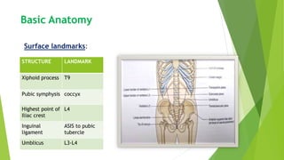
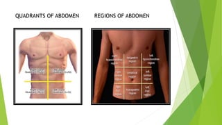
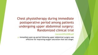
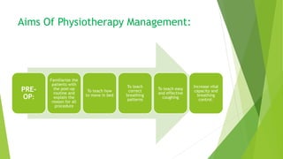
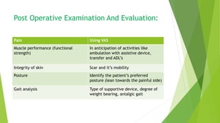
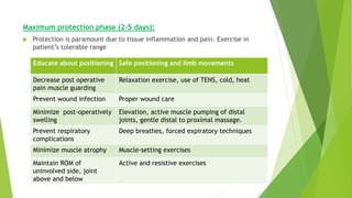
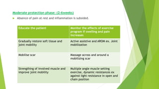
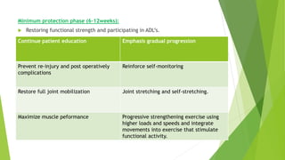
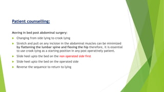
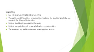
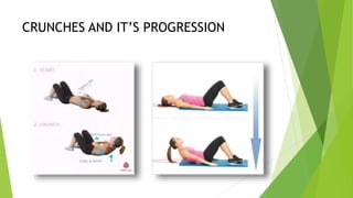
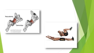
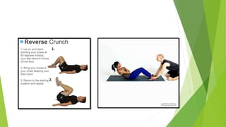
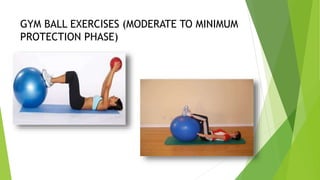
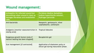
2. Basic Anatomy
Surface landmarks:
STRUCTURE LANDMARK
Xiphoid process T9
Pubic symphysis coccyx
Highest point of
Iliac crest
L4
Inguinal
ligament
ASIS to pubic
tubercle
Umblicus L3-L4
3. SURFACE LANDMARKS:
Supero-lateral margins of the anterior abdominal wall are formed by right
and left 7,8,9,10 costal cartilages.
Posteriorly and laterally the abdominal wall is much less as it is replaced by
thoracic cage in upper part and gluteal region in lower part.
The anterior abdominal wall is divided into right and left halves by linea
alba.
Lateral to linea alba there is linea semilunaris which corresponds to lateral
margin of rectus abdominis.
SKIN :
capable of undergoing enormous stretching as seen in pregnancy, ascites or
obesity.
4. UMBLICUS:
Normal scar in the anterior abdominal wall.
Lymph and venous blood which flow upward above the plane of the umbilicus
and downwards below this plane.
In portal hypertension, dilated vein radiating from the umbilicus is seen called
caput medusa.
5. SUPERFICIAL FASCIA:
Divided into superficial fatty (fascia of Camper) and deep (fascia of Scarpa)
CUTANEOUS NERVE:
T7- T11, ilio-inguinal and ilio-hypogastric (T12-L1) enter the abdominal wall
through inter-coastal spaces supplies front of abdomen.
LYMPH NODES:
Above the watershed lines lymphatic run upward to drain into axillary lymph
nodes and below the level of umbilicus drain into inguinal lymph nodes.
MUSCLES:
Abdominal muscle provide a firm but elastic support for the viscera against
gravity.
It helps in all expulsive and expiratory acts.
6. MUSCLE ORIGIN INSERTION ACTION
External Oblique Shafts of lower
eight ribs
Xiphoid, Linea
alba, pubis
Forceful
expiration
Internal Oblique Inguinal ligament,
iliac crest
7-10 ribs, xiphoid,
linea alba, pubis
Ipsilateral trunk
rotation
Transversus
Abdominis
Inguinal ligament,
iliac crest,
thoracolumbar
fascia
Xiphoid, Linea
alba, pubis
Corset like effect-
compress
abdominal
structures
Rectus Abdominis Pubic crest and
ligament
Tendinous
insertion to
xiphoid
Flexion of trunk
Quadratus
Lumborum
Iliac crest 12th rib Stabilize pelvis,
lateral flexion,
accessory muscle
to expiration
7.
8. Quadrants And Regions Of Abdomen
Abdomen is divided into four quadrants to allow the localization
of pain and tenderness, scars or lumps.
Nine regions can be marked using two horizontal (subcoastal and
intertubercular line) and two vertical dividing lines (midclavicular)
QUADRANTS STRUCTURE
Left upper Stomach, spleen, pancrease,
left kidney, colon
Left lower Descending colon, left ovary,
left ureter
Right upper Right kidney, liver, gall bladder,
duodenum
Right lower Right ovary, right ureter,
appendix, ascending colon
10. Abdominal Incision:
A wisely chosen incision and correct method of making and closing the wound
is really important.
Care is taken to avoid tram line incision and acute angle incision as it could
lead to devascularisation of tissue causing delayed healing and incisional
hernia.
Physiotherapist has to juggle intra-abdominal and intra thoracic pressure to
maintain maximum air entry and clearance of secretion without creating a
rupture of the suture.
14. INCISION ADVANTAGES DISADVANTADES MUSCLE
CUT
MUSCLE
SPARED
MARKINGS
MIDLINE Bloodless,
good
accessibility,
can be
extended,no
nerve injury
Slow healing,
Incisional hernia
none all Follows
linea alba
PARAMEDIAN Good access to
lateral
structures,
more secure
Muscle atrophy,
more bleeding,
difficult to extend
Rectus 2-5cm
lateral to
umblicus
KOCHER’S Lesser post-op
pain,
early recovery
Poor exposure Rectus,
internal
oblique,
transversus
abdominis
External
oblique
2-5cm below
xiphoid
which
extends
outward and
downwards
to costal
margin
15. INCISION ADVANTAGES DISADVANTAGES MUSCLE
CUT
MUSCLE
SPARED
MARKINGS
McBURNEY’S Good healing
and cosmesis
Accidental injury
to iliohypogastric
and ilio-inguinal
nerve
External
oblique
Rectus,
internal
oblique
Medial 2/3rd
and lateral
1/3rd of a line
running from
umbilicus to
ASIS
Transverse
muscle
cutting
incision
(Maryland
incision)
Reduced rate
of incisional
hernia,
cosmetic
appeal
Painful, not offer
sufficient
exposure to
Rectus and
all the
muscle are
incised
5-8cm above
pubic
symphysis
Pfannestial
incision
Heals faster,
best cosmetic
results
More
hemorrhagic
Rectus
muscle is
not cut
12cm long
and 2cm
above the
pubic
symphysis.
16. History And Examination
Common complaints:
Pain, dyspepsia, anorexia, weight loss, jaundice, altered bowel habit, blood
loss and fatigue.
History:
Medical history- for e.g. person with β-blocking drug will not have
tachycardia proportionate to internal hemorrhage.
Surgical history- previous abdominal surgeries and it’s complication.
Family history- evident particularly in cancer, inflammatory bowel disease,
endocrine disease.
Personal history- smoking, drinking or nicotine.
17. Type of injury-
Closed Abdominal Injury-
Closed injuries are due to waves of shock or direct compression of a viscera
against a bony prominence.
If a large segment of the abdomen or abdomino-thoracic wall is compressed it
may burst or split organs like liver and spleen.
A similar force may split the diaphragm if the breath is held and the
diaphragm is tense.
Open Abdominal Injury-
With a severe open abdominal injury, abdominal organs sometimes protrude
through the wound.
18. General Examination:
Vitals: Tachycardia and tachypnea are noticed in internal hemorrhage.
Subnormal temperature and low blood pressure are features of shock.
Fever
Weight loss
Dehydration
Anemia
Jaundice or pale skin should be ascertain at sclera, skin, nail bed, under
surface of the tongue, soft palate.
Vomiting or constipation
Loss of appetite is an early feature of carcinoma affecting any part of the
gastro-intestinal tract.
Ankle odema if bilateral suggests gallstones.
19. Pain:
Abdominal pain may be due to inflammatory, infection or obstructive
pathology.
Pain from the viscera is principally due to ischaemia, muscle spasm or
stretching of the visceral peritoneum. Unlike somatic pain, autonomic pain is
deep and poorly localized.
Pathology involving diaphragm or phrenic nerve (C4) pain arising in this region
is referred to the tip of the shoulder.
Pancreatitis often has an abrupt onset of severe epigastric pain radiating to
the back.
20. Biliary colic will classically results pain in the right upper quadrant of the
abdomen which radiates to the angle of the scapula.
Murphy’s sign- It is performed by asking the patient to breathe out and then
gently placing the hand below the costal margin on the right side at the mid-
clavicular line.
If the patient stops breathing in as the gallbladder is tender and winces with a
"catch" in breath, the test is considered positive
21. Review Of System:
Gastrointestinal System:
In gastric ulcer intake of food brings pain, whereas in duodenal ulcer food
relieves pain.
belching and heart-burn occurs in patients with oesophageal hiatus hernia.
Hematemesis- vomiting of blood
Black tarry (melena) — which indicates hemorrhage in the upper G.I. tract or
ingestion of large doses of iron or bismuth
whitish or clay colored indicates biliary obstruction.
large, fatty and offensive stool suggests chronic pancreatitis.
22. Inspection
Examiner should kneels by the patient’s bed so that the his eye is at the level
of the patient’s anterior abdominal wall to examine for abdominal masses,
scars, mobility on respiration, visible peristalsis, dilated veins, or swelling.
Generalized distension of the abdomen occurs in internal hemorrhage.
Localized distension may be due to localized internal hemorrhage or
peritonitis.
Abdominal distention may cause impairment of excursion of the diaphragm,
may also inhibit the patient from coughing and deep breathing and thus,
contribute to respiratory failure.
An enlarged liver may be found when chronic right-sided heart failure has
occurred as a consequence of chronic respiratory disease.
There will be absence of abdominal movements in respiration due to
peritonitis.
23. Bruise with hematoma affecting lumbar region which should arouse suspicion
of renal injury and bruising with hematoma affecting lower ribs indicates liver
or splenic injury.
Grey Turner’s sign – skin discoloration of the flanks due to retroperitoneal
haemorrhage in severe acute pancreatitis and leaking abdominal aortic
aneurysm
Cullen’s sign – discoloration around the umbilicus – may indicate severe
acute pancreatitis, ruptured ectopic pregnancy or trauma to the liver.
24. Scar- linear scar indicates healing by first intention and broad or irregular
scar suggestive of wound infection.
Ask the patient to lift his or her legs with the knees extended, or perform
Valsalva’s maneuvre for laterally placed swellings. Lumps superficial to the
abdominal wall muscles will become more obvious and those arising within
the muscle layer will become fixed and remain unchanged in size.
An intra-peritoneal mass in contact with the diaphragm will move on
respiration.
25. Palpation:
Patient should lie flat on his back with knee flexed (to relax abdominal
muscle) taking deep breaths.
Palpation should start in the region furthest away from the site of pain.
Under no circumstances he should be hurt. Otherwise abdominal muscles
will go into spasm and important findings may be missed.
Muscle Guarding represents contraction of the abdominal wall muscles
over the area of pain due to peritonitis or presence of internal bleeding.
Liver, spleen, gall bladder and kidneys are best palpated during inspiration.
26. Liver- Normally, the liver spans approximately 10 cm at the right
midclavicular line.
If the liver extends more than 10 cm, it is considered enlarged.
To palpate for an enlarged liver one should place the hand on the right iliac
fossa with the fingers pointing towards the left axilla (that means parallel to
the right costal margin)
Spleen- Normally 7-14 cm but when enlarged it extends from the left costal
margin to the right iliac fossa. It moves freely with respiration.
Spleen is palpation with the tips of the fingers pointing upwards and pressed
inwards
Left supraclavicular lymph nodes- indicates carcinoma of the stomach and
other abdominal organs.
27. Percussion:
Percussion helps to distinguish whether distension is due to bowel gas from
solid masses or free fluid in the abdomen.
If the patient winces with pain on abdominal percussion it denotes underlying
peritonitis
The liver extends from the 6th rib to the costal margin on the right mid-
axillary line.
The spleen extends from the 9th to the 11th rib on the left mid-axillary line.
When there is free fluid in the abdomen shifting dullness can be obtained.
28. Auscultation:
The bell of the stethoscope is placed below and to the left of the xiphi-
sternum.
The point at which the sound changes is the boundary line of the stomach.
Normal bowel sound almost excludes any serious injury to the abdominal
viscera.
Auscultation of the chest may indicate presence of bowel sound in case of
rupture of the diaphragm.
29. Investigation:
Blood- ESR
Urine-haemteuria
X-ray chest and abdomen
Endoscpoy
Sonography
Peritoneocentesis
Cystoscopy is of high value in diagnosis of urinary bladder injury.
31. Physiotherapy Complications:
Hemorrahage: palor, restlessness, thirst and anxiety.
Pulmonary complication:
Retention of secretions which may lead to pneumonia.
secretions greater in volume due to irritation by the anaesthesia and
sedatives, plus decreased depth of pulmonary ventilation.
Secretions become more sticky and tenacious due to dehydration post
operatively.
The lying position creates low lung volume. Therefore airway early closure
occurs early and atelectasis may occur.
32. Cough reflex suppressed due to anesthesia, sedation and inhibiting effect of
pain.
Rarely there may be inhalation of foreign matter.
Breathing during anesthesia, or under anesthesia is very even with no periodic
sighs, this leads to low lung volumes and reduced surfactants and atelectasis.
Vital Capacities can be reduced by >50%
Atelectasis:
Threatened collapse due to low lung volumes.
Decreased rib cage movement
Decreased breath sound,late inspiratory crackles occurs in first 24-48 hours
33. Complete obstruction due to thick secretions:
collapse of an area of lung seen on x-ray
Fever.37.7
Increased pulse rate
Decreased movement of the rib cage over involved area
Dyspnea if extensive
Absent breath sounds
Occurs in first 24-48 hours
Retention of secretions often in the large airway which may lead to broncho-
pneumonia and bubbling sound may felt on palpation and auscultation during
expiration
34. High risk factors:
High incision causing splinting and reflex inhibition of lower rib cage and
hemi- diaphragm on the same side.
Elderly (over 50) due to normal effects of aging on the lungs
Smokers
Tendency to have mild bronchitis cough with cold
Previous COPD and Bronchiectasis
History of repeated attacks of pneumonia
Obesity
35. The ability of prehabilitation to
influence post-operatively outcome
after an intra-abdominal
operation(2016)
moran2016.pdf
Post-op complications are more than mortality with a rate of about
35% after an abdominal operation.
Prehab given are inspiratory muscle training, musculoskeletal and
aerobic exercise.
Results showed significant decreased in PPCs.
36. Physiotherapeutic approaches and the effects
on inspiratory muscle force in patients with
chronic obstructive pulmonary disease in the
pre-operative preparation for abdominal
surgical procedures
complication2019.pdf
The modern and traditional bronchial clearance techniques associated
with inspiratory muscle training were equally effective in gaining
inspiratory muscle strength with increased Pmax.
37. Complication involving peripheral vascular system:
Phlebitis- inflammation of vein
Thrombosis- formation of clot in the blood vessel
Phlebothrombosis- thrombosis without much inflammation
Thrombophlebitis - usually accompanied by a certain degree of clot
formation due to walls becoming rough as a result of inflammation.
Thrombosis of leg veins which may lead to pulmonary embolism
Physiotherapy is contra-indicated until danger of emboli is over.
Immediate- elevation, supportive bandage, cradle, heparin and later
calmodulin is given.
Later- Elevation, mild heat infra-red, exercise should be gentle at first
38. Unhealed hernia:
Healing is slowed down by several factors and when it occurs it leads to greater
scar formation
Causes:
Infection
Excessive strain
Abdominal distention
Presence of malignancies
Diabetic patients
Weak abdominals pre-op
Hernia weakens anterior support of lumbar vertebre leading to increased lordosis
and possible back problems.
Weakness may lead to visceroptosis {abdominal organ fall to the lower part of
abdomen}
Pelvic floor weakness may lead to stress incontinence
39. Infections:
Wound is tender and increased temperature
Hyperpyrexia
Redness and swelling around the wound
If infection occurs through the full thickness of the wall then rupture of the
wound leads to Dehiscence
Muscle atrophy:
Muscles which are cut.
40. ICEAGE (Incidence of Complications following
Emergency Abdominal surgery: Get Exercising):
study protocol of a pragmatic, multicentre,
randomised controlled trial testing
physiotherapy for the prevention of
complications and improved physical recovery
after emergency abdominal surgery
13017_2018_Article_189.pdf
PPC diagnostic criteria
ICEAGE exercise protocol reps and repitition
41. Chest physiotherapy during immediate
postoperative period among patients
undergoing upper abdominal surgery:
Randomized clinical trial
manzanoSPMJ2008.pdf
Immediate post-op period following upper abdominal surgery was
effective for improving oxygen saturation that last longer.
42. Aims Of Physiotherapy Management:
PRE-
OP:
Familiarize the
patients with
the post-op
routine and
explain the
reason for all
procedure
To teach how
to move in bed
To teach
correct
breathing
patterns
To teach easy
and effective
coughing
Increase vital
capacity and
breathing
control.
43. Post-op:
EARLY-
To ensure full
inflation of
the basal
lobes.
To aid
expectoration
of mucus
To maintain
adequate
speed of
blood flow in
lower
extremities.
LONG TERM-
To ensure
strength to
the muscle
cut.
To reduce
gastric
distress or gas
pains
44. Post Operative Examination And Evaluation:
Pain Using VAS
Muscle performance (functional
strength)
In anticipation of activities like
ambulation with assistive device,
transfer and ADL’s
Integrity of skin Scar and it’s mobility
Posture Identify the patient’s preferred
posture (lean towards the painful side)
Gait analysis Type of supportive device, degree of
weight bearing, antalgic gait
45. Maximum protection phase (2-5 days):
Protection is paramount due to tissue inflammation and pain. Exercise in
patient’s tolerable range
Educate about positioning Safe positioning and limb movements
Decrease post operative
pain muscle guarding
Relaxation exercise, use of TENS, cold, heat
Prevent wound infection Proper wound care
Minimize post-operatively
swelling
Elevation, active muscle pumping of distal
joints, gentle distal to proximal massage.
Prevent respiratory
complications
Deep breathes, forced expiratory techniques
Minimize muscle atrophy Muscle-setting exercises
Maintain ROM of
uninvolved side, joint
above and below
Active and resistive exercises
46. Moderate protection phase: (2-6weeks)
Absence of pain at rest and inflammation is subsided.
Educate the patient Monitor the effects of exercise
program if swelling and pain
increases
Gradually restore soft tissue and
joint mobility
Active assistive and AROM ex. Joint
mobilization
Mobilise scar Massage across and around a
mobilizing scar
Strengthing of involved muscle and
improve joint mobility
Multiple angle muscle setting
exercise, dynamic resistances ex
against light resistance in open and
chain position
47. Minimum protection phase (6-12weeks):
Restoring functional strength and participating in ADL’s.
Continue patient education Emphasis gradual progression
Prevent re-injury and post operatively
complications
Reinforce self-monitoring
Restore full joint mobilization Joint stretching and self-stretching.
Maximize muscle peformance Progressive strengthening exercise using
higher loads and speeds and integrate
movements into exercise that stimulate
functional activity.
48. Patient counselling:
Moving in bed post abdominal surgery:
Changing from side lying to crook lying
Stretch and pull on any incision in the abdominal muscles can be minimized
by flattening the lumbar spine and flexing the hip therefore, it is essential
to use crook lying as a starting position in any post operatively patient.
Slide heel upto the bed on the non operated side first
Slide heel upto the bed on the operated side
Reverse the sequence to return to lying
49. Log rolling:
Log roll to crook lying to side crook lying
Therapist assist the patient by supporting head and the shoulder girdle by one
arm and the thighs with the other
Patient should roll towards the therapist.
Patient instructed to roll in one whole piece onto the sides.
The shoulder, hip and knees should move together as one.
50.
51. To sit up on the side of bed:
Straighten the knees out so that the lower legs are resting parallel to close to the
edge of the bed.
Push trunk upwards with both hands in steps.
Once the patient is sitting stay close to the patient.
To assist patient upto long sitting from side lying in order to cough:
If there is time, roll the head end of the bed up
Place one hand under the patients head, brace the other hand and arm on the bed
besides the patient upper thigh to protect your back
Instruct the patient to keep the neck and upper back rigid
Lift the patient up by using transference of your body weight
To return the patient to lying hand position as above instruct the patient to push
back very strongly against the resistance of the therapist.
The back and hip extensors work very strongly and abdominals remain relaxed due
to reciprocal innervation.
52. Respiratory exercises:
Breathing exercise:
Time visit with analgesics, nebulization.
Focus on both lateral costal and diaphragmatic breathing pattern.
Half lying to enhance lateral costal expansion.
Crook lying to enhance diaphragmatic expansion.
Side lying to lateral costal excursion in upper lung and diaphragmatic excursion in lower lung
Slow sustained inspiratory breathing to increase Vital Capacity.
Clearance of secreation:
vibration, percussion and suctioning.
Mobilization:
Alter angle of head end of bed
Moving around in bed
Bending knees
Turning onto sides
53. Ambulation:
Getting out of bed by getting up from crook lying position using arms.
Encourage ambulation
Resisted foot exercises.
Effective coughing :
With knees well bent up in half lying or side lying except for pelvic surgeries
legs adducted in lying and knees extended.
Support incision-manually or using towel and pillow.
Abdominal exercises:
Should start with static contraction and pelvic tilt
Progression should be gentle and should not produce pain
Work on the upper and lower abdominals in all phases of their actions in
rotation, forward flexion and side flexion.
Donot breath hold its important to breathe normally while contracting.
54. Principle of exercise after abdominal
surgeries:
Recruit core muscle so that the trunk and pelvis has much better support
which helps to counteract the shearing forces during activities such as lifting,
coughing straining at stool.
Two important muscles to be recruited is
Transeversus abdominis and
Mutifidus
Core strengthening programme is an exercise program that improves trunk
stability and support the spine.
55. Progression of abdominal exercise:
Contract deep
muscles
Increase the
endurance capacity
of core muscle by
contracting them in
different positions
and as long as they
can
Begin arm and leg
movements while
contracting core
muscle.
56. Moving the pelvis and legs are mostly concentric in middle range therefore,
suitable for strengthening exercise.
Movement of head, arms and legs in the supine position automatically will
involve static work for the abdominals even no actual trunk movement as
occurred. Resistance to these movements will increase the strength of the
isometric contraction of the abdominals.
Concentric work of the abdominal muscle is only obtained when pelvis and or
the ribs move towards one another.
60. Functions:
To support abdominal viscera
To act as a synergic muscle for the diaphragm
To fixate the thorax during head and arm movements.
To fixate pelvis during leg movements
Prevents loss of lumbar lordosis
Assists in expulsive acts coughing defecation, childbirth
To reduce gas pain:
Abdominal contraction followed by bulging the stomach
Alternate hip and knee flexion
Knee to chest
61. POST-OP DAY EXERCISES
1 Check wound, upright sitting, isometric abdominal exercise,
knee to chest, knee roll
2-5 Previous exercise three times a day with increased reps, start
walking(cardiovascular training) Alternate hip and knee flexion
6 Initiate core exercise (1*6), static cycling, core exercise
7-8 Increase sets (2*8), start aqua jogging wearing buoyancy belt if
wound is closed and stitches are removed
9-12 Increased sets (3*10), continue cardiovascular exercises
13-15 Initiate walking on treadmill
16-18 Add squats, dynamic core strengthening
19-21 Increase speed in treadmill, progress to slow paced running,
increased sets (4*10), gym ball exercises
71. Wound management:
Antiseptics for infected wound but
discontinue when wound is clean as
damages fibroblast and endothelial
cells.
Povidone-idodine (betadine),
Sodium hypochlorite solution,
Hydrogen peroxide
Anti-bacterials Neosporin, gentamycin, silver
sulphadiazine, sulfamylon
Analgesics {monitor vasoconstriction in
nearby area}
Tropical lidocaine
Exogenous growth factors {only if
natural healing has been stopped}
Becaplermin gel
Scar management {if contracted} Application of elastomer putty or
silicon gel during maturation phase.
72. Mechanical Modalities:
Ultra sound Increase cell activity, use hydrogel
sheet for open wound
Ultaviolet radiations Bactericidal effects, enhance
granulation tissue.
Hyperbaric oxygen therapy Increases oxygen available for cell
metabolism, improving oxygen to
hypoxic tissue.
Negative Pressure wound therapy A suction tube is connected from the
foam to a portable pump usually till 48
hours, it removes excess wound fluid
and bacteria from wound bed.
Cold laser therapy Low energy level infrared lasers,
reduce pain by releasing nitric oxide
into micro circulation.