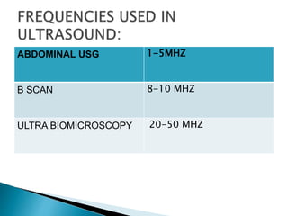
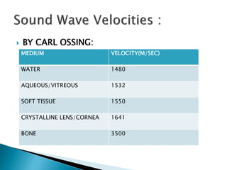
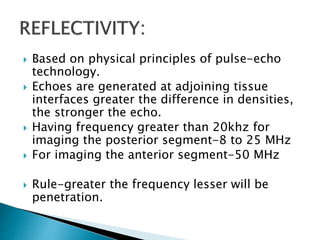
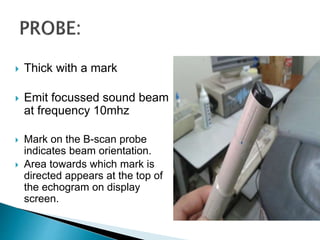
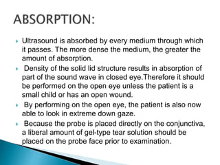
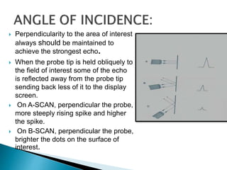
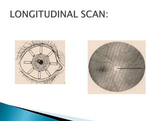
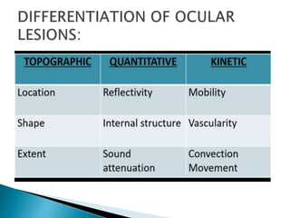
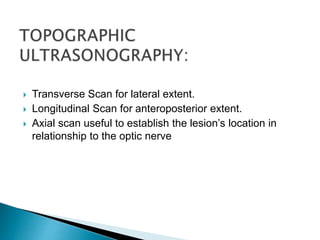
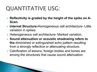
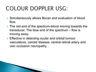
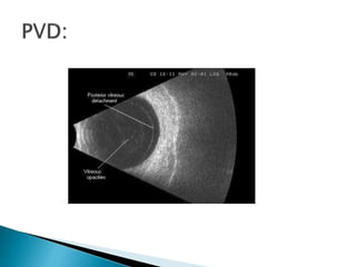
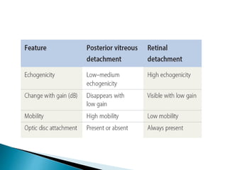
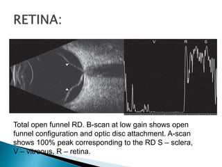
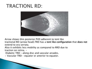
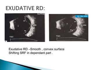
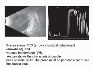
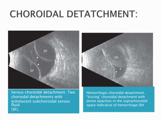
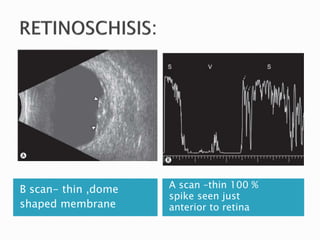
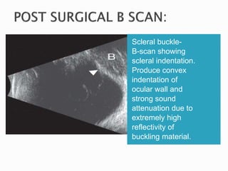
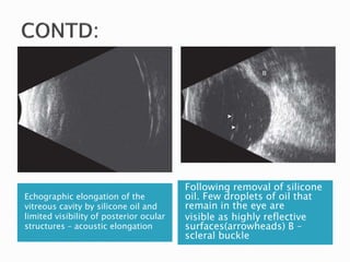
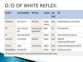
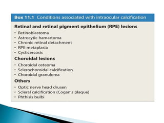
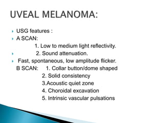
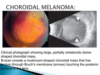
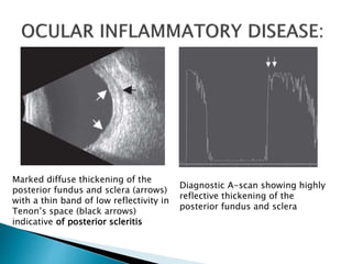
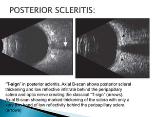
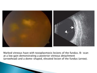
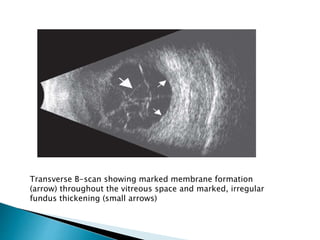
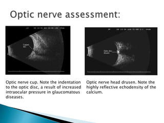
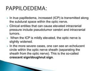
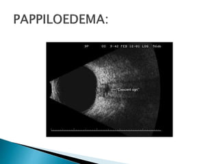
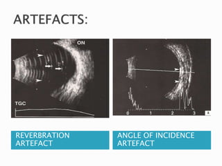
DR SHREYA BHARGAVA
PG2ND YEAR
 B- SCAN is a two dimensional imaging system which
utilizes high frequency sound waves ranging from 8-10
MHz.
 B stands for bright echoes
 1793: Lazzaro Spallanzani(Italy) discovered that bats
orient themselves with the help of sound whistles while
flying in darkness. This was the basis of modern
ultrasound application.
 1880-Curie brothers first demonstrated that difference
in electric potential could be created by mechanically
rubbing tourmaline crystals this is called as piezoelectric
effect .
 This was first applied in World war 2 for underwater
sonar system .
 B Scan was first introduced by Baum and Greenwood
in 1958
 First commercially available B scan is developed by
Coleman et al in seventies
 The importance of the instrument and technique is
emphasised by Karl Ossoinig
 Contact B-scan was introduced by Bronson(1975) and
it being portable, become a part of everyday use in
ophthalmology.
 Longitudinal sound waves
 Greater than the upper limit of human hearing that is
20,000Hz
 Alternate compressions and rarefactions.
 Just like sound waves – reflected and refracted.
 PHYSICS:
 Frequency of the wave is inversely proportional to its
wavelength.
 Wavelength is directly proportional to its penetration.
 So larger the frequency ,less is the penetration and more
is the resolution of the resultant echograph
ABDOMINAL USG 1-5MHZ
B SCAN 8-10 MHZ
ULTRA BIOMICROSCOPY 20-50 MHZ
 BY CARL OSSING:
MEDIUM VELOCITY(M/SEC)
WATER 1480
AQUEOUS/VITREOUS 1532
SOFT TISSUE 1550
CRYSTALLINE LENS/CORNEA 1641
BONE 3500
 Based on physical principles of pulse-echo
technology.
 Echoes are generated at adjoining tissue
interfaces greater the difference in densities,
the stronger the echo.
 Having frequency greater than 20khz for
imaging the posterior segment-8 to 25 MHz
 For imaging the anterior segment-50 MHz
 Rule-greater the frequency lesser will be
penetration.
•A thin, parallel sound beam is emitted, which passes
through the eye and images one small axis of tissue; the
echoes of which are represented as spikes arising from a
baseline.
•The stronger the echo, the higher the spike.
 Thick with a mark
 Emit focussed sound beam
at frequency 10mhz
 Mark on the B-scan probe
indicates beam orientation.
 Area towards which mark is
directed appears at the top of
the echogram on display
screen.
 Ultrasound is absorbed by every medium through which
it passes. The more dense the medium, the greater the
amount of absorption.
 Density of the solid lid structure results in absorption of
part of the sound wave in closed eye.Therefore it should
be performed on the open eye unless the patient is a
small child or has an open wound.
 By performing on the open eye, the patient is also now
able to look in extreme down gaze.
 Because the probe is placed directly on the conjunctiva,
a liberal amount of gel-type tear solution should be
placed on the probe face prior to examination.
 In a dense cataract more of the sound is absorbed and
less is able to pass through to the next medium,
resulting in weaker echoes and images on both A-scan
and B-scan.
 Thus the best images of the posterior segment are
obtained when the probe is in contact with the
conjunctiva rather than the corneal surface, bypassing
the crystalline lens or intraocular lens implant.
 Finally, when calcification of tissue is present, there is so
much absorption and such a strong reflection of the echo
back to the probe that there is no signal posterior to that
medium. This is referred to as shadowing.
 Perpendicularity to the area of interest
always should be maintained to
achieve the strongest echo.
 When the probe tip is held obliquely to
the field of interest some of the echo
is reflected away from the probe tip
sending back less of it to the display
screen.
 On A-SCAN, perpendicular the probe,
more steeply rising spike and higher
the spike.
 On B-SCAN, perpendicular the probe,
brighter the dots on the surface of
interest.
 This is the important part of signal processing that
occurs before the sound wave reached the display
monitor.
 Three types:
1. Linear: limited range of echo densities, but can
show minor difference within this range
2. Logarithmic: wider range of echo densities ,but
does not show minor difference between the echo
intensity.
3. S Curve: combine benefit of both the above .
 The ability to distinguish between adjacent echoes-both
axial and lateral.
 This is enhanced by use of focused sound beam.
 Axial resolution is related to frequency, piezoelectric
crystal shape and dampening material attached to
crystal.
 Shorter the pulse better the axial resolution.
 The concave shape of the crystal focuses the sound.
The focused sound increases axial as well as lateral
resolution.
 It is used for increasing or decreasing the amplitude of
echoes that are displayed on the screen.
 Measured in decibels.
 Does not change the frequency or velocity of the sound
.
 Changes the sensitivity of the instrument’s display
screen .
 Higher gain display weaker echoes like vitreous
opacities.
 Lower gain displays stronger echoes like retina and
sclera
 Typically all examination begin with highest gain, so that
no weaker signal is missed ,and then the gain is reduced
as necessary for good resolution of stronger signals.
TIME GAIN COMPENSATION
 This technique is used to enhance the echoes returning
from deeper structures by reducing those from the
structures closer to the surface, which is typically utilized
in studying the orbit.
 DISPLAY OF SIGNALS: In three ways : A mode , B
mode or a combination of the above.
 Other modification includes:
 three dimensional ultrasound
 a combination of colour doppler with the BSCAN
 Transverse
 Longitudinal
 Axial
 Mark is kept parallel to the limbus
 Movement of transducer parallel to the limbus .
 Probe shifted from the limbus to the fornix and sideways.
 Produce a circumferential slice through several
meridian.
 To detect lateral extent of lesion.
 Transducer –perpendicular to the limbus
 Probe marker -towards centre of cornea
 Antero posterior extent of the lesion
 Optic disc and posterior aspect of the globe –lower
portion of screen
 Best –demonstrating the insertion of membranes to optic
disc.
 The LMAC view allows for proper visualization of the
macula and optic nerve.
 Gently place the probe on the nasal aspect of the eye
with the patient's gaze directed temporally.
 Note: For this position, the marker of the probe should
be directed toward the pupil, instead of superiorly.
 A longitudinal scan is the only scan where this occurs!
 In this view, the optic nerve will be below the macula.
Maneuver the probe to bring the macula into the center
of the image to obtain the best resolution.
 Probe centered on the cornea
 Easiest to understand (displays lens & optic nerve)
 Documenting lesions & membranes in relation to
optic disc
 Evaluates macular region
 Hinder resolution of posterior portion of globe (Sound
attenuation and refraction )
CLOCK HOUR POSITION AREA SCANNED
3-LIMBUS 9-POSTERIOR
3-EQUATOR 9-EQUATOR
3-FORNIX 9-ANTERIOR
6-LIMBUS 12- POSTERIOR
6-EQUATOR 12-EQUATOR
6-FORNIX 12-ANTERIOR
 Six scan screening – four transverse ,one
axial and one longitudinal B-scan – the
entire posterior segment can be well imaged.
 Media opacity
 Corneal opacity
 Hyphaema
 Small pupil
 Pupillary membrane
 Dense cataract
 Dense vitreous hemorrhage
 Dense vitreous exudates
 Differentiation of solid from cystic and homogenous from
heterogeneous masses.
 Examination of retrobulbar soft tissue masses and
normally present orbital structures (to differentiate
proptosis from exophthalmos).
 Identification, localization of foreign bodies.
 Assessment of collateral damage in trauma cases.
 Vitreous
 Retina
 Choroid
 Optic nerve
 Transverse Scan for lateral extent.
 Longitudinal Scan for anteroposterior extent.
 Axial scan useful to establish the lesion’s location in
relationship to the optic nerve
 Reflectivity is graded by the height of the spike on A-
Scan.
 Internal Structure-Homogeneous cell architecture- Little
variation in spikes.
 Heterogeneous cell architecture- Marked variation.
 Sound attenuation or acoustic shadowing refers to
the diminished or extinguished echo pattern resulting
from a strongly reflective or attenuating structure.
 Calcification of lesions, foreign bodies and bones are
among the structures that cause sound attenuation.
 Mobility- Movement of a membrane or opacity
following a change in gaze.
 Vascularity-Fast, low-amplitude Blood flow within an
flickering intraocular solid lesion.
 Convection Movement-
 Slow, continuous movement of blood, layered
inflammatory cells, or cholesterol debris.
 Occurs secondary to convection currents.
 Seen in eyes with long-standing vitreous haemorrhage
 Simultaneously allows Bscan and evaluation of blood
flow.
 The red end of the spectrum-blood moving towards the
transducer. The blue end of the spectrum – flow is
moving away.
 Effective in detecting ocular and orbital tumour
vasculature, carotid disease, central retinal artery and
vein occlusion neuropathy .
 Multiple consecutive Bscan are utilized to create 3 D
block.
 The transducer rotates.
 Useful in evaluating volume of intraocular lesions and for
evaluation of retrobulbar optic nerve.
 Done using the B-Scan Probe with an immersion scleral
shell or a small water filled balloon.
 Contact B-scan is of little use in evaluating anterior eye
structures because there is a 5-mm area directly in front
of the probe known as the “dead zone”
 Patient is asked to fixate in primary gaze when doing
axial scan.
 It allows display of cornea, anterior chamber, iris, lens
and retrolental space along the visual axis.
 35-100 MHZ
 Anatomy of anterior segment, as well as associated
pathologies, including angle closure glaucoma, ciliary
body cysts, cyclodialysis, foreign bodies in angle
neoplasm and angle trauma.
 VITREOUS HAEMORRHAGE
 Most common causes:
I. posterior vitreous detachment with or without retinal
tear
II. proliferative diabetic retinopathy
III. ocular trauma
IV.neovascularisation secondary to retinal vein
occlusion.
Fresh vitreous
hemorrhage showing
diffuse low to medium
echoes
Pseudomembrane
representing the
organization of blood
moderately dense
vitreous hemorrhage
SUBHYALOID
HAEMOHHRAGE
LAYERED VH MIMICKS RD
MIMICKING CONDITIONS:
 Asteroid hyalosis- echoluscent gap between echoes
and posterior globe wall.
 Inflammatory echoes- other signs present such as
retinochoroidal thickening, exudative RD, optic disc
elevation or Tenon’s space widening seen.
 PVD appears as a thin, smooth membrane that may
retain its attachment to the retina at sites of retinal tears,
areas of neovascularization, the optic disc, or the
vitreous base.
 PVD demonstrates significant movement and
after movements on dynamic B-scan
Total open funnel RD. B-scan at low gain shows open
funnel configuration and optic disc attachment. A-scan
shows 100% peak corresponding to the RD S – sclera,
V – vitreous, R – retina.
Arrow shows thin posterior PVD adherent to tent like
tractional RD (arrow head).TRD has a tent like configuration that does not
extend to ora serrata.
Also it exhibits less mobility as compared to RRD due to
traction on retina. •
Diabetic TRD – along disc and vascular arcades.
• Vascular TRD – equator or anterior to equator.
Exudative RD –Smooth , convex surface
Shifting SRF in dependant part .
 In exudative RD caused by photocoagulation –peripheral
detachment or isolated pockets,choroidal detachment
may also be present.
 Inflammatory etiology – retinochoroidal
thickening.
 Look for tumour mass or granuloma.
FEATURES CHOROID
DETATCHMENT
RETINAL
DETATCHMENT
PVD
SHAPE DOME LINEAR -
MOBILITY MINIMAL MODERATE MARKED
ATTATCHMENT
TO ONH
NO YES VARIABLE
A-SCAN SPIKE% 90-100 80-100 40-90
LOCATION PERIPHERY VARIABLE VARIABLE
AFTER
MOVEMENTS
- MINIMAL MARKED
OTHER
FINDINGS
KISSING
CHOROIDALS
FOLDS,BREAKS,
PVR CHANGES
PROMINENT
INFERIORLY
B-scan shows PVD (arrow), choroidal detachment
(arrowhead), and
vitreous hemorrhage (VH).
A-scan shows the characteristic double
peak on initial spike The probe must be perpendicular to see
the double peak.
Serous choroidal detachment. Two
choroidal detachments with
echolucent subchoroidal serous
fluid
(SF).
Hemorrhagic choroidal detachment.
“kissing” choroidal detachment with
dense opacities in the suprachoroidal
space indicative of hemorrhage (SH
B scan- thin ,dome
shaped membrane
A scan –thin 100 %
spike seen just
anterior to retina
Scleral buckle-
B-scan showing
scleral indentation.
Produce convex
indentation of
ocular wall and
strong sound
attenuation due to
extremely high
reflectivity of
buckling material.
Echographic elongation of the
vitreous cavity by silicone oil and
limited visibility of posterior ocular
structures – acoustic elongation
Following removal of silicone
oil. Few droplets of oil that
remain in the eye are
visible as highly reflective
surfaces(arrowheads) B –
scleral buckle
RETINOBLASTOMA:
 Solid tumor arising from the retinal layer obliterating the
vitreous cavity.
 Calcification within the tumor mass is typical of
retinoblastoma
 Shadowing effect behind the lesion in the
orbital mass.
 Concomitant RD may be present.
 A scan- high reflectivity, vascularity and
absence of after movements.
 First few weeks of life
 Unilateral
 Associated ocular anomalies - microphthalmos, shallow
AC, axial length shortening.
 Retrolental fibrovascular mass that cause ciliary body
process to rotate inwards.
 But unlike retinoblastoma, no discrete mass visualised.
Persistent fetal vasculature (PFV). Taut, thickened
vitreous band adherent to the slightly elevated optic
disc.
 USG features :
 A SCAN:
1. Low to medium light reflectivity.
 2. Sound attenuation.
 Fast, spontaneous, low amplitude flicker.
B SCAN: 1. Collar button/dome shaped
2. Solid consistency
3.Acoustic quiet zone
4. Choroidal excavation
5. Intrinsic vascular pulsations
Clinical photograph showing large, partially amelanotic dome-
shaped choroidal mass.
B-scan reveals a mushroom-shaped choroidal mass that has
broken through Bruch’s membrane (arrows) touching the posterior
surface of the lens.
Clinical photograph of peripapillary calcified astrocytic
hamartoma. B-scan demonstrating calcification near optic
disc (arrow).
Marked diffuse thickening of the
posterior fundus and sclera (arrows)
with a thin band of low reflectivity in
Tenon’s space (black arrows)
indicative of posterior scleritis
Diagnostic A-scan showing highly
reflective thickening of the
posterior fundus and sclera
“T-sign” in posterior scleritis. Axial B-scan shows posterior scleral
thickening and low reflective infiltrate behind the peripapillary
sclera and optic nerve creating the classical “T-sign” (arrows).
Axial B-scan showing marked thickening of the sclera with only a
very thin band of low reflectivity behind the peripapillary sclera
(arrows)
 Intravitreal punctiform echoes.
 Thickening of posterior hyaloid.
 Partial or total PVD.
 Focal retinochoroidal thickening.
Marked vitreous haze with toxoplasmosis lesions of the fundus. B- scan
at a low gain demonstrating a posterior vitreous detachment
(arrowhead) and a dome-shaped, elevated lesion of the fundus (arrow).
 Low reflective vitreous echoes- dot or cobweb shaped.
 More severe cases- thick membrane like echoes.
 PVD may or may not be present.
 As PVD develops in eye with endophthalmitis, TRD like
picture may develop - this is due to thickened inflammed
posterior hyaloid.
Transverse B-scan showing marked membrane formation
(arrow) throughout the vitreous space and marked, irregular
fundus thickening (small arrows)
Dropped intraocular lens (IOL)
IOL is identified by high amplitude
spike which is associated with acoustic
reverberations.
Lens matter drop (posterior dislocation
of lens matter)
. The lens matter appears as a lesion in
the vitreous cavity (usually inferiorly)
with moderate to high amplitude which
moves on ocular movement.
 Optic disc cupping usually can be seen on clinical
examination. if media opacities prevent examination,
the contour (including the degree of cupping) can be
detected with ultrasound. Similarly, optic nerve
colobomas are imaged easily with ultrasound.
Optic nerve cup. Note the indentation
to the optic disc, a result of increased
intraocular pressure in glaucomatous
diseases.
Optic nerve head drusen. Note the
highly reflective echodensity of the
calcium.
 In true papilledema, increased (ICP) is transmitted along
the subdural space within the optic nerve.
 Clinical entities that can cause elevated intracranial
pressure include pseudotumor cerebri and intracranial
tumors.
 When the ICP is mildly elevated, the optic nerve is
slightly widened.
 In the more severe cases, one can see an echolucent
circle within the optic nerve sheath (separating the
sheath from the optic nerve). This is the so-called
crescent sign/doughnut sign.
 The presence of increased fluid within the sheath is
confirmed best with the 30-degree test , a dynamic A-
scan test that measures the width of the optic nerve in
primary gaze and again after the patient shifts gaze 30
degrees from primary.
 In cases of increased ICP, the nerve and sheath are
stretched as the globe turns 30 degrees, and the
subarachnoid fluid is distributed over the extent of the
nerve, resulting in measurements less than when in
primary gaze.
 If nerve enlargement is due to parenchymal infiltration or
thickening of the optic nerve sheath, then the
measurement will not change as the globe turns from
primary.
 Insufficient fluid coupling ( i.e., lack of methyl cellulose)
cause entrapment of air between the probe and eye
leading to display of bright echos which represent
multiple signals.
REVERBRATION
ARTEFACT
ANGLE OF INCIDENCE
ARTEFACT
B scan

B scan

  • 1.
  • 2.
     B- SCANis a two dimensional imaging system which utilizes high frequency sound waves ranging from 8-10 MHz.  B stands for bright echoes
  • 3.
     1793: LazzaroSpallanzani(Italy) discovered that bats orient themselves with the help of sound whistles while flying in darkness. This was the basis of modern ultrasound application.  1880-Curie brothers first demonstrated that difference in electric potential could be created by mechanically rubbing tourmaline crystals this is called as piezoelectric effect .  This was first applied in World war 2 for underwater sonar system .
  • 4.
     B Scanwas first introduced by Baum and Greenwood in 1958  First commercially available B scan is developed by Coleman et al in seventies  The importance of the instrument and technique is emphasised by Karl Ossoinig  Contact B-scan was introduced by Bronson(1975) and it being portable, become a part of everyday use in ophthalmology.
  • 5.
     Longitudinal soundwaves  Greater than the upper limit of human hearing that is 20,000Hz  Alternate compressions and rarefactions.  Just like sound waves – reflected and refracted.  PHYSICS:  Frequency of the wave is inversely proportional to its wavelength.  Wavelength is directly proportional to its penetration.  So larger the frequency ,less is the penetration and more is the resolution of the resultant echograph
  • 6.
    ABDOMINAL USG 1-5MHZ BSCAN 8-10 MHZ ULTRA BIOMICROSCOPY 20-50 MHZ
  • 7.
     BY CARLOSSING: MEDIUM VELOCITY(M/SEC) WATER 1480 AQUEOUS/VITREOUS 1532 SOFT TISSUE 1550 CRYSTALLINE LENS/CORNEA 1641 BONE 3500
  • 8.
     Based onphysical principles of pulse-echo technology.  Echoes are generated at adjoining tissue interfaces greater the difference in densities, the stronger the echo.  Having frequency greater than 20khz for imaging the posterior segment-8 to 25 MHz  For imaging the anterior segment-50 MHz  Rule-greater the frequency lesser will be penetration.
  • 9.
    •A thin, parallelsound beam is emitted, which passes through the eye and images one small axis of tissue; the echoes of which are represented as spikes arising from a baseline. •The stronger the echo, the higher the spike.
  • 10.
     Thick witha mark  Emit focussed sound beam at frequency 10mhz  Mark on the B-scan probe indicates beam orientation.  Area towards which mark is directed appears at the top of the echogram on display screen.
  • 11.
     Ultrasound isabsorbed by every medium through which it passes. The more dense the medium, the greater the amount of absorption.  Density of the solid lid structure results in absorption of part of the sound wave in closed eye.Therefore it should be performed on the open eye unless the patient is a small child or has an open wound.  By performing on the open eye, the patient is also now able to look in extreme down gaze.  Because the probe is placed directly on the conjunctiva, a liberal amount of gel-type tear solution should be placed on the probe face prior to examination.
  • 12.
     In adense cataract more of the sound is absorbed and less is able to pass through to the next medium, resulting in weaker echoes and images on both A-scan and B-scan.  Thus the best images of the posterior segment are obtained when the probe is in contact with the conjunctiva rather than the corneal surface, bypassing the crystalline lens or intraocular lens implant.  Finally, when calcification of tissue is present, there is so much absorption and such a strong reflection of the echo back to the probe that there is no signal posterior to that medium. This is referred to as shadowing.
  • 13.
     Perpendicularity tothe area of interest always should be maintained to achieve the strongest echo.  When the probe tip is held obliquely to the field of interest some of the echo is reflected away from the probe tip sending back less of it to the display screen.  On A-SCAN, perpendicular the probe, more steeply rising spike and higher the spike.  On B-SCAN, perpendicular the probe, brighter the dots on the surface of interest.
  • 14.
     This isthe important part of signal processing that occurs before the sound wave reached the display monitor.  Three types: 1. Linear: limited range of echo densities, but can show minor difference within this range 2. Logarithmic: wider range of echo densities ,but does not show minor difference between the echo intensity. 3. S Curve: combine benefit of both the above .
  • 15.
     The abilityto distinguish between adjacent echoes-both axial and lateral.  This is enhanced by use of focused sound beam.  Axial resolution is related to frequency, piezoelectric crystal shape and dampening material attached to crystal.  Shorter the pulse better the axial resolution.  The concave shape of the crystal focuses the sound. The focused sound increases axial as well as lateral resolution.
  • 16.
     It isused for increasing or decreasing the amplitude of echoes that are displayed on the screen.  Measured in decibels.  Does not change the frequency or velocity of the sound .  Changes the sensitivity of the instrument’s display screen .
  • 17.
     Higher gaindisplay weaker echoes like vitreous opacities.  Lower gain displays stronger echoes like retina and sclera  Typically all examination begin with highest gain, so that no weaker signal is missed ,and then the gain is reduced as necessary for good resolution of stronger signals.
  • 18.
    TIME GAIN COMPENSATION This technique is used to enhance the echoes returning from deeper structures by reducing those from the structures closer to the surface, which is typically utilized in studying the orbit.  DISPLAY OF SIGNALS: In three ways : A mode , B mode or a combination of the above.  Other modification includes:  three dimensional ultrasound  a combination of colour doppler with the BSCAN
  • 19.
  • 20.
     Mark iskept parallel to the limbus  Movement of transducer parallel to the limbus .  Probe shifted from the limbus to the fornix and sideways.  Produce a circumferential slice through several meridian.  To detect lateral extent of lesion.
  • 22.
     Transducer –perpendicularto the limbus  Probe marker -towards centre of cornea  Antero posterior extent of the lesion  Optic disc and posterior aspect of the globe –lower portion of screen  Best –demonstrating the insertion of membranes to optic disc.
  • 24.
     The LMACview allows for proper visualization of the macula and optic nerve.  Gently place the probe on the nasal aspect of the eye with the patient's gaze directed temporally.  Note: For this position, the marker of the probe should be directed toward the pupil, instead of superiorly.  A longitudinal scan is the only scan where this occurs!  In this view, the optic nerve will be below the macula. Maneuver the probe to bring the macula into the center of the image to obtain the best resolution.
  • 26.
     Probe centeredon the cornea  Easiest to understand (displays lens & optic nerve)  Documenting lesions & membranes in relation to optic disc  Evaluates macular region  Hinder resolution of posterior portion of globe (Sound attenuation and refraction )
  • 28.
    CLOCK HOUR POSITIONAREA SCANNED 3-LIMBUS 9-POSTERIOR 3-EQUATOR 9-EQUATOR 3-FORNIX 9-ANTERIOR 6-LIMBUS 12- POSTERIOR 6-EQUATOR 12-EQUATOR 6-FORNIX 12-ANTERIOR
  • 29.
     Six scanscreening – four transverse ,one axial and one longitudinal B-scan – the entire posterior segment can be well imaged.
  • 30.
     Media opacity Corneal opacity  Hyphaema  Small pupil  Pupillary membrane  Dense cataract  Dense vitreous hemorrhage  Dense vitreous exudates
  • 31.
     Differentiation ofsolid from cystic and homogenous from heterogeneous masses.  Examination of retrobulbar soft tissue masses and normally present orbital structures (to differentiate proptosis from exophthalmos).  Identification, localization of foreign bodies.  Assessment of collateral damage in trauma cases.
  • 32.
     Vitreous  Retina Choroid  Optic nerve
  • 34.
     Transverse Scanfor lateral extent.  Longitudinal Scan for anteroposterior extent.  Axial scan useful to establish the lesion’s location in relationship to the optic nerve
  • 35.
     Reflectivity isgraded by the height of the spike on A- Scan.  Internal Structure-Homogeneous cell architecture- Little variation in spikes.  Heterogeneous cell architecture- Marked variation.  Sound attenuation or acoustic shadowing refers to the diminished or extinguished echo pattern resulting from a strongly reflective or attenuating structure.  Calcification of lesions, foreign bodies and bones are among the structures that cause sound attenuation.
  • 36.
     Mobility- Movementof a membrane or opacity following a change in gaze.  Vascularity-Fast, low-amplitude Blood flow within an flickering intraocular solid lesion.  Convection Movement-  Slow, continuous movement of blood, layered inflammatory cells, or cholesterol debris.  Occurs secondary to convection currents.  Seen in eyes with long-standing vitreous haemorrhage
  • 37.
     Simultaneously allowsBscan and evaluation of blood flow.  The red end of the spectrum-blood moving towards the transducer. The blue end of the spectrum – flow is moving away.  Effective in detecting ocular and orbital tumour vasculature, carotid disease, central retinal artery and vein occlusion neuropathy .
  • 38.
     Multiple consecutiveBscan are utilized to create 3 D block.  The transducer rotates.  Useful in evaluating volume of intraocular lesions and for evaluation of retrobulbar optic nerve.
  • 39.
     Done usingthe B-Scan Probe with an immersion scleral shell or a small water filled balloon.  Contact B-scan is of little use in evaluating anterior eye structures because there is a 5-mm area directly in front of the probe known as the “dead zone”  Patient is asked to fixate in primary gaze when doing axial scan.  It allows display of cornea, anterior chamber, iris, lens and retrolental space along the visual axis.
  • 40.
     35-100 MHZ Anatomy of anterior segment, as well as associated pathologies, including angle closure glaucoma, ciliary body cysts, cyclodialysis, foreign bodies in angle neoplasm and angle trauma.
  • 41.
     VITREOUS HAEMORRHAGE Most common causes: I. posterior vitreous detachment with or without retinal tear II. proliferative diabetic retinopathy III. ocular trauma IV.neovascularisation secondary to retinal vein occlusion.
  • 42.
    Fresh vitreous hemorrhage showing diffuselow to medium echoes Pseudomembrane representing the organization of blood moderately dense vitreous hemorrhage
  • 43.
  • 44.
    MIMICKING CONDITIONS:  Asteroidhyalosis- echoluscent gap between echoes and posterior globe wall.  Inflammatory echoes- other signs present such as retinochoroidal thickening, exudative RD, optic disc elevation or Tenon’s space widening seen.
  • 45.
     PVD appearsas a thin, smooth membrane that may retain its attachment to the retina at sites of retinal tears, areas of neovascularization, the optic disc, or the vitreous base.  PVD demonstrates significant movement and after movements on dynamic B-scan
  • 48.
    Total open funnelRD. B-scan at low gain shows open funnel configuration and optic disc attachment. A-scan shows 100% peak corresponding to the RD S – sclera, V – vitreous, R – retina.
  • 49.
    Arrow shows thinposterior PVD adherent to tent like tractional RD (arrow head).TRD has a tent like configuration that does not extend to ora serrata. Also it exhibits less mobility as compared to RRD due to traction on retina. • Diabetic TRD – along disc and vascular arcades. • Vascular TRD – equator or anterior to equator.
  • 50.
    Exudative RD –Smooth, convex surface Shifting SRF in dependant part .
  • 51.
     In exudativeRD caused by photocoagulation –peripheral detachment or isolated pockets,choroidal detachment may also be present.  Inflammatory etiology – retinochoroidal thickening.  Look for tumour mass or granuloma.
  • 52.
    FEATURES CHOROID DETATCHMENT RETINAL DETATCHMENT PVD SHAPE DOMELINEAR - MOBILITY MINIMAL MODERATE MARKED ATTATCHMENT TO ONH NO YES VARIABLE A-SCAN SPIKE% 90-100 80-100 40-90 LOCATION PERIPHERY VARIABLE VARIABLE AFTER MOVEMENTS - MINIMAL MARKED OTHER FINDINGS KISSING CHOROIDALS FOLDS,BREAKS, PVR CHANGES PROMINENT INFERIORLY
  • 53.
    B-scan shows PVD(arrow), choroidal detachment (arrowhead), and vitreous hemorrhage (VH). A-scan shows the characteristic double peak on initial spike The probe must be perpendicular to see the double peak.
  • 54.
    Serous choroidal detachment.Two choroidal detachments with echolucent subchoroidal serous fluid (SF). Hemorrhagic choroidal detachment. “kissing” choroidal detachment with dense opacities in the suprachoroidal space indicative of hemorrhage (SH
  • 55.
    B scan- thin,dome shaped membrane A scan –thin 100 % spike seen just anterior to retina
  • 56.
    Scleral buckle- B-scan showing scleralindentation. Produce convex indentation of ocular wall and strong sound attenuation due to extremely high reflectivity of buckling material.
  • 57.
    Echographic elongation ofthe vitreous cavity by silicone oil and limited visibility of posterior ocular structures – acoustic elongation Following removal of silicone oil. Few droplets of oil that remain in the eye are visible as highly reflective surfaces(arrowheads) B – scleral buckle
  • 58.
    RETINOBLASTOMA:  Solid tumorarising from the retinal layer obliterating the vitreous cavity.  Calcification within the tumor mass is typical of retinoblastoma  Shadowing effect behind the lesion in the orbital mass.  Concomitant RD may be present.  A scan- high reflectivity, vascularity and absence of after movements.
  • 62.
     First fewweeks of life  Unilateral  Associated ocular anomalies - microphthalmos, shallow AC, axial length shortening.  Retrolental fibrovascular mass that cause ciliary body process to rotate inwards.  But unlike retinoblastoma, no discrete mass visualised.
  • 63.
    Persistent fetal vasculature(PFV). Taut, thickened vitreous band adherent to the slightly elevated optic disc.
  • 64.
     USG features:  A SCAN: 1. Low to medium light reflectivity.  2. Sound attenuation.  Fast, spontaneous, low amplitude flicker. B SCAN: 1. Collar button/dome shaped 2. Solid consistency 3.Acoustic quiet zone 4. Choroidal excavation 5. Intrinsic vascular pulsations
  • 65.
    Clinical photograph showinglarge, partially amelanotic dome- shaped choroidal mass. B-scan reveals a mushroom-shaped choroidal mass that has broken through Bruch’s membrane (arrows) touching the posterior surface of the lens.
  • 66.
    Clinical photograph ofperipapillary calcified astrocytic hamartoma. B-scan demonstrating calcification near optic disc (arrow).
  • 67.
    Marked diffuse thickeningof the posterior fundus and sclera (arrows) with a thin band of low reflectivity in Tenon’s space (black arrows) indicative of posterior scleritis Diagnostic A-scan showing highly reflective thickening of the posterior fundus and sclera
  • 68.
    “T-sign” in posteriorscleritis. Axial B-scan shows posterior scleral thickening and low reflective infiltrate behind the peripapillary sclera and optic nerve creating the classical “T-sign” (arrows). Axial B-scan showing marked thickening of the sclera with only a very thin band of low reflectivity behind the peripapillary sclera (arrows)
  • 69.
     Intravitreal punctiformechoes.  Thickening of posterior hyaloid.  Partial or total PVD.  Focal retinochoroidal thickening.
  • 70.
    Marked vitreous hazewith toxoplasmosis lesions of the fundus. B- scan at a low gain demonstrating a posterior vitreous detachment (arrowhead) and a dome-shaped, elevated lesion of the fundus (arrow).
  • 71.
     Low reflectivevitreous echoes- dot or cobweb shaped.  More severe cases- thick membrane like echoes.  PVD may or may not be present.  As PVD develops in eye with endophthalmitis, TRD like picture may develop - this is due to thickened inflammed posterior hyaloid.
  • 72.
    Transverse B-scan showingmarked membrane formation (arrow) throughout the vitreous space and marked, irregular fundus thickening (small arrows)
  • 73.
    Dropped intraocular lens(IOL) IOL is identified by high amplitude spike which is associated with acoustic reverberations. Lens matter drop (posterior dislocation of lens matter) . The lens matter appears as a lesion in the vitreous cavity (usually inferiorly) with moderate to high amplitude which moves on ocular movement.
  • 74.
     Optic disccupping usually can be seen on clinical examination. if media opacities prevent examination, the contour (including the degree of cupping) can be detected with ultrasound. Similarly, optic nerve colobomas are imaged easily with ultrasound.
  • 75.
    Optic nerve cup.Note the indentation to the optic disc, a result of increased intraocular pressure in glaucomatous diseases. Optic nerve head drusen. Note the highly reflective echodensity of the calcium.
  • 76.
     In truepapilledema, increased (ICP) is transmitted along the subdural space within the optic nerve.  Clinical entities that can cause elevated intracranial pressure include pseudotumor cerebri and intracranial tumors.  When the ICP is mildly elevated, the optic nerve is slightly widened.  In the more severe cases, one can see an echolucent circle within the optic nerve sheath (separating the sheath from the optic nerve). This is the so-called crescent sign/doughnut sign.
  • 78.
     The presenceof increased fluid within the sheath is confirmed best with the 30-degree test , a dynamic A- scan test that measures the width of the optic nerve in primary gaze and again after the patient shifts gaze 30 degrees from primary.  In cases of increased ICP, the nerve and sheath are stretched as the globe turns 30 degrees, and the subarachnoid fluid is distributed over the extent of the nerve, resulting in measurements less than when in primary gaze.  If nerve enlargement is due to parenchymal infiltration or thickening of the optic nerve sheath, then the measurement will not change as the globe turns from primary.
  • 79.
     Insufficient fluidcoupling ( i.e., lack of methyl cellulose) cause entrapment of air between the probe and eye leading to display of bright echos which represent multiple signals.
  • 80.