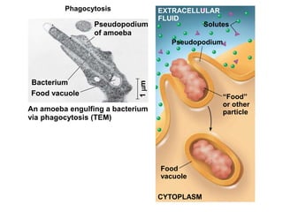
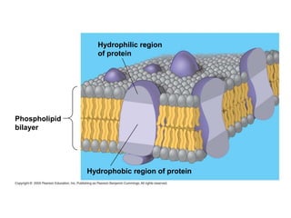
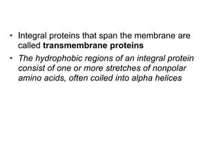
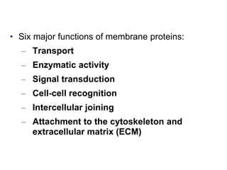
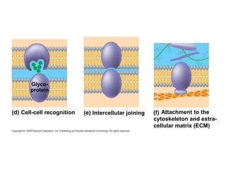
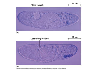
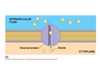
The plasma membrane serves as a selective barrier for cells, facilitating the movement of substances through a fluid mosaic structure composed of lipids and proteins. Membrane proteins perform various functions, including transport, enzymatic activity, and cell recognition, while membrane carbohydrates play a key role in cell-cell interactions. The document also discusses mechanisms of transport including passive transport, osmosis, facilitated diffusion, and active transport, as well as bulk transport processes like exocytosis and endocytosis.


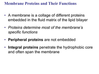


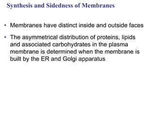


![Cytoplasmic Na+ bonds to
the sodium-potassium pump
CYTOPLASM
Na+
[Na+] low
[K+] high
Na+
Na+
EXTRACELLULAR
FLUID
[Na+] high
[K+] low
Na+
Na+
Na+
ATP
ADP
P
Na+ binding stimulates
phosphorylation by ATP.
Na+
Na+
Na+
Phosphorylation causes
the protein to change its
conformation, expelling Na+
to the outside.
P
Extracellular K+ binds
to the protein, triggering
release of the phosphate
group.
P
P
Loss of the phosphate
restores the protein’s
original conformation.
K+ is released and Na+
sites are receptive again;
the cycle repeats.](https://image.slidesharecdn.com/cellmembrane-240628055753-c7c24e0a/85/As-level-biology-cell-membrane-chapter-notes-45-320.jpg)