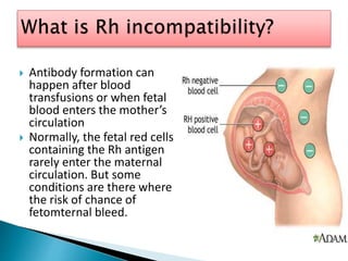
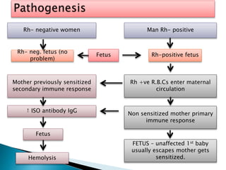
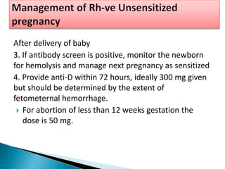
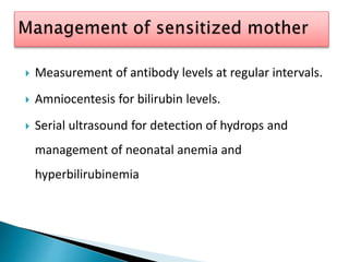
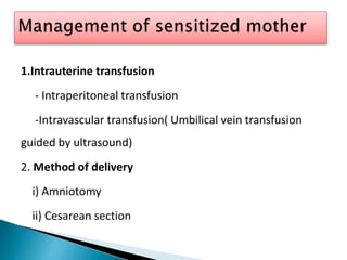
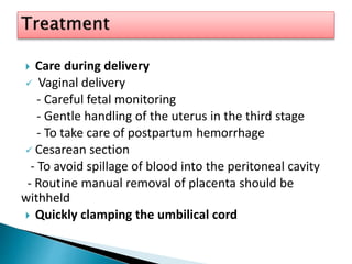
Rh-Incompatibility
Presented by
Sujata sahu
Msc tutor,SNC,Odisha
 This protein does not affect your overall health, but it
is important to know your Rh status if you are
pregnant. Rh factor can cause complications during
pregnancy if you are Rh-negative and your child is Rh-
positive.
 Antibody formation can
happen after blood
transfusions or when fetal
blood enters the mother’s
circulation
 Normally, the fetal red cells
containing the Rh antigen
rarely enter the maternal
circulation. But some
conditions are there where
the risk of chance of
fetomternal bleed.
1. Ectopic pregnancy
2. Placenta previa
3. Placental abruptio
4. Abdominal/pelvic trauma
5. In –uterofetal death
6. Any invasive obstetric procedure
7. Lack of prenatal care
8. Spontaneous abortion
9. Antepartum bleeding
10. Platelet transfusion
Rh- negative women Man Rh- positive
Mother previously sensitized
secondary immune response
Fetus
Fetus
Rh +ve R.B.Cs enter maternal
circulation
Hemolysis
FETUS – unaffected 1st baby
usually escapes mother gets
sensitized.
↑ ISO antibody IgG
Rh- neg. fetus (no
problem)
Rh-positive fetus
Non sensitized mother primary
immune response
Rh- incompatibility can cause symptoms ranging
from very mild to fatal.
 Mildest from
1. Hemolysis with the release of free hemoglobin
into the infant’s circulation.
2. Jaundice (hb is converted into, bilirubin which
cause an infant to became yellow)
 Severe form
1. Hydrops fetalis
2. Icterus gravis neonatrum
3. Congenital anemia
4. kernicterus (bilirubin induced brain dysfunction)
 Affection of the mother
The mother may also be affected somewhat,
there is increased incidence of :
a) Pre-eclampsia
b) Polyhydramnios
c) Big size baby with its hazard
d) Postpartum hemorrhage due to big placenta
e) Maternal syndrome- features are generalized
edema & proteinuria
 Obstetrics history
a) In a parous woman, a detailed obstetric history has
to be taken.
b) History of prophylactic administration of anti-D
immunoglobulin following abortion or delivery.
 Physical examination
There are no any physical examination seen in this.
 Also known as Anti globulin test (AGT)
 Two coomb’s test are:
i) Direct coomb’s test (DCT)
ii) Indirect coomb’s test (ICT)
1. DIRECT COOMB’s TEST
To detect the antibodies or complement
protein that are bound to the surface of RBCs.
2. INDIRECT COOMB’s TEST
It detect antibodies against RBCs that are
present unbound in the patient’s serum.
 Antibody detection
- Detected by indirect coomb’s test
- If test negative at 12th week, it repeated at 28th week
and 36th week in primi gravida.
- If positive then screening of patient.
- Quantitative estimation of IgG antibody at weekly
intervals.
 Doppler ultrasound
- A value > 1.5 multiples of the median (MOMs) for
the corresponding gestational age, predicts moderate
to severe fetal anemia.
 Amniocentesis
Amniocentesis and estimation of bilirubin in
the amniotic fluid by spectrophotometry are
indicated in :
a) Previous history of severely affected baby
b) Father is heterozygous to determine whether
the particular baby will be affected or not.
Management
Identification
Sensitized
pregnancy
Unsensitized
pregnancy
Management
Management
1.Identification of pregnancies at risk at the initial ANC
visit
- determine blood group, Rh factor & ICT
2. Repeat ICT at 28 weeks and at 36 weeks, if negative
300 micrograms anti-D at 28 weeks or after any
procedure (external version, amniocentesis).
.
After delivery of baby
3. If antibody screen is positive, monitor the newborn
for hemolysis and manage next pregnancy as sensitized
4. Provide anti-D within 72 hours, ideally 300 mg given
but should be determined by the extent of
fetometernal hemorrhage.
 For abortion of less than 12 weeks gestation the
dose is 50 mg.
 Measurement of antibody levels at regular intervals.
 Amniocentesis for bilirubin levels.
 Serial ultrasound for detection of hydrops and
management of neonatal anemia and
hyperbilirubinemia
1.Intrauterine transfusion
- Intraperitoneal transfusion
-Intravascular transfusion( Umbilical vein transfusion
guided by ultrasound)
2. Method of delivery
i) Amniotomy
ii) Cesarean section
 Care during delivery
 Vaginal delivery
- Careful fetal monitoring
- Gentle handling of the uterus in the third stage
- To take care of postpartum hemorrhage
 Cesarean section
- To avoid spillage of blood into the peritoneal cavity
- Routine manual removal of placenta should be
withheld
 Quickly clamping the umbilical cord
3. Resuscitation
In an anemic and premature infant, lung diseases is
common, due to:
- Surfactant deficiency
- Pulmonary edema
 Exchange transfusion
Indications- early : CHb <12 g/dl
: Cord bilirubin > 85 mmol/L
: Strong +ve coomb’s test
4. Phototherapy
- Placing newborn baby under a halogen or
fluorescent lamp with their eyes covered.
- During phototherapy, intravenous hydration is
required.
 To avoid mismatched transfusion
 To prevent or minimize fetomaternal bleed
 Precautions during cesarean section
 Carefully amniocentesis
 Gently Manual removal of placenta
 Avoid forcible attempt to perform external
version.
 During pregnancy
- Mild anemia, hyperbilirubinemia and jaundice.
- Severe anemia with enlargement of the liver and
spleen.
- Hydrops fetalis
 After birth
- Severe hyperbilirubinemia and jaundice
- kernicterus
SUMMARY
 Kamini R, “Textbook of midwifery obstetrics for
nurses” 2011, ELSEVIER, p.p. 243-244.
 D.C. dutta, “ A textbook of obstetrics“, 8th
edition, 2015, Jaypee brothers Medical
publishers (p) ltd, p.p 386-388.
 Jacob, A. “Manual of midwifery and
gynecological nursing, first edition, 2009, New
Delhi , Japee brother Medical publication (p)ltd.
P.447.
6. rh &amp; abo incompatibility

6. rh &amp; abo incompatibility

  • 2.
  • 4.
     This proteindoes not affect your overall health, but it is important to know your Rh status if you are pregnant. Rh factor can cause complications during pregnancy if you are Rh-negative and your child is Rh- positive.
  • 5.
     Antibody formationcan happen after blood transfusions or when fetal blood enters the mother’s circulation  Normally, the fetal red cells containing the Rh antigen rarely enter the maternal circulation. But some conditions are there where the risk of chance of fetomternal bleed.
  • 6.
    1. Ectopic pregnancy 2.Placenta previa 3. Placental abruptio 4. Abdominal/pelvic trauma 5. In –uterofetal death 6. Any invasive obstetric procedure 7. Lack of prenatal care 8. Spontaneous abortion 9. Antepartum bleeding 10. Platelet transfusion
  • 7.
    Rh- negative womenMan Rh- positive Mother previously sensitized secondary immune response Fetus Fetus Rh +ve R.B.Cs enter maternal circulation Hemolysis FETUS – unaffected 1st baby usually escapes mother gets sensitized. ↑ ISO antibody IgG Rh- neg. fetus (no problem) Rh-positive fetus Non sensitized mother primary immune response
  • 8.
    Rh- incompatibility cancause symptoms ranging from very mild to fatal.  Mildest from 1. Hemolysis with the release of free hemoglobin into the infant’s circulation. 2. Jaundice (hb is converted into, bilirubin which cause an infant to became yellow)
  • 9.
     Severe form 1.Hydrops fetalis 2. Icterus gravis neonatrum 3. Congenital anemia 4. kernicterus (bilirubin induced brain dysfunction)
  • 10.
     Affection ofthe mother The mother may also be affected somewhat, there is increased incidence of : a) Pre-eclampsia b) Polyhydramnios c) Big size baby with its hazard d) Postpartum hemorrhage due to big placenta e) Maternal syndrome- features are generalized edema & proteinuria
  • 11.
     Obstetrics history a)In a parous woman, a detailed obstetric history has to be taken. b) History of prophylactic administration of anti-D immunoglobulin following abortion or delivery.  Physical examination There are no any physical examination seen in this.
  • 12.
     Also knownas Anti globulin test (AGT)  Two coomb’s test are: i) Direct coomb’s test (DCT) ii) Indirect coomb’s test (ICT)
  • 13.
    1. DIRECT COOMB’sTEST To detect the antibodies or complement protein that are bound to the surface of RBCs. 2. INDIRECT COOMB’s TEST It detect antibodies against RBCs that are present unbound in the patient’s serum.
  • 14.
     Antibody detection -Detected by indirect coomb’s test - If test negative at 12th week, it repeated at 28th week and 36th week in primi gravida. - If positive then screening of patient. - Quantitative estimation of IgG antibody at weekly intervals.  Doppler ultrasound - A value > 1.5 multiples of the median (MOMs) for the corresponding gestational age, predicts moderate to severe fetal anemia.
  • 15.
     Amniocentesis Amniocentesis andestimation of bilirubin in the amniotic fluid by spectrophotometry are indicated in : a) Previous history of severely affected baby b) Father is heterozygous to determine whether the particular baby will be affected or not.
  • 16.
  • 17.
    1.Identification of pregnanciesat risk at the initial ANC visit - determine blood group, Rh factor & ICT 2. Repeat ICT at 28 weeks and at 36 weeks, if negative 300 micrograms anti-D at 28 weeks or after any procedure (external version, amniocentesis). .
  • 18.
    After delivery ofbaby 3. If antibody screen is positive, monitor the newborn for hemolysis and manage next pregnancy as sensitized 4. Provide anti-D within 72 hours, ideally 300 mg given but should be determined by the extent of fetometernal hemorrhage.  For abortion of less than 12 weeks gestation the dose is 50 mg.
  • 19.
     Measurement ofantibody levels at regular intervals.  Amniocentesis for bilirubin levels.  Serial ultrasound for detection of hydrops and management of neonatal anemia and hyperbilirubinemia
  • 20.
    1.Intrauterine transfusion - Intraperitonealtransfusion -Intravascular transfusion( Umbilical vein transfusion guided by ultrasound) 2. Method of delivery i) Amniotomy ii) Cesarean section
  • 21.
     Care duringdelivery  Vaginal delivery - Careful fetal monitoring - Gentle handling of the uterus in the third stage - To take care of postpartum hemorrhage  Cesarean section - To avoid spillage of blood into the peritoneal cavity - Routine manual removal of placenta should be withheld  Quickly clamping the umbilical cord
  • 22.
    3. Resuscitation In ananemic and premature infant, lung diseases is common, due to: - Surfactant deficiency - Pulmonary edema  Exchange transfusion Indications- early : CHb <12 g/dl : Cord bilirubin > 85 mmol/L : Strong +ve coomb’s test
  • 23.
    4. Phototherapy - Placingnewborn baby under a halogen or fluorescent lamp with their eyes covered. - During phototherapy, intravenous hydration is required.
  • 24.
     To avoidmismatched transfusion  To prevent or minimize fetomaternal bleed  Precautions during cesarean section  Carefully amniocentesis  Gently Manual removal of placenta  Avoid forcible attempt to perform external version.
  • 25.
     During pregnancy -Mild anemia, hyperbilirubinemia and jaundice. - Severe anemia with enlargement of the liver and spleen. - Hydrops fetalis  After birth - Severe hyperbilirubinemia and jaundice - kernicterus
  • 26.
  • 28.
     Kamini R,“Textbook of midwifery obstetrics for nurses” 2011, ELSEVIER, p.p. 243-244.  D.C. dutta, “ A textbook of obstetrics“, 8th edition, 2015, Jaypee brothers Medical publishers (p) ltd, p.p 386-388.  Jacob, A. “Manual of midwifery and gynecological nursing, first edition, 2009, New Delhi , Japee brother Medical publication (p)ltd. P.447.