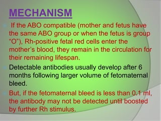
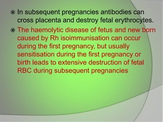
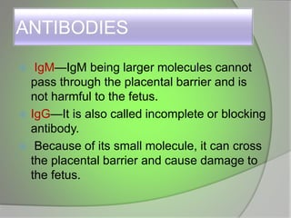
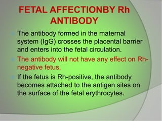
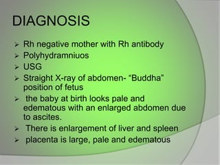
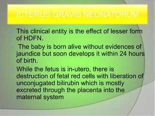
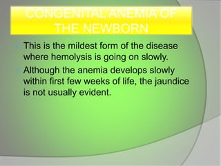
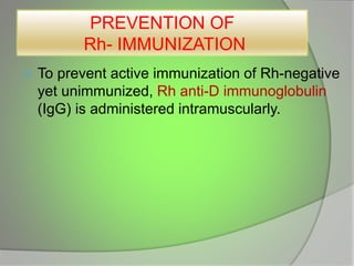
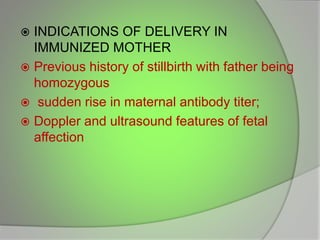
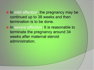
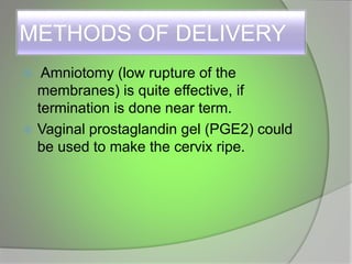
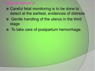
(👑VVIP ISHAAN ) Russian Call Girls Service Navi Mumbai🖕9920874524🖕Independent...
Rh incompatibility
1. By – Mrs. Aparnna Moharana
Msc Tutor
Kalinga Institute of Nursing Sciences,KIIT
University, (DU), Bhubaneswar, Odisha
2. INTRODUCTION
The Rhesus factor gets its name from
experiments conducted in 1937 by
scientists Karl Landsteiner and
Alexander S. Weiner.
The genotype is determined by the
inheritance of 3 pairs of closely linked
allelic genes situated on chromosome 9
named as D/d, C/c, E/e .
Rh-positive is taken to mean D-positive
and Rh-negative to mean its absence.
3. A person's Rh type is generally most relevant with
respect to pregnancies
If the pregnant woman and her husband are Rh
negative, there is no reason to worry about Rh
incompatibility
If she is Rh negative and her husband is Rh
positive,the baby will inherit the father's blood
type ,creating incompatibility between mother and
her fetus.
If some of the fetal blood gets into mother's blood
stream, her body will produce antibodies.
These antibodies could pass back through the
placenta and harm the developing baby's red
blood cells, causing very mild to very serious
anemia in the fetus.
4.
5. DEFINITION
Rh incompatibility is a condition which develops
when there is a difference in Rh blood type
between that of the pregnant mother (Rh
negative) and that of the fetus (Rh positive)
Usually placenta acts as barrier to fetal blood
entering maternal circulation.
How ever, sometimes during pregnancy or birth,
fetomaternal haemorrhage (FMH) can occur.
The woman’s immune system reacts by
producing anti-D antibodies that cause
sensitisation.
6. MECHANISM
If the ABO compatible (mother and fetus have
the same ABO group or when the fetus is group
“O”), Rh-positive fetal red cells enter the
mother’s blood, they remain in the circulation for
their remaining lifespan.
Detectable antibodies usually develop after 6
months following larger volume of fetomaternal
bleed.
But, if the fetomaternal bleed is less than 0.1 ml,
the antibody may not be detected until boosted
by further Rh stimulus.
7. In subsequent pregnancies antibodies can
cross placenta and destroy fetal erythrocytes.
The haemolytic disease of fetus and new born
caused by Rh isoimmunisation can occur
during the first pregnancy, but usually
sensitisation during the first pregnancy or
birth leads to extensive destruction of fetal
RBC during subsequent pregnancies
8. ANTIBODIES
IgM—IgM being larger molecules cannot
pass through the placental barrier and is
not harmful to the fetus.
IgG—It is also called incomplete or blocking
antibody.
Because of its small molecule, it can cross
the placental barrier and cause damage to
the fetus.
9. FETAL AFFECTIONBY Rh
ANTIBODY
The antibody formed in the maternal
system (IgG) crosses the placental barrier
and enters into the fetal circulation.
The antibody will not have any effect on Rh-
negative fetus.
If the fetus is Rh-positive, the antibody
becomes attached to the antigen sites on
the surface of the fetal erythrocytes.
10. The affected cells are rapidly removed from
the circulation.
Depending upon the degree of agglutination
and destruction of the fetal red cells, various
types of fetal hemolytic diseases appear.
Conditions which affect 1st pregnancy are:
Miscarriage
Abortion
Feto-maternal haemorrage
11. MANIFESTATIONS OF FETAL
AFFECTION
Clinical manifestations of the hemolytic
disease of the fetus and newborn are :
Hydrops fetalis
Icterus gravis neonatorum
Congenital anemia of the newborn
12. HYDROPS FETALIS
This is the most serious form of Rh
hemolytic disease (HDFN).
Excessive destruction of the fetal red
cells leads to severe anemia, tissue
anoxemia and metabolic acidosis.
These have got adverse effects on the
fetal heart and brain and on the
placenta.
13. As a result of fetal anoxemia, there is
damage to the liver leading to
hypoproteinemia which is responsible
for generalized edema (hydrops fetalis),
ascites and hydrothorax.
Fetal death occurs sooner or later due to
cardiac failure.
14.
15. DIAGNOSIS
Rh negative mother with Rh antibody
Polyhydramniuos
USG
Straight X-ray of abdomen- “Buddha”
position of fetus
the baby at birth looks pale and
edematous with an enlarged abdomen due
to ascites.
There is enlargement of liver and spleen
placenta is large, pale and edematous
16. ICTERUS GRAVIS NEONATORUM
This clinical entity is the effect of lesser form
of HDFN.
The baby is born alive without evidences of
jaundice but soon develops it within 24 hours
of birth.
While the fetus is in-utero, there is
destruction of fetal red cells with liberation of
unconjugated bilirubin which is mostly
excreted through the placenta into the
maternal system
17. If the bilirubin rises to the critical level of
20 mg per 100 ml, the bilirubin crosses
the blood-brain barrier to damage the
basal nuclei of the brain permanently
producing the clinical manifestation of
kernicterus.
18. CONGENITAL ANEMIA OF
THE NEWBORN
This is the mildest form of the disease
where hemolysis is going on slowly.
Although the anemia develops slowly
within first few weeks of life, the jaundice
is not usually evident.
19. PREVENTION OF
Rh- IMMUNIZATION
To prevent active immunization of Rh-negative
yet unimmunized, Rh anti-D immunoglobulin
(IgG) is administered intramuscularly.
20. IN PREGNANCY-
All Rh-negative unsensitized women should
receive 50 µg of Rh-immune globulin IM
within 72 hours of induced or spontaneous
abortion, ectopic or molar pregnancy in the
first trimester.
Women with pregnancy beyond 12 weeks
should have full dose of 300 µg.
Generally 300 µg dose will protect a woman
from fetal hemorrhage of upto 30 mL of fetal
whole blood.
21. If the woman is Rh- negative and has no
antibody, she should have one dose of
300 µg Rh immune globulin as
prophylaxis at around 28 weeks (ACOG–
1999) and again after birth (within 72
hours).
Women with multiple pregnancy may
need more than usual 300 µg of anti-D
immunoglobulin
22. PLAN OF DELIVERY
UNIMMUNIZED MOTHERS: In cases
where there is no detectable antibody
found during pregnancy, an expectant
attitude is followed till term.
Tendency of post term pregnancy to
should not be allowed.
23. IMMUNIZED MOTHERS: As mentioned
previously, whenever there is evidence of
hemolytic process in the fetus in utero, the
patient should be shifted to an equipped
center specialized to deal with Rh problems.
An intensive neonatal care unit, arrangements
for exchange transfusion and an expert
neonatologist are the basic requirements to
tackle the affected babies.
24. INDICATIONS OF DELIVERY IN
IMMUNIZED MOTHER
Previous history of stillbirth with father being
homozygous
sudden rise in maternal antibody titer;
Doppler and ultrasound features of fetal
affection
25. In mild affection, the pregnancy may be
continued up to 38 weeks and then
termination is to be done.
In severe affection: It is reasonable to
terminate the pregnancy around 34
weeks after maternal steroid
administration.
26. METHODS OF DELIVERY
Amniotomy (low rupture of the
membranes) is quite effective, if
termination is done near term.
Vaginal prostaglandin gel (PGE2) could
be used to make the cervix ripe.
27. Vaginal delivery:
Careful fetal monitoring is to be done to
detect at the earliest, evidences of distress
Gentle handling of the uterus in the third
stage
To take care of postpartum hemorrhage.
28. Caesarean section
In cases when termination has to be
done prematurely (say 34–37 weeks),
the cervix will be unfavorable and
considering the severity of affection and
urgency of termination, cesarean section
is a safe procedure
29. Clamping the umbilical cord
In either methods, the cord is to be clamped
as quickly as possible to minimize even
minute amount of antibody to cross to the
fetus from the mother.
The cord should be kept long (15–20 cm) for
exchange transfusion, if required.