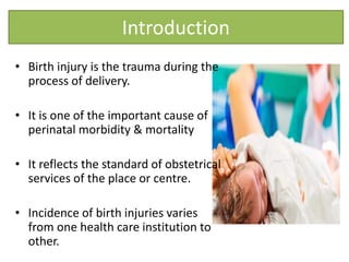
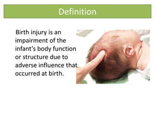
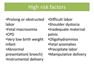
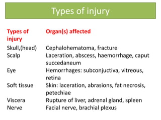
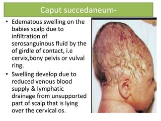
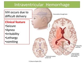
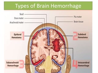
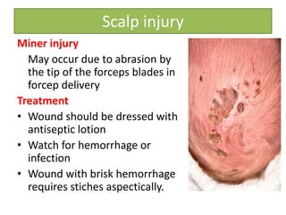
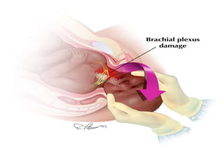
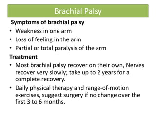
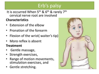
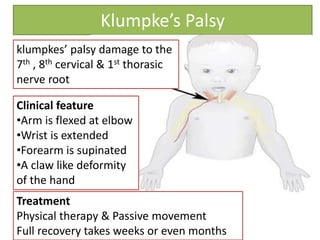
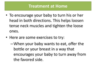
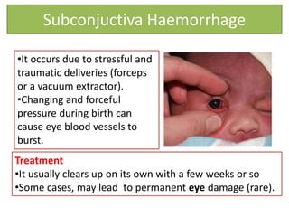
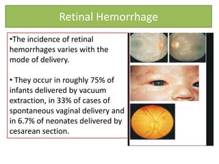
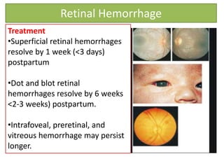
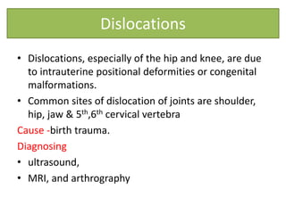
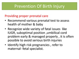
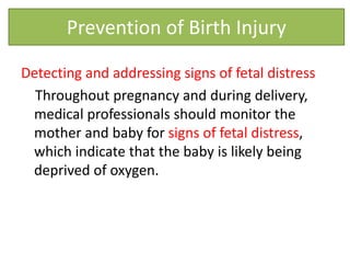
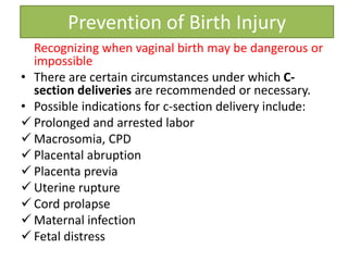
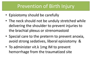
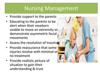
Presented by
Sujata Sahu
OBJECTIVES
At the end of the class, students will be able to–
• Define the birth injury
• Explain the high risk factors of birth injury
• Discuss the types of birth
injury
• Discuss prevention
on birth injury
Introduction
• Birth injury is the trauma during the
process of delivery.
• It is one of the important cause of
perinatal morbidity & mortality
• It reflects the standard of obstetrical
services of the place or centre.
• Incidence of birth injuries varies
from one health care institution to
other.
Definition
Birth injury is an
impairment of the
infant’s body function
or structure due to
adverse influence that
occurred at birth.
High risk factors
•Prolong or obstructed
labor
•Fetal macrosomia
•CPD
•Very low birth weight
infant
•Abnormal
presentation( breech)
•Instrumental delivery
•Difficult labor
•Shoulder dystocia
•Inadequate maternal
pelvis
•Oligohydramnios
•Fetal anomalies
•Precipitate labor
•Manipulative delivery
Types of injury
Types of
injury
Organ(s) affected
Skull,(head)
Scalp
Eye
Soft tissue
Viscera
Nerve
Cephalohematoma, fracture
Laceration, abscess, haemorrhage, caput
succedaneum
Hemorrhages: subconjuctiva, vitreous,
retina
Skin: laceration, abrasions, fat necrosis,
petechiae
Rupture of liver, adrenal gland, spleen
Facial nerve, brachial plexus
SKULL INJURY
Caput succedaneum-
• Edematous swelling on the
babies scalp due to
infiltration of
serosanguinous fluid by the
of girdle of contact, i.e
cervix,bony pelvis or vulval
ring.
• Swelling develop due to
reduced venous blood
supply & lymphatic
drainage from unsupported
part of scalp that is lying
over the cervical os.
Cont…
• Area becomes congested &
oedematous & present as
caput at birth
• It may cross the suture line
• Pits on pressure, non
fluctuant & diffuse in nature
Treatment
• No treatment require
• It usually disappear within 36
hour
• Maternal anxiety should be
reduced by reassurance.
Cephalohematoma
Collection of blood in
between the periosteum
& flat bone of skull
Causes
• Friction between fetal
skull & pelvis leads to
rupture of small emissary
veins from skull
• Found in forcep delivery
• Fracture of skull bone
Cephalohematoma
Characteristics
• Never present at birth
• Gradually develops a few hours after birth
• Incompressible, cystic, circumscribed,
fluctuant.
• Limited by suture line
• Unilateral over parietal bone
Cephalohematoma
Treatment
• Most cephalohematoma disappear
spontaneously after few days or weeks
• No active treatment is necessary unless it
becomes complicated
• Give injection vit k 1 to 2 mg IM
Infected hematoma
• Incision & drainage done
• Antibiotic
• Monitoring hematocrit & bilirubin level
• Explained details to mother
Skull Fracture
• Common in frontal bone or anterior
part of the parietal bone
• It is associated with
cephalohematoma
• 2 types
Causes
• Difficult forcep delivery
• Projected sacral promontory of the
flat bone
Treatment
• No management for linear
or fissure fracture
• Surgical treatment is
required for depressed
fracture with neurological
manifestations
Skull Fracture
INTRACRANIAL INJURY
Intraventricular Hemorrhage
IVH occurs due to
difficult delivery
Clinical feature
•Seizure
•Apnea
•Irritability
•Lethargy
•vomiting
Types of Brain Hemorrhage
Scalp injury
Miner injury
May occur due to abrasion by
the tip of the forceps blades in
forcep delivery
Treatment
• Wound should be dressed with
antiseptic lotion
• Watch for hemorrhage or
infection
• Wound with brisk hemorrhage
requires stiches aspectically.
Facial Palsy
• Known as bell’s palsy
• Injured of facial nerve by direct
pressure of forcep blades
Clinical feature
• Inability to close the eye.
• Absent of rooting reflex on the affected side.
• On crying, the angle of mouth is drawn over the
unaffected side.
• Sucking reflex remains unaffected.
Treatment
• Eye should be protected with antiseptic ointment, as
they remain open, even during sleep
• Brachial plexus is a group
of nerves that comes the
spinal cord in the neck &
travel down the arm
• These nerves control the
muscles of the shoulder,
elbow, wrist & hand as well
as provides feeling in the
arm.
Brachial palsy
Brachial Palsy
Cause of Brachial palsy
• Stretch injuries usually occur during a difficult
delivery, such as with a large baby, a breech
presentation, or a prolonged labor.
• The person assisting the delivery must deliver
the baby quickly and exert some force to pull
the baby from the birth canal.
• If one side of the baby's neck is stretched, the
nerves may also be stretched, and injury may
result.
Brachial Palsy
Symptoms of brachial palsy
• Weakness in one arm
• Loss of feeling in the arm
• Partial or total paralysis of the arm
Treatment
• Most brachial palsy recover on their own, Nerves
recover very slowly; take up to 2 years for a
complete recovery.
• Daily physical therapy and range-of-motion
exercises, suggest surgery if no change over the
first 3 to 6 months.
Erb’s palsy
It is occurred When 5th & 6th & rarely 7th
cervical nerve root are involved
Characteristics
• Extension of the elbow
• Pronation of the forearm
• Flexion of the wrist( waiter’s tip)
• Moro reflex is absent
Treatment
• Gentle massage,
• Strength exercises,
• Range of motion movements,
stimulation exercises, and
• Gentle stretching.
Klumpke’s Palsy
klumpkes’ palsy damage to the
7th , 8th cervical & 1st thorasic
nerve root
Clinical feature
•Arm is flexed at elbow
•Wrist is extended
•Forearm is supinated
•A claw like deformity
of the hand
Treatment
Physical therapy & Passive movement
Full recovery takes weeks or even months
MUSCLE INJURY
Torticolis
• Torticollis, or wryneck, literally
means "twisted neck" in Latin.
• In newborns, torticollis can happen
due to positioning in the womb or
after a difficult childbirth. This is
called infant torticollis or congenital
muscular torticollis.
• Boys and girls are equally likely to
develop the head tilt. It can be
present at birth or take up to 3
months to develop.
Torticolis
Causes of Torticolis
• Cramping of a fetus inside the uterus or
• Abnormal positioning (breech position).
• Forceps or vacuum delivery
• These factors put pressure on a baby's
sternocleidomastoid muscle (SCM).
• This large, rope-like muscle runs on both sides of the
neck from the back of the ears to the collarbone.
• Extra pressure on one side of the SCM can cause it
to tighten, making it hard for a baby to turn his or
her neck.
Torticolis
Symptoms
Head tilted
towards
affected muscle
Contracted
SCM muscle
Chin pointed away
from contrcred
muscle
Difficulty B/F on
one side
Treatment at Home
• To encourage your baby to turn his or her
head in both directions. This helps loosen
tense neck muscles and tighten the loose
ones.
• Here are some exercises to try:
–When your baby wants to eat, offer the
bottle or your breast in a way that
encourages your baby to turn away from
the favored side.
Cont…
–When putting baby down to sleep, position
him or her to face the wall.
– Since babies prefer to look out onto the
room, baby will actively turn away from the
wall and this will stretch the tightened
muscles of the neck.
–During play, draw baby's attention with
toys and sounds to make him or her turn in
both directions.
Subconjuctiva Haemorrhage
•It occurs due to stressful and
traumatic deliveries (forceps
or a vacuum extractor).
•Changing and forceful
pressure during birth can
cause eye blood vessels to
burst.
Treatment
•It usually clears up on its own with a few weeks or so
•Some cases, may lead to permanent eye damage (rare).
Retinal Hemorrhage
•The incidence of retinal
hemorrhages varies with the
mode of delivery.
• They occur in roughly 75% of
infants delivered by vacuum
extraction, in 33% of cases of
spontaneous vaginal delivery and
in 6.7% of neonates delivered by
cesarean section.
Retinal Hemorrhage
Treatment
•Superficial retinal hemorrhages
resolve by 1 week (<3 days)
postpartum
•Dot and blot retinal
hemorrhages resolve by 6 weeks
<2-3 weeks) postpartum.
•Intrafoveal, preretinal, and
vitreous hemorrhage may persist
longer.
Dislocations
• Dislocations, especially of the hip and knee, are due
to intrauterine positional deformities or congenital
malformations.
• Common sites of dislocation of joints are shoulder,
hip, jaw & 5th,6th cervical vertebra
Cause -birth trauma.
Diagnosing
• ultrasound,
• MRI, and arthrography
Prevention Of Birth Injury
Providing proper prenatal care
• Recommend various prenatal test to assess
health of mother & baby
• Recognize wide variety of fatal issues like
IUGR, suboptimal position ,umbilical cord
problem early & managed properly , it is often
possible to avoid serious birth injuries
• Identify high risk pregnancies , refer to
maternal- fetal specialist.
Detecting and addressing signs of fetal distress
Throughout pregnancy and during delivery,
medical professionals should monitor the
mother and baby for signs of fetal distress,
which indicate that the baby is likely being
deprived of oxygen.
Prevention of Birth Injury
Recognizing when vaginal birth may be dangerous or
impossible
• There are certain circumstances under which C-
section deliveries are recommended or necessary.
• Possible indications for c-section delivery include:
 Prolonged and arrested labor
 Macrosomia, CPD
 Placental abruption
 Placenta previa
 Uterine rupture
 Cord prolapse
 Maternal infection
 Fetal distress
Prevention of Birth Injury
• Episiotomy should be carefully
• The neck should not be unduly stretched while
delivering the shoulder to prevent injuries to
the brachial plexus or strenomastoid
• Special care to the preterm to prevent anoxia,
avoid strong sedatives, liberal episiotomy &
• To administer vit.k 1mg IM to prevent
hemorrhage from the traumatized site
Prevention of Birth Injury
• Provide support to the parents
• Educating to the parents to be
alert when their newborn
unable to move an extremity or
demonstrate asymmetric facial
movements
• Assess the resolution of trauma
• Provide reassurance that some
injuries resolve with minimal or
no treatment
• Provide realistic picture of
situation to gain their
understanding & trust
Nursing Management
Birth injury
Birth injury

Birth injury

  • 2.
  • 3.
    OBJECTIVES At the endof the class, students will be able to– • Define the birth injury • Explain the high risk factors of birth injury • Discuss the types of birth injury • Discuss prevention on birth injury
  • 4.
    Introduction • Birth injuryis the trauma during the process of delivery. • It is one of the important cause of perinatal morbidity & mortality • It reflects the standard of obstetrical services of the place or centre. • Incidence of birth injuries varies from one health care institution to other.
  • 5.
    Definition Birth injury isan impairment of the infant’s body function or structure due to adverse influence that occurred at birth.
  • 6.
    High risk factors •Prolongor obstructed labor •Fetal macrosomia •CPD •Very low birth weight infant •Abnormal presentation( breech) •Instrumental delivery •Difficult labor •Shoulder dystocia •Inadequate maternal pelvis •Oligohydramnios •Fetal anomalies •Precipitate labor •Manipulative delivery
  • 7.
    Types of injury Typesof injury Organ(s) affected Skull,(head) Scalp Eye Soft tissue Viscera Nerve Cephalohematoma, fracture Laceration, abscess, haemorrhage, caput succedaneum Hemorrhages: subconjuctiva, vitreous, retina Skin: laceration, abrasions, fat necrosis, petechiae Rupture of liver, adrenal gland, spleen Facial nerve, brachial plexus
  • 8.
  • 9.
    Caput succedaneum- • Edematousswelling on the babies scalp due to infiltration of serosanguinous fluid by the of girdle of contact, i.e cervix,bony pelvis or vulval ring. • Swelling develop due to reduced venous blood supply & lymphatic drainage from unsupported part of scalp that is lying over the cervical os.
  • 10.
    Cont… • Area becomescongested & oedematous & present as caput at birth • It may cross the suture line • Pits on pressure, non fluctuant & diffuse in nature Treatment • No treatment require • It usually disappear within 36 hour • Maternal anxiety should be reduced by reassurance.
  • 11.
    Cephalohematoma Collection of bloodin between the periosteum & flat bone of skull Causes • Friction between fetal skull & pelvis leads to rupture of small emissary veins from skull • Found in forcep delivery • Fracture of skull bone
  • 12.
    Cephalohematoma Characteristics • Never presentat birth • Gradually develops a few hours after birth • Incompressible, cystic, circumscribed, fluctuant. • Limited by suture line • Unilateral over parietal bone
  • 13.
    Cephalohematoma Treatment • Most cephalohematomadisappear spontaneously after few days or weeks • No active treatment is necessary unless it becomes complicated • Give injection vit k 1 to 2 mg IM Infected hematoma • Incision & drainage done • Antibiotic • Monitoring hematocrit & bilirubin level • Explained details to mother
  • 14.
    Skull Fracture • Commonin frontal bone or anterior part of the parietal bone • It is associated with cephalohematoma • 2 types Causes • Difficult forcep delivery • Projected sacral promontory of the flat bone
  • 15.
    Treatment • No managementfor linear or fissure fracture • Surgical treatment is required for depressed fracture with neurological manifestations Skull Fracture
  • 16.
  • 17.
    Intraventricular Hemorrhage IVH occursdue to difficult delivery Clinical feature •Seizure •Apnea •Irritability •Lethargy •vomiting
  • 18.
    Types of BrainHemorrhage
  • 19.
    Scalp injury Miner injury Mayoccur due to abrasion by the tip of the forceps blades in forcep delivery Treatment • Wound should be dressed with antiseptic lotion • Watch for hemorrhage or infection • Wound with brisk hemorrhage requires stiches aspectically.
  • 21.
    Facial Palsy • Knownas bell’s palsy • Injured of facial nerve by direct pressure of forcep blades Clinical feature • Inability to close the eye. • Absent of rooting reflex on the affected side. • On crying, the angle of mouth is drawn over the unaffected side. • Sucking reflex remains unaffected. Treatment • Eye should be protected with antiseptic ointment, as they remain open, even during sleep
  • 22.
    • Brachial plexusis a group of nerves that comes the spinal cord in the neck & travel down the arm • These nerves control the muscles of the shoulder, elbow, wrist & hand as well as provides feeling in the arm. Brachial palsy
  • 23.
    Brachial Palsy Cause ofBrachial palsy • Stretch injuries usually occur during a difficult delivery, such as with a large baby, a breech presentation, or a prolonged labor. • The person assisting the delivery must deliver the baby quickly and exert some force to pull the baby from the birth canal. • If one side of the baby's neck is stretched, the nerves may also be stretched, and injury may result.
  • 25.
    Brachial Palsy Symptoms ofbrachial palsy • Weakness in one arm • Loss of feeling in the arm • Partial or total paralysis of the arm Treatment • Most brachial palsy recover on their own, Nerves recover very slowly; take up to 2 years for a complete recovery. • Daily physical therapy and range-of-motion exercises, suggest surgery if no change over the first 3 to 6 months.
  • 26.
    Erb’s palsy It isoccurred When 5th & 6th & rarely 7th cervical nerve root are involved Characteristics • Extension of the elbow • Pronation of the forearm • Flexion of the wrist( waiter’s tip) • Moro reflex is absent Treatment • Gentle massage, • Strength exercises, • Range of motion movements, stimulation exercises, and • Gentle stretching.
  • 27.
    Klumpke’s Palsy klumpkes’ palsydamage to the 7th , 8th cervical & 1st thorasic nerve root Clinical feature •Arm is flexed at elbow •Wrist is extended •Forearm is supinated •A claw like deformity of the hand Treatment Physical therapy & Passive movement Full recovery takes weeks or even months
  • 28.
  • 29.
    Torticolis • Torticollis, orwryneck, literally means "twisted neck" in Latin. • In newborns, torticollis can happen due to positioning in the womb or after a difficult childbirth. This is called infant torticollis or congenital muscular torticollis. • Boys and girls are equally likely to develop the head tilt. It can be present at birth or take up to 3 months to develop.
  • 30.
    Torticolis Causes of Torticolis •Cramping of a fetus inside the uterus or • Abnormal positioning (breech position). • Forceps or vacuum delivery • These factors put pressure on a baby's sternocleidomastoid muscle (SCM). • This large, rope-like muscle runs on both sides of the neck from the back of the ears to the collarbone. • Extra pressure on one side of the SCM can cause it to tighten, making it hard for a baby to turn his or her neck.
  • 31.
    Torticolis Symptoms Head tilted towards affected muscle Contracted SCMmuscle Chin pointed away from contrcred muscle Difficulty B/F on one side
  • 32.
    Treatment at Home •To encourage your baby to turn his or her head in both directions. This helps loosen tense neck muscles and tighten the loose ones. • Here are some exercises to try: –When your baby wants to eat, offer the bottle or your breast in a way that encourages your baby to turn away from the favored side.
  • 33.
    Cont… –When putting babydown to sleep, position him or her to face the wall. – Since babies prefer to look out onto the room, baby will actively turn away from the wall and this will stretch the tightened muscles of the neck. –During play, draw baby's attention with toys and sounds to make him or her turn in both directions.
  • 34.
    Subconjuctiva Haemorrhage •It occursdue to stressful and traumatic deliveries (forceps or a vacuum extractor). •Changing and forceful pressure during birth can cause eye blood vessels to burst. Treatment •It usually clears up on its own with a few weeks or so •Some cases, may lead to permanent eye damage (rare).
  • 35.
    Retinal Hemorrhage •The incidenceof retinal hemorrhages varies with the mode of delivery. • They occur in roughly 75% of infants delivered by vacuum extraction, in 33% of cases of spontaneous vaginal delivery and in 6.7% of neonates delivered by cesarean section.
  • 36.
    Retinal Hemorrhage Treatment •Superficial retinalhemorrhages resolve by 1 week (<3 days) postpartum •Dot and blot retinal hemorrhages resolve by 6 weeks <2-3 weeks) postpartum. •Intrafoveal, preretinal, and vitreous hemorrhage may persist longer.
  • 37.
    Dislocations • Dislocations, especiallyof the hip and knee, are due to intrauterine positional deformities or congenital malformations. • Common sites of dislocation of joints are shoulder, hip, jaw & 5th,6th cervical vertebra Cause -birth trauma. Diagnosing • ultrasound, • MRI, and arthrography
  • 38.
    Prevention Of BirthInjury Providing proper prenatal care • Recommend various prenatal test to assess health of mother & baby • Recognize wide variety of fatal issues like IUGR, suboptimal position ,umbilical cord problem early & managed properly , it is often possible to avoid serious birth injuries • Identify high risk pregnancies , refer to maternal- fetal specialist.
  • 39.
    Detecting and addressingsigns of fetal distress Throughout pregnancy and during delivery, medical professionals should monitor the mother and baby for signs of fetal distress, which indicate that the baby is likely being deprived of oxygen. Prevention of Birth Injury
  • 40.
    Recognizing when vaginalbirth may be dangerous or impossible • There are certain circumstances under which C- section deliveries are recommended or necessary. • Possible indications for c-section delivery include:  Prolonged and arrested labor  Macrosomia, CPD  Placental abruption  Placenta previa  Uterine rupture  Cord prolapse  Maternal infection  Fetal distress Prevention of Birth Injury
  • 41.
    • Episiotomy shouldbe carefully • The neck should not be unduly stretched while delivering the shoulder to prevent injuries to the brachial plexus or strenomastoid • Special care to the preterm to prevent anoxia, avoid strong sedatives, liberal episiotomy & • To administer vit.k 1mg IM to prevent hemorrhage from the traumatized site Prevention of Birth Injury
  • 42.
    • Provide supportto the parents • Educating to the parents to be alert when their newborn unable to move an extremity or demonstrate asymmetric facial movements • Assess the resolution of trauma • Provide reassurance that some injuries resolve with minimal or no treatment • Provide realistic picture of situation to gain their understanding & trust Nursing Management