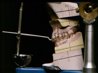
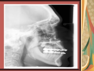
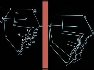
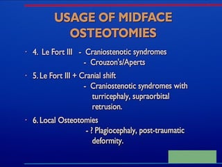
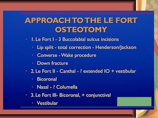
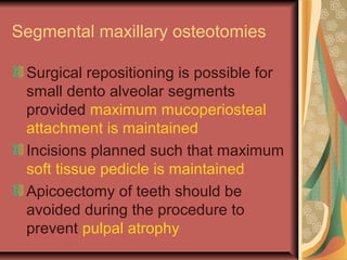
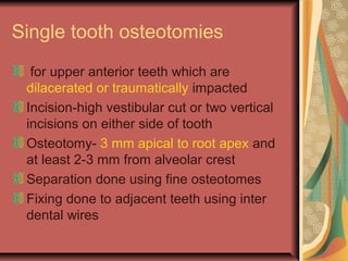
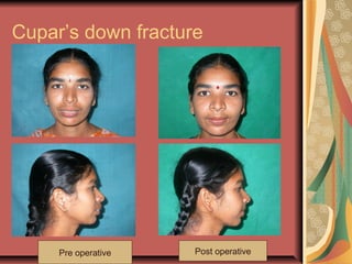
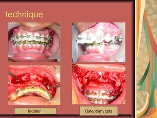
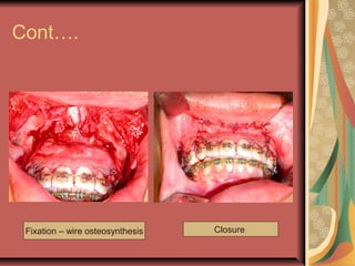
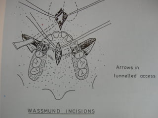
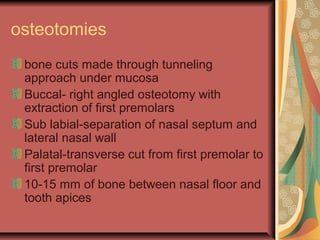
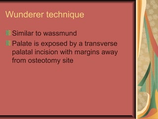
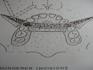
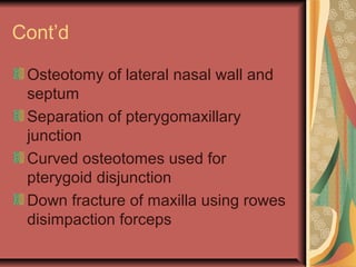
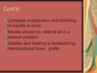
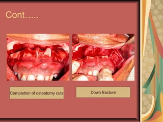
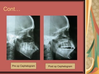
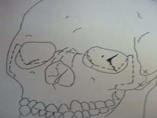
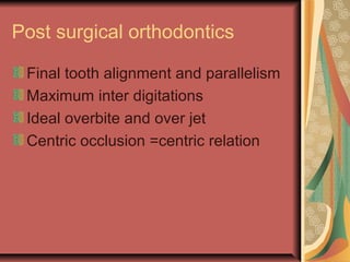
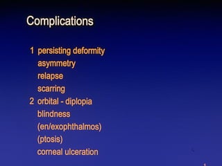
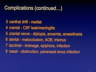
Maxillary osteotomies 
Dr V.RAMKUMAR 
CONSULTANT DENTAL&FACIOMAXILLARY 
SURGEON 
REG NO:4118-TAMILNADU-INDIA(ASIA)
Common maxillary deformities 
Maxillary anteroposterior excess 
Maxillary anteroposterior deficiency 
Vertical maxillary excess 
Vertical maxillary deficiency 
Alveolar clefts 
Craniosynostoses (aperts,crouzon) 
Binders syndrome 
achondroplasia
Patient evaluation and diagnosis 
a. Patient concerns 
b. Clinical evaluation 
1. Facial form-frontal and profile 
long, short, convex, concave, flat 
2. Relation ship of facial thirds 
3. Soft tissue –dentition relations 
smile line, Occlusal cant, dental 
midlines
Orthognathic planning 
To get an optimal balance between 
1.Aesthetics 
2.Function 
3.Stability 
4.Clinical measurements 
a. Vertical dimensions 
b. Antero posterior dimensions 
c. Transverse dimensions 
d. Intra arch dimensions
Cont’d 
5.Radiographic analysis 
a. Cephalometric 
b. Orthopantomogram 
6.Dental study models 
7.Speech 
8.Audiometry 
9.Medical and psychological
Sequence of treatment planning 
Dental and periodontal 
Extractions 
Presurgical orthodontics 
Orthognathic surgery 
Post surgical orthodontics 
Maintenance 
Others
Presurgical orthodontics 
Position the teeth over their 
respective basal bone 
Align and level the teeth 
Adjust for tooth size discrepancies 
Correct rotated teeth 
Co-ordinate upper and lower arch 
widths
Types 
Segmental maxillary surgery 
1. Single tooth osteotomy 
2. Corticotomy 
3. Anterior segmental osteotomy 
a.wassmund-1935 
b.wunderer-1963 
c. Cupar’s down fracture
Cont’d 
4.Posterior segmental osteotomy 
Schuchardt 
Kufner 
Perko & Bell 
5.Horse shoe osteotomy 
Wolford and epker
Total maxillary surgery 
Le fort I osteotomy 
a. Classic down fracture 
b. Buttress release (surgically assisted 
maxillary expansion) 
c. Quadrangular 
Le fort II osteotomy 
a. Anterior 
b. Pyramidal 
c. quadrangular
Cont’d 
Le fort III osteotomy 
Gillies 
Tessier 
4.Other midface osteotomies 
a. Zygomatic osteotomies 
b. Malar –maxillary osteotomy
Segmental maxillary osteotomies 
Surgical repositioning is possible for 
small dento alveolar segments 
provided maximum mucoperiosteal 
attachment is maintained 
Incisions planned such that maximum 
soft tissue pedicle is maintained 
Apicoectomy of teeth should be 
avoided during the procedure to 
prevent pulpal atrophy
Single tooth osteotomies 
for upper anterior teeth which are 
dilacerated or traumatically impacted 
Incision-high vestibular cut or two vertical 
incisions on either side of tooth 
Osteotomy- 3 mm apical to root apex and 
at least 2-3 mm from alveolar crest 
Separation done using fine osteotomes 
Fixing done to adjacent teeth using inter 
dental wires
Corticotomy 
To permit surgically assisted 
retraction of upper anterior teeth in 
class II div I mal occlusions 
Vestibular incision from premolar to 
premolar is used 
Cortical bone removed labially and 
palatally 
Bone also removed from 5mm above 
the teeth
Posterior segmental maxillary 
osteotomy 
Correction of anterior or posterior 
open bite 
Correction of posterior cross bites 
Closure of edentulous spaces as in 
cleft cases
Horse shoe osteotomy 
Palate remains in original position 
Dento alveolar complex alone is 
repositioned 
Aim is to minimize the size of the 
reduction of the nasal cavity 
Technically difficult since multiple 
areas of bony contacts are there
Anterior segmental maxillary 
osteotomy 
Used when alteration of premaxilla in 
the vertical plane is required as in 
anterior open bite or deep over bite 
Three techniques are usually 
described 
Down fracture technique preferred 
when vertical movement is required
Cupar’s down fracture 
Pre operative Post operative
technique 
Incision Osteotomy cuts
Cont…. 
Fixation – wire osteosynthesis Closure
Wassmund technique 
Incisions 
vertical incisions in premolar region 
and along frenum 
Midline sagittal section along hard 
palate
osteotomies 
bone cuts made through tunneling 
approach under mucosa 
Buccal- right angled osteotomy with 
extraction of first premolars 
Sub labial-separation of nasal septum and 
lateral nasal wall 
Palatal-transverse cut from first premolar to 
first premolar 
10-15 mm of bone between nasal floor and 
tooth apices
Wunderer technique 
Similar to wassmund 
Palate is exposed by a transverse 
palatal incision with margins away 
from osteotomy site
Le fort I osteotomy 
Classic le fort I down fracture (bell) 
1.Allows full mobilization of maxilla 
2.Permits bone surgery under direct 
vision 
3.Reduced risk of relapse
incision Osteotomy cuts 
Pterygoid dysjunction
Surgical technique 
Vestibular incision from first molar to 
first molar 
Osteotomy at least 5 mm above 
apices of the teeth 
Anterior cut-4-5 mm above canine 
vertical cut-zygomatic buttress region 
Posterior cuts-4-5 mm above molar 
apices
Cont’d 
Osteotomy of lateral nasal wall and 
septum 
Separation of pterygomaxillary 
junction 
Curved osteotomes used for 
pterygoid disjunction 
Down fracture of maxilla using rowes 
disimpaction forceps
Cont’d 
Complete mobilization and trimming 
of maxilla is done 
Maxilla should be able to sit in a 
passive position 
Stability and healing is facilitated by 
interpositional bone grafts
Case -1 
Incision Osteotomy cuts
Cont….. 
Completion of osteotomy cuts Down fracture
Cont… 
Pre op Cephelogram Post op Cephelogram
Le fort II osteotomy 
For correction of nasomaxillary 
hypoplasia 
It is a pyramidal naso-orbital maxillary 
osteotomy
Le fort III osteotomy 
Total midface osteotomy 
For correction of various craniofacial 
syndromes like aperts crouzon etc
Post surgical orthodontics 
Final tooth alignment and parallelism 
Maximum inter digitations 
Ideal overbite and over jet 
Centric occlusion =centric relation
THANK YOU

6 maxillary osteotomies

  • 1.
    Maxillary osteotomies DrV.RAMKUMAR CONSULTANT DENTAL&FACIOMAXILLARY SURGEON REG NO:4118-TAMILNADU-INDIA(ASIA)
  • 2.
    Common maxillary deformities Maxillary anteroposterior excess Maxillary anteroposterior deficiency Vertical maxillary excess Vertical maxillary deficiency Alveolar clefts Craniosynostoses (aperts,crouzon) Binders syndrome achondroplasia
  • 3.
    Patient evaluation anddiagnosis a. Patient concerns b. Clinical evaluation 1. Facial form-frontal and profile long, short, convex, concave, flat 2. Relation ship of facial thirds 3. Soft tissue –dentition relations smile line, Occlusal cant, dental midlines
  • 5.
    Orthognathic planning Toget an optimal balance between 1.Aesthetics 2.Function 3.Stability 4.Clinical measurements a. Vertical dimensions b. Antero posterior dimensions c. Transverse dimensions d. Intra arch dimensions
  • 6.
    Cont’d 5.Radiographic analysis a. Cephalometric b. Orthopantomogram 6.Dental study models 7.Speech 8.Audiometry 9.Medical and psychological
  • 10.
    Sequence of treatmentplanning Dental and periodontal Extractions Presurgical orthodontics Orthognathic surgery Post surgical orthodontics Maintenance Others
  • 11.
    Presurgical orthodontics Positionthe teeth over their respective basal bone Align and level the teeth Adjust for tooth size discrepancies Correct rotated teeth Co-ordinate upper and lower arch widths
  • 12.
    Types Segmental maxillarysurgery 1. Single tooth osteotomy 2. Corticotomy 3. Anterior segmental osteotomy a.wassmund-1935 b.wunderer-1963 c. Cupar’s down fracture
  • 13.
    Cont’d 4.Posterior segmentalosteotomy Schuchardt Kufner Perko & Bell 5.Horse shoe osteotomy Wolford and epker
  • 14.
    Total maxillary surgery Le fort I osteotomy a. Classic down fracture b. Buttress release (surgically assisted maxillary expansion) c. Quadrangular Le fort II osteotomy a. Anterior b. Pyramidal c. quadrangular
  • 15.
    Cont’d Le fortIII osteotomy Gillies Tessier 4.Other midface osteotomies a. Zygomatic osteotomies b. Malar –maxillary osteotomy
  • 19.
    Segmental maxillary osteotomies Surgical repositioning is possible for small dento alveolar segments provided maximum mucoperiosteal attachment is maintained Incisions planned such that maximum soft tissue pedicle is maintained Apicoectomy of teeth should be avoided during the procedure to prevent pulpal atrophy
  • 20.
    Single tooth osteotomies for upper anterior teeth which are dilacerated or traumatically impacted Incision-high vestibular cut or two vertical incisions on either side of tooth Osteotomy- 3 mm apical to root apex and at least 2-3 mm from alveolar crest Separation done using fine osteotomes Fixing done to adjacent teeth using inter dental wires
  • 21.
    Corticotomy To permitsurgically assisted retraction of upper anterior teeth in class II div I mal occlusions Vestibular incision from premolar to premolar is used Cortical bone removed labially and palatally Bone also removed from 5mm above the teeth
  • 22.
    Posterior segmental maxillary osteotomy Correction of anterior or posterior open bite Correction of posterior cross bites Closure of edentulous spaces as in cleft cases
  • 23.
    Horse shoe osteotomy Palate remains in original position Dento alveolar complex alone is repositioned Aim is to minimize the size of the reduction of the nasal cavity Technically difficult since multiple areas of bony contacts are there
  • 24.
    Anterior segmental maxillary osteotomy Used when alteration of premaxilla in the vertical plane is required as in anterior open bite or deep over bite Three techniques are usually described Down fracture technique preferred when vertical movement is required
  • 25.
    Cupar’s down fracture Pre operative Post operative
  • 26.
  • 27.
    Cont…. Fixation –wire osteosynthesis Closure
  • 28.
    Wassmund technique Incisions vertical incisions in premolar region and along frenum Midline sagittal section along hard palate
  • 30.
    osteotomies bone cutsmade through tunneling approach under mucosa Buccal- right angled osteotomy with extraction of first premolars Sub labial-separation of nasal septum and lateral nasal wall Palatal-transverse cut from first premolar to first premolar 10-15 mm of bone between nasal floor and tooth apices
  • 31.
    Wunderer technique Similarto wassmund Palate is exposed by a transverse palatal incision with margins away from osteotomy site
  • 35.
    Le fort Iosteotomy Classic le fort I down fracture (bell) 1.Allows full mobilization of maxilla 2.Permits bone surgery under direct vision 3.Reduced risk of relapse
  • 36.
    incision Osteotomy cuts Pterygoid dysjunction
  • 37.
    Surgical technique Vestibularincision from first molar to first molar Osteotomy at least 5 mm above apices of the teeth Anterior cut-4-5 mm above canine vertical cut-zygomatic buttress region Posterior cuts-4-5 mm above molar apices
  • 38.
    Cont’d Osteotomy oflateral nasal wall and septum Separation of pterygomaxillary junction Curved osteotomes used for pterygoid disjunction Down fracture of maxilla using rowes disimpaction forceps
  • 39.
    Cont’d Complete mobilizationand trimming of maxilla is done Maxilla should be able to sit in a passive position Stability and healing is facilitated by interpositional bone grafts
  • 40.
    Case -1 IncisionOsteotomy cuts
  • 41.
    Cont….. Completion ofosteotomy cuts Down fracture
  • 42.
    Cont… Pre opCephelogram Post op Cephelogram
  • 43.
    Le fort IIosteotomy For correction of nasomaxillary hypoplasia It is a pyramidal naso-orbital maxillary osteotomy
  • 46.
    Le fort IIIosteotomy Total midface osteotomy For correction of various craniofacial syndromes like aperts crouzon etc
  • 48.
    Post surgical orthodontics Final tooth alignment and parallelism Maximum inter digitations Ideal overbite and over jet Centric occlusion =centric relation
  • 53.