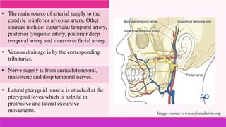
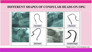
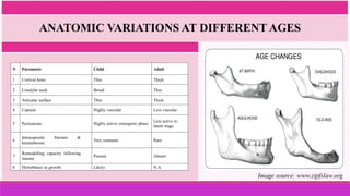
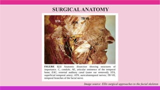
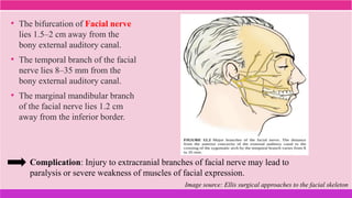
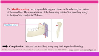
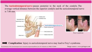
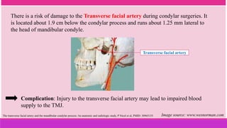
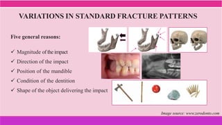
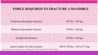
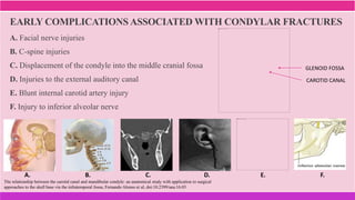
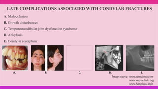
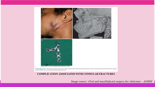
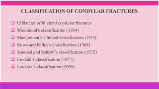
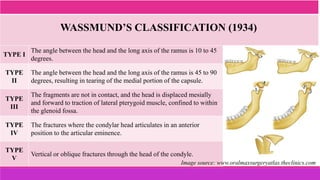
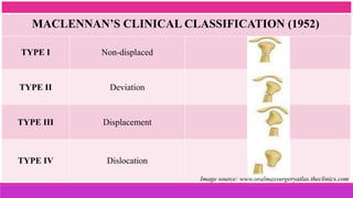
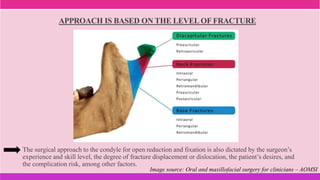
This document discusses mandibular condylar fractures, including:
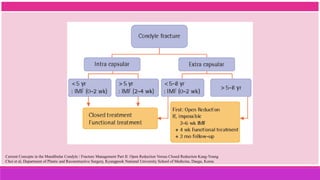
1. It provides an overview of the etiology, classification, clinical features, diagnosis, and management of mandibular condylar fractures.
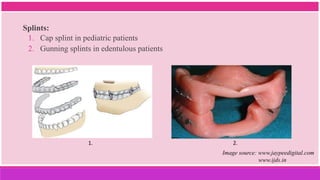
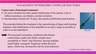
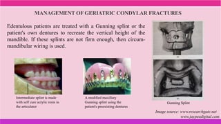
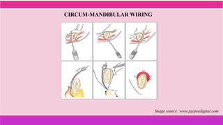
2. Treatment protocols for geriatric and pediatric patients are also discussed.
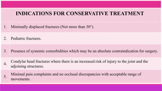
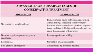
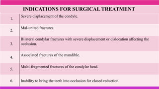
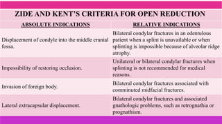
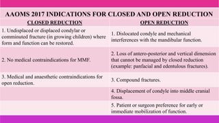
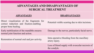
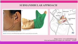
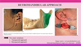
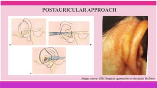
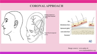
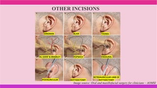
3. The indications and contraindications of closed and open reduction and fixation techniques for condylar fractures are analyzed in detail.
This document provides an overview of zygomaticomaxillary complex fractures, including:
- The anatomy and biomechanics that make these fractures common.
- Various classification systems used to describe fracture patterns.
- The signs and symptoms seen with these injuries, such as swelling, ecchymosis, and eye problems.
- The importance of a thorough physical exam and imaging for diagnosis.
The document provides information on condylar fractures, including:
1. Condylar fractures account for 26-40% of all mandible fractures and can result in pain, dysfunction and deformity if not treated properly.
2. The condyle has a unique anatomy and is an important growth center for the mandible. Fractures can occur in the condylar head, neck or subcondylar region.
3. Various classification systems are described that categorize fractures by location, degree of displacement, and direction of forces involved. Accurate classification is important for determining appropriate treatment.
This document provides an overview of Lefort I osteotomy, including:
- A brief history describing the development of maxillary osteotomy techniques.
- Anatomical considerations and the biologic basis for maxillary osteotomies, which have shown adequate blood supply can be maintained.
- Indications for Lefort I osteotomy include altering vertical dimension, anteroposterior movements, and surgical expansion of the maxilla.
- Types of Lefort I osteotomies are described, including classic, quadrangular, and segmental variations. Postoperative management and potential complications are also outlined.
This document discusses fractures of the zygoma bone. It begins with an introduction and overview of fracture patterns, classification, clinical features, investigations, management approaches, reduction techniques, fixation methods, and complications. Key points include that zygoma fractures often involve adjacent structures like the maxilla and orbit. Fracture lines typically extend from the inferior orbital fissure in three directions. Clinical features may include facial deformity, diplopia, and neurological symptoms. Investigations include radiography and CT scanning. Surgical approaches to reduction include temporal, intraoral, and endoscopic methods. Fixation often utilizes miniplates applied at one to four points depending on the fracture pattern and displacement.
This document provides an overview of reconstruction flaps in oral and maxillofacial surgery. It begins with an introduction discussing the challenges of reconstructing maxillofacial defects. The history of flap surgery is then reviewed from 600 BC to modern developments. Flaps are defined as tissues containing a blood vessel network to support survival when transferred. The document outlines classifications of flaps by movement, blood supply, composition, and other characteristics. Specific local and regional flap types are described in detail, including forehead, submental island, and pectoralis major flaps. Factors in planning reconstruction with flaps and evaluating defects are also discussed.
Condylar sag is defined as an immediate or late change in the position of the condyle in the glenoid fossa after orthognathic surgery, leading to an undesirable change in occlusion. It can occur after procedures like BSSO, IVRO, and Lefort I osteotomy. Risk factors include incorrect vectoring during condylar positioning and incomplete bone splits. Condylar sag is classified as central or peripheral, and can be diagnosed intraoperatively by examining changes in occlusion. Preventative measures include rigid fixation and intraoperative patient awakening to detect changes.
orthognathic surgery is very intresting and well knowing branch in oral surgery ....this presentation is dealing with jaw correction surgery in upper jaw.
The document discusses maxillary orthognathic procedures, including common dentofacial deformities involving the maxilla, diagnosis and treatment planning, surgical anatomy and approaches, types of mid-face osteotomies, and potential complications. It describes techniques for anterior and posterior segmental maxillary osteotomies, as well as total maxillary osteotomies including LeFort I, II, and III procedures. Complications addressed include vascular compromise, hemorrhage, and infection.
Local and regional flaps in head and neck reconstructionSaleh Bakry
Local and regional flaps are used for head and neck reconstruction. There are several types of flaps classified by blood supply, location, configuration, tissue content, and transfer method. Local flaps use adjacent tissue while regional flaps are near but not adjacent. Free flaps completely detach tissue and reconnect blood vessels microsurgically. Examples of local and regional flaps discussed are advancement, rotation, and transposition flaps as well as pedicled and free myocutaneous flaps.
This document provides an overview of genioplasty procedures. It begins with an introduction to genioplasty and anatomy. It then discusses preoperative evaluation including facial analysis, cephalometric evaluation, and chin classifications. Next, it covers various techniques for correcting chin deformities including osseous genioplasty procedures like horizontal osteotomy with advancement or reduction, and alloplastic genioplasty. It concludes with a brief discussion of complications. The document provides detailed information on evaluating patients, planning procedures, and technical aspects of different genioplasty techniques.
The document discusses various surgical approaches for the facial skeleton, including extraoral and intraoral incisions. It covers key principles for placing incisions such as following natural lines and avoiding vital structures. Specific approaches are described for the mandible, condyle, orbit, maxilla, and nasal skeleton. Factors to consider for any facial incision include scar visibility, underlying anatomy, and adequate surgical access. Both open and endoscopic techniques are presented.
The document discusses zygomatic complex fractures, including:
1) Zygomatic fractures are common facial injuries that frequently involve fractures of the frontal, orbital, maxillary, and zygomatic processes.
2) Clinical examination involves inspection for asymmetries and palpation of the zygomatic area to assess for posterior displacement.
3) Diagnosis is aided by CT scan, which provides detailed images of fracture patterns and displacement of the zygomatic bone and surrounding structures.
Fractures of the zygomatic complex are common facial injuries that often involve displacement of the zygomatic bone from its normal position. Clinical examination involves inspection for deformities and palpation of the zygomatic bone and arch. Radiographic evaluation with CT scanning is important to fully assess the fracture pattern and displacement. Successful management requires accurate reduction and fixation of the zygomatic bone to restore facial contour and function.
This document provides information about genioplasty surgery. It begins with an introduction and overview of genioplasty. It then discusses the history, indications, contraindications, preoperative evaluation including cephalometric and soft tissue analysis, surgical anatomy, classification of chin deformities, and surgical procedure. The surgical procedure section provides a step-by-step explanation of genioplasty surgery from incision and osteotomy to fixation and closure. Key steps include marking reference points, performing the osteotomy, mobilizing and repositioning the chin segment, and securing it with either screws or bone plates. Attention to details like reference marks, osteotomy angle and position, and bone contouring help achieve the planned aesthetic results of
This document discusses various surgical approaches to the temporomandibular joint (TMJ). It begins by outlining important anatomical structures in the region, including nerves, arteries and layers of fascia. It then describes several common approaches - preauricular, endaural, postauricular, submandibular, retromandibular and intraoral. For each approach, it provides details on the surgical technique, indications, advantages and disadvantages. References are also provided at the end for further reading on the surgical anatomy of the cervical and mandibular distributions of the facial nerve.
Apertognathia and its surgical managementHimanshu Soni
Hullihen performed the first "V" shaped osteotomy of the mandible to correct anterior open bites. Kole modified this procedure by excising a wedge of bone from the mandible's symphysis and inferior border to shorten facial height. Thoma suggested Y-shaped and trapezoid mandibular body osteotomies to correct open bites associated with prognathism.
This document discusses various complications that can occur with orthognathic surgery, including:
- Common intraoperative complications are nerve injuries, most often to the inferior alveolar nerve during mandibular surgery, and hemorrhage, usually from the maxillary arteries.
- Frequent postoperative issues involve neurosensory deficits, nonunion or delayed bone healing, and infections, which have been reported in up to 9.7% of patients.
- Other risks include loss of vascularity leading to aseptic necrosis of the maxilla or mandible, nasal deformities, malocclusion, and TMJ dysfunction. Careful planning and technique aim to minimize complications while pursuing the benefits of orthognathic
This seminar explains about the development, relations, ligaments, various attachments, vascular and nervous supply and various surgical approaches and its modifications to TMJ
This document discusses several controversies in the management of maxillofacial trauma, including:
1) The management of fractures through the angle of the mandible, regarding factors like the location and extension of the fracture line and whether teeth in the line require extraction.
2) The management of condylar process fractures, debating whether closed or open reduction is preferred based on factors like displacement, facial contour, and risk of malocclusion.
3) The techniques for managing comminuted mandible fractures, including traditional conservative approaches versus open reduction and internal fixation using reconstruction plates.
It provides an overview of key points of debate and considerations in the treatment of various types of maxillofacial fractures
This document provides an overview of various osteotomy approaches for accessing lesions in the skull base and neck. It discusses the history, classification, advantages, and disadvantages of different osteotomies. Key approaches mentioned include fronto-nasal-orbital osteotomy, Lefort I osteotomy, zygomatic osteotomy, and mandibulotomy. The document emphasizes that the choice of osteotomy depends on factors like the location and extent of the lesion as well as involvement of surrounding structures. Modifications to standard approaches are also described to optimize exposure and resection of different pathologies.
1. The document describes various surgical incision techniques for accessing and removing impacted third molars, including envelope flap, three-cornered flap, and lingual split bone techniques.
2. It also discusses methods for bone removal like guttering using a bur or chiseling techniques to create space for tooth extraction.
3. Tooth sectioning is described as an option when the path of removal is hindered to reduce the risk of injury to surrounding structures.
The document discusses condylar fractures, including:
- Anatomy of the condyle and temporomandibular joint
- Various classifications of condylar fractures
- Clinical features like swelling, pain, and limited jaw movement
- Diagnosis using radiographs like panoramic x-rays and CT scans
- Treatment approaches like closed or open reduction
- Indications for non-surgical versus surgical management
In 3 sentences it summarizes that the document discusses the anatomy, classifications, diagnosis, and treatment approaches like closed or open reduction for condylar fractures of the temporomandibular joint.
This study compared a supratemporalis approach to the traditional preauricular approach for treating intracapsular condylar fractures. 84 patients were treated with one of the two approaches. The supratemporalis approach provided excellent exposure while preventing facial nerve injury, which has a risk of 1-32% with the preauricular approach. No additional complications occurred with the supratemporalis approach. The authors conclude it is an effective routine approach for intracapsular condylar fractures due to its safety advantages over the traditional method.
This document describes surgical techniques for treating TMJ ankylosis in children, including:
1. Excising the ankylotic mass through a preauricular incision and reconstructing the ramus condyle unit with either a costochondral graft or distraction osteogenesis.
2. Lining the glenoid fossa with a vascularized temporalis fascia flap to prevent reankylosis.
3. An intensive post-operative physical therapy regimen to regain jaw mobility.
This document discusses the classification and treatment of mandibular condylar fractures. It notes that condylar fractures account for 25-35% of all mandibular fractures and discusses various classification systems over time, from Brophy in 1915 to more recent subclassifications. Treatment options discussed include maxillomandibular fixation, functional therapy without fixation, and open reduction with or without internal fixation. Factors favoring nonsurgical treatment and potential complications of both early/concurrent and late management are also summarized.
This document discusses reduction techniques for zygomatic bone fractures. It begins by describing the anatomy of the zygomatic bone and common types of zygomatic fractures. It then outlines various surgical approaches that can be used for open reduction of zygomatic fractures, including the temporal fossa, intraoral, percutaneous, malar hook, Carroll Girard screw, and lateral eyebrow approaches. Indications for surgery include depressed malar eminence, enophthalmos, infraorbital paresthesia, and inability to open the mouth. The conclusion recommends that technique choice depends on the location and degree of displacement of the fracture.
Mandibular fractures are common facial injuries, often caused by motor vehicle accidents or assaults, especially in males aged 21-30. The mandible lacks strong support, so its prominent position makes it vulnerable to fractures. Common fracture sites are the body, condyle, and angle of the mandible. Associated injuries are also common, such as head injuries. A thorough examination is needed to properly diagnose and classify the fracture.
The document discusses injuries to the spine. It covers the epidemiology, anatomy, classification of injuries as stable or unstable, and mechanisms of injury. It then describes specific cervical and thoracolumbar spine injuries, including fractures, dislocations, and treatment approaches which may involve immobilization, traction, or surgery.
Dr. Ahmed M. Adawy, Professor Emeritus, Dep. Oral & Maxillofacial Surgery. Former Dean, Faculty of Dental Medicine, Al-Azhar University. Condylar fractures represent one of the most controversial issues in maxillofacial traumatology regarding classification, diagnoses and therapeutic management. Classification systems of condylar fracture is discussed. Diagnosis is usually based on history clinical examination and radiographic finding. Treatment ranges from observation, jaw exercises to closed or opened interventions. For years closed reduction was thought to be essentially complication-free. Several serious complications however have been reported including temporomandibular joint ankyloses, malocclusion, mandibular deviation and the generative joint pathology. The absolute and relative indications for open reduction is given. The debate between supporters of open or closed reduction is still continuing and the issue has not been resolved. However, the final choice treatment modality should takes into account the location of the fracture, age of the patient, presence or absence of other associated injuries, cosmetic impact of the surgery and presence of other systemic medical conditions.
This document discusses condylar fractures of the mandible. It begins with an introduction and overview of condylar fracture classification systems. It then covers the etiology, clinical examination, principles of treatment, and treatment options for condylar fractures, including closed and open reduction techniques. Complications of treatment are also outlined. The document emphasizes that the treatment approach depends on factors like the patient's age, fracture characteristics, and whether other injuries are present. The goal of treatment is to achieve a stable occlusion and restore function through both surgical and non-surgical means.
Mandibular fractures
Dr. Ahmed M. Adawy
Professor Emeritus, Dept. Oral & Maxillofacial Surg.
Former Dean, Faculty of Dental Medicine
Al-Azhar University
Fractures of the mandible are a common form of facial injury in adults and occur most frequently in males during the third decade of life. The main causes of mandibular fractures are road traffic accidents, interpersonal violence, falls and sport injuries. Mandibular fractures are classified according to various criteria. The three main factors to consider are the cause of the fracture, the type of fracture and the site of the fracture. Clinical diagnosis as well as radiographic examinations are presented. Treatment modalities are discussed. Moreover, treatment-related complications are given.
Leonel Martinez presented on basilar skull fractures. The incidence of skull base fractures is 2-24% of head injuries. Evaluation includes physical exam signs like periorbital ecchymosis, CSF rhinorrhea, and cranial nerve deficits. Imaging like CT is important to classify the fracture. Management depends on the fracture type and symptoms, with observation for asymptomatic cases and surgery to repair CSF leaks or decompress neural structures.
This document provides an overview of condylar fractures, including:
- The surgical anatomy of the temporomandibular joint region.
- Common mechanisms of injury that can cause condylar fractures.
- Several classification systems used to describe different types of condylar fractures.
- Clinical examination findings and various radiologic imaging modalities useful for diagnosing condylar fractures.
- Considerations for treatment and complications that can arise from condylar fractures.
Is Medial Ridge Sign a Reliable Indicator Glenoid Bone Loss-Dr. Dhanasekarapr...TheRightDoctors
The document discusses the "medial ridge sign" seen on CT scans of patients with recurrent shoulder dislocations as an indicator of glenoid bone loss. The study aimed to evaluate if the medial ridge sign reliably indicates significant bone loss. It found the sign had high sensitivity but low specificity for significant loss. While the sign suggested some bone loss, glenoid bone loss measurements were still needed to determine if augmentation was required rather than just Bankart repair. So the medial ridge sign is not reliable in deciding between the two surgical procedures.
Leiomyoma is a benign tumor that originates from smooth
muscle cell. The most common sites are the uterus, gastrointestinal tract & skin. Leiomyoma is a relatively uncommon smooth muscle tumor rarely found in the head and neck. Enzinger and Weiss (1995), analyzed a total of 7748 leiomyomas, 95% of the tumors occurred in the female genitalia (uterus), 3% in the skin, 0.9% in the gastrointestinal tract and the remainder at various sites including skull base.
Mandibular fractures are common injuries that result from facial trauma. The document discusses the history, anatomy, classification, examination, and treatment of mandibular fractures. Key points include that mandibular fractures were first described in ancient Egyptian medical texts, occur most often in males ages 20-30 from vehicular accidents or assaults, and can be classified based on location, number of fragments, involvement of teeth, and direction of the fracture. Radiographic examination including panoramic x-rays are important for diagnosis.
This document provides information on cervical spine injuries, including:
- Upper cervical injuries involve C1-C2 and lower cervical injuries involve C3-C7.
- Common upper cervical injuries include fractures of the atlas and axis as well as occipital condyle fractures and occipitoatlantal dislocations.
- Lower cervical spine injuries include fractures of the vertebral bodies and posterior elements like the lamina.
- Detailed classifications and treatment recommendations are provided for various cervical fractures and dislocations. Imaging like CT and MRI play an important role in evaluation of these injuries.
Facial trauma is without doubt a most challenging area within the specialty of oral and maxillofacial surgery. Trauma with all its aspects has great importance, being the main cause of morbidity and mortality with rising frequency worldwide, especially in recent decades. Traumatic facial injuries are often associated with high mortality and varying degrees of physical, functional, psychological damage, cosmetic disfigurement, and concomitant injuries to other organs that may be added complicating factors. Road traffic accidents represent the main cause of facial trauma. According to WHO, Egypt leads the Middle East when it comes to road accidents, with an average of 12,000 people killed annually. Interpersonal violence is the second most prevalent etiologic factor. Our society is progressively becoming more and more violent and impatient, perhaps due to overcrowding, so the frequency of patients reporting in emergency with facial bones fracture is increasing.
During the last three decades, significant advances have occurred in the methods of fixation used for facial bone fractures, resulting in improved functional and aesthetic outcomes. Surgical techniques have been moving away from delayed closed reduction with internal wires suspension to early open reduction and internal plate fixation. The transition from wire osteosynthesis to rigid internal fixation in facial bone fractures using different micro or mini-plates and screw systems is regarded as one of the greatest advances in the field of maxillofacial surgery. I hope this book reflects the latest trends, concepts and innovations in the care of patients with facial trauma.
For convenience, the text is divided into 3 sections. Section 1 deals with primary care of the patients. Section 2 is concerned with midface fractures. In section 3 management of trauma to the lower face is discussed. Upper face injuries are not included and the reader could find the subject elsewhere under the topic of craniofacial traumatology. From the basic to the most complex, readers will find that each chapter is sequentially organized to provide a concise, and practical description of the operative details. The goal was to provide the reader with a fully comprehensive, yet highly illustrated text on the subject of facial trauma.
This article analyzes 175 mandibular condyle fractures treated over 4 years at a hospital in southern India. It finds that condyle fractures made up 18.39% of all mandibular fractures and were most common in patients over 16 years old. Most condyle fractures were unilateral and associated with other mandible fractures. Non-surgical treatment was used for undisplaced fractures in younger patients, while open reduction and internal fixation was performed for displaced fractures in older patients. The study aims to establish treatment guidelines for condyle fractures based on a patient's age, fracture pattern, and level of displacement.
This document summarizes principles and concepts in fixing proximal humerus fractures with a locking plate. It describes the anatomy of the proximal humerus, classification systems for these fractures, considerations for surgical treatment, and techniques for open reduction and internal fixation using a locking plate. Key points covered include applied anatomy of the fracture deforming forces, blood supply to the humeral head, Neer's classification system, indications for operative vs non-operative treatment, surgical approach and techniques for placing a locking plate and securing it with screws including the importance of a calcar screw.
Unlocking the Secrets to Safe Patient Handling.pdfLift Ability
Furthermore, the time constraints and workload in healthcare settings can make it challenging for caregivers to prioritise safe patient handling Australia practices, leading to shortcuts and increased risks.
Let's Talk About It: Breast Cancer (What is Mindset and Does it Really Matter?)bkling
Your mindset is the way you make sense of the world around you. This lens influences the way you think, the way you feel, and how you might behave in certain situations. Let's talk about mindset myths that can get us into trouble and ways to cultivate a mindset to support your cancer survivorship in authentic ways. Let’s Talk About It!
We are one of the top Massage Spa Ajman Our highly skilled, experienced, and certified massage therapists from different corners of the world are committed to serving you with a soothing and relaxing experience. Luxuriate yourself at our spas in Sharjah and Ajman, which are indeed enriched with an ambiance of relaxation and tranquility. We could confidently claim that we are one of the most affordable Spa Ajman and Sharjah as well, where you can book the massage session of your choice for just 99 AED at any time as we are open 24 hours a day, 7 days a week.
Visit : https://massagespaajman.com/
Call : 052 987 1315
INFECTION OF THE BRAIN -ENCEPHALITIS ( PPT)blessyjannu21
Neurological system includes brain and spinal cord. It plays an important role in functioning of our body. Encephalitis is the inflammation of the brain. Causes include viral infections, infections from insect bites or an autoimmune reaction that affects the brain. It can be life-threatening or cause long-term complications. Treatment varies, but most people require hospitalization so they can receive intensive treatment, including life support.
Feeding plate for a newborn with Cleft Palate.pptxSatvikaPrasad
A feeding plate is a prosthetic device used for newborns with a cleft palate to assist in feeding and improve nutrition intake. From a prosthodontic perspective, this plate acts as a barrier between the oral and nasal cavities, facilitating effective sucking and swallowing by providing a more normal anatomical structure. It helps to prevent milk from entering the nasal passage, thereby reducing the risk of aspiration and enhancing the infant's ability to feed efficiently. The feeding plate also aids in the development of the oral muscles and can contribute to better growth and weight gain. Its custom fabrication and proper fitting by a prosthodontist are crucial for ensuring comfort and functionality, as well as for minimizing potential complications. Early intervention with a feeding plate can significantly improve the quality of life for both the infant and the parents.
Can coffee help me lose weight? Yes, 25,422 users in the USA use it for that ...nirahealhty
The South Beach Coffee Java Diet is a variation of the popular South Beach Diet, which was developed by cardiologist Dr. Arthur Agatston. The original South Beach Diet focuses on consuming lean proteins, healthy fats, and low-glycemic index carbohydrates. The South Beach Coffee Java Diet adds the element of coffee, specifically caffeine, to enhance weight loss and improve energy levels.
Hypertension and it's role of physiotherapy in it.Vishal kr Thakur
This particular slides consist of- what is hypertension,what are it's causes and it's effect on body, risk factors, symptoms,complications, diagnosis and role of physiotherapy in it.
This slide is very helpful for physiotherapy students and also for other medical and healthcare students.
Here is summary of hypertension -
Hypertension, also known as high blood pressure, is a serious medical condition that occurs when blood pressure in the body's arteries is consistently too high. Blood pressure is the force of blood pushing against the walls of blood vessels as the heart pumps it. Hypertension can increase the risk of heart disease, brain disease, kidney disease, and premature death.
The best massage spa Ajman is Chandrima Spa Ajman, which was founded in 2023 and is exclusively for men 24 hours a day. As of right now, our parent firm has been providing massage services to over 50,000+ clients in Ajman for the past 10 years. It has about 8+ branches. This demonstrates that Chandrima Spa Ajman is among the most reasonably priced spas in Ajman and the ideal place to unwind and rejuvenate. We provide a wide range of Spa massage treatments, including Indian, Pakistani, Kerala, Malayali, and body-to-body massages. Numerous massage techniques are available, including deep tissue, Swedish, Thai, Russian, and hot stone massages. Our massage therapists produce genuinely unique treatments that generate a revitalized sense of inner serenely by fusing modern techniques, the cleanest natural substances, and traditional holistic therapists.
Empowering ACOs: Leveraging Quality Management Tools for MIPS and BeyondHealth Catalyst
Join us as we delve into the crucial realm of quality reporting for MSSP (Medicare Shared Savings Program) Accountable Care Organizations (ACOs).
In this session, we will explore how a robust quality management solution can empower your organization to meet regulatory requirements and improve processes for MIPS reporting and internal quality programs. Learn how our MeasureAble application enables compliance and fosters continuous improvement.
Dr. David Greene R3 stem cell Breakthroughs: Stem Cell Therapy in CardiologyR3 Stem Cell
Dr. David Greene, founder and CEO of R3 Stem Cell, is at the forefront of groundbreaking research in the field of cardiology, focusing on the transformative potential of stem cell therapy. His latest work emphasizes innovative approaches to treating heart disease, aiming to repair damaged heart tissue and improve heart function through the use of advanced stem cell techniques. This research promises not only to enhance the quality of life for patients with chronic heart conditions but also to pave the way for new, more effective treatments. Dr. Greene's work is notable for its focus on safety, efficacy, and the potential to significantly reduce the need for invasive surgeries and long-term medication, positioning stem cell therapy as a key player in the future of cardiac care.
Dr. David Greene R3 stem cell Breakthroughs: Stem Cell Therapy in Cardiology
Condyle Fractures.pptx
1. MANDIBULAR CONDYLAR FRACTURES
Animation source: www.wikimedia.org
Presenter – Dr. Harjeet Yadav
PG III, Dept. of OMFS
Moderator – Dr. Ajay Das
Reader, Dept. of OMFS
2. Animation source: www.wikimedia.org
This seminar deals with the etiology, classification, clinical
features, diagnosis, and contemporary management of
mandibular condylar fractures.
Along with the regular management strategies, treatment
protocols for geriatric and pediatric patients have also been
discussed.
The indications and contraindications of closed as well as
open reduction and fixation of condylar fractures are
analyzed in detail.
CONTENTS
3. • The topic of mandibular condylar fracture has generated more discussion and controversy than
any other in the field of maxillofacial trauma.
• Direct or indirect trauma can lead to fracture of the condyle. The degree of displacement of
fractured condyle depends on the direction and magnitude of force.
• In spite of a common occurrence, the management of condylar fractures has been
controversial as there is no established consensus. Traditionally closed reduction has been the
treatment of choice for condylar fractures and have been treated by various forms of
intermaxillary fixation. With the improvement in radiographic imaging and biomaterials used
in the fixation, surgical management has gradually found acceptance as it restores early
function.
INTRODUCTION
4. Image source: www.researchgate.net
Elliptical
Shape
• Condyle is a knuckle like structure. It is
a strong upward projection from the
postero-superior part of the ramus.
• The condyle has a backward angulation
of 15–33° to the frontal plane and is
elliptical in shape.
• The mesio-lateral width is 15–20 mm
and the antero-posterior width is 8–
10 mm.
• The condyle articulates with the
mandibular fossa in the temporal bone,
as part of the TMJ.
ANATOMY OF CONDYLE
5. • The main source of arterial supply to the
condyle is inferior alveolar artery. Other
sources include: superficial temporal artery,
posterior tympanic artery, posterior deep
temporal artery and transverse facial artery.
• Venous drainage is by the corresponding
tributaries.
• Nerve supply is from auriculotemporal,
masseteric and deep temporal nerves.
• Lateral pterygoid muscle is attached at the
pterygoid fovea which is helpful in
protrusive and lateral excursive
movements. Image source: www.aofoundation.org
7. Image source: www.zipfslaw.org
ANATOMIC VARIATIONS AT DIFFERENT AGES
N Parameter Child Adult
1 Cortical bone Thin Thick
2 Condylar neck Broad Thin
3 Articular surface Thin Thick
4 Capsule Highly vascular Less vascular
5 Periosteum Highly active osteogenic phase
Less active in
latent stage
6
Intracapsular fracture &
hemarthrosis.
Very common Rare
7
Remodelling capacity following
trauma
Present Absent
8 Disturbance in growth Likely N.A
8. Image source: Ellis surgical approaches to the facial skeleton
SURGICALANATOMY
9. • The bifurcation of Facial nerve
lies 1.5–2 cm away from the
bony external auditory canal.
• The temporal branch of the facial
nerve lies 8–35 mm from the
bony external auditory canal.
• The marginal mandibular branch
of the facial nerve lies 1.2 cm
away from the inferior border.
Complication: Injury to extracranial branches of facial nerve may lead to
paralysis or severe weakness of muscles of facial expression.
Image source: Ellis surgical approaches to the facial skeleton
10. The Maxillary artery can be injured during procedures in the subcondylar portion
of the mandible. The mean distance of the branching point of the maxillary artery
to the tip of the condyle is 22.4 mm.
Maxillary artery
Maxillary artery: anatomical landmarks and relationship with the mandibular subcondyle, Hakan Orbay et al, PMID: 18090748 Image source: www.researchgate.net
Complication: Injury to the maxillary artery may lead to profuse bleeding.
11. The Auriculotemporal nerve passes posterior to the neck of the condyle. The
average vertical distance between the superior condyle and the auriculotemporal nerve
is 7.06 mm
Auriculotemporal nerve
The distribution of the auriculotemporal nerve around the temporomandibular joint, B L Schmidt et al, PMID: 9720090 Image source: www.springer.com
Complication: Injury to auriculotemporal nerve may lead to Frey’s syndrome.
Frey’s syndrome
12. There is a risk of damage to the Transverse facial artery during condylar surgeries. It
is located about 1.9 cm below the condylar process and runs about 1.25 mm lateral to
the head of mandibular condyle.
Transverse facial artery
The transverse facial artery and the mandibular condylar process: An anatomic and radiologic study, P Nicol et al, PMID: 30965155 Image source: www.wesnorman.com
Complication: Injury to the transverse facial artery may lead to impaired blood
supply to the TMJ.
13. The mean distance between the Middle meningeal artery and the apex of the
condyle is 18.8 mm.
Middle meningeal artery
Proximity of the middle meningeal artery and maxillary artery to the mandibular head and mandibular neck as
revealed by three-dimensional time-of-flight magnetic resonance angiography, Daphne Schönegg, PMID: 34024006 Image source: www.worldofmedicalsaviours.com
Complication: Injury to middle meningeal artery may lead to epidural hematoma.
14. The mean distance between medial margin of mandibular condyle to Internal carotid artery
is 11.2 mm ±0.6.
Surgical importance of distance from mandibular condyle to carotid canal and foramen spinosum: an anatomical study, November 2019, International Journal of Research in Medical
Sciences 7(12):4733, DOI:10.18203/2320-6012.ijrms20195547
Internal carotid artery
Complication: Injury to internal carotid artery may lead to thrombosis and
neurological deficit.
15. The Masseteric nerve is about 16 mm superior to the lowest point on the
mandibular notch.
Masseteric nerve
Topographical Landmarks for the Identification of Branches of Mandibular Nerve and Its Surgical Implications: A Cadaveric
Study, Ariyanachi Kaliappan et al, PMID: 34984156 Image source: www.pocketdentistry.com
Complication: Injury to masseteric nerve may lead to weakness in masseter
muscle and sensory deficit to the TMJ.
16. The location of Superficial temporal artery bifurcation is about 9.5 ± 5.3 mm
anterior to the posterior margin of the condyle.
Superficial temporal artery
The Anatomy of the Superficial Temporal Artery in Adult Koreans Using 3-Dimensional Computed Tomographic
Angiogram: Clinical Research, Byung Soo Kim et al, PMID: 24167792 Image source: www.musculoskeletalkey.com
Complication: Injury to superficial temporal artery may lead to profuse bleeding.
18. INCIDENCE
About 30% of all mandibular fractures.
Male predilection – About 80% cases. Image source: www.researchgate.net
28%
19. BLOW FALL RTA
Kinetic energy imported to the
static individual by a moving
object.
Lindahl (1977) proposed 3 mechanisms of injury to the condyle
MECHANISM OF INJURY
Kinetic energy derived from the
movement of individual and
extended upon a static object.
Kinetic energy which is
summation of forces derived
from a combination of 1. & 2
1 2 3
20. BONE BIOMECHANICS
Mandible functions as a class III lever system, where the
muscle force is between the TMJ & the occlusal load.
Sources: www.oralmaxsurgeryatlas.theclinics.com
www.springer.com
21. BIOMECHANICS OF INJURY
(HUNTING BOW CONCEPT)
• The Mandible resembles a Hunting bow
which is weak at the ends and strong in
the midline. Its ends (the condyles) are
enclosed by the glenoid fossa.
• So any blow to the midline (symphysis
region) of the mandible can cause
bilateral condylar fracture and any blow
to the parasymphysis region may cause a
contralateral condylar fracture.
• This is based on the impact of the force.
Condyle
Tension
Rotational movement
permitted
Tension
Condyle
Symphysis
Blow
Image source: www.researchgate.net
T
T T
C C
C
22. VARIATIONS IN STANDARD FRACTURE PATTERNS
Five general reasons:
Magnitude oftheimpact
Direction of the impact
Position of the mandible
Condition of the dentition
Shape of the object delivering the impact
Image source: www.zerodonto.com
23. Unilateral subcondylar fractures 425 lbs / 193 kgs
Bilateral subcondylar fractures 550 lbs / 220 kgs
Symphyseal fractures 924 lbs / 419 kgs
Lateral impact for body fractures 300 to 700 lbs / 136 to 317 kgs
Biomechanics of cranio-maxillofacial trauma – Biju Pappachan & Mohan Alexander 2012 June PMID 23730074
FORCE REQUIRED TO FRACTURE A MANDIBLE
24. A. Facial nerve injuries
B. C-spine injuries
C. Displacement of the condyle into the middle cranial fossa
D. Injuries to the external auditory canal
E. Blunt internal carotid artery injury
F. Injury to inferior alveolar nerve
EARLY COMPLICATIONS ASSOCIATED WITH CONDYLAR FRACTURES
A. B. C. D. E. F.
The relationship between the carotid canal and mandibular condyle: an anatomical study with application to surgical
approaches to the skull base via the infratemporal fossa, Fernando Alonso et al, doi:10.2399/ana.16.03
GLENOID FOSSA
CAROTID CANAL
25. LATE COMPLICATIONS ASSOCIATED WITH CONDYLAR FRACTURES
A. Malocclusion
B. Growth disturbances
C. Temporomandibular joint dysfunction syndrome
D. Ankylosis
E. Condylar resorption
A. B. C. D. E.
Image source: www.zerodonto.com
www.mayoclinic.org
www.banglajol.info
26. COMPLICATION ASSOCIATED WITH CONDYLAR FRACTURES
Image source: Oral and maxillofacial surgery for clinicians – AOMSI
27. CLASSIFICATION OF CONDYLAR FRACTURES
Unilateral or bilateral condylar fractures
Wassmund's classification (1934)
MacLennan’s Clinical classification (1952)
Rowe and Killey's classification (1968)
Spiessel and Schroll’s classification (1972)
Lindahl’s classification (1977)
Loukota’s classification (2005)
28. TYPE I
The angle between the head and the long axis of the ramus is 10 to 45
degrees.
TYPE
II
The angle between the head and the long axis of the ramus is 45 to 90
degrees, resulting in tearing of the medial portion of the capsule.
TYPE
III
The fragments are not in contact, and the head is displaced mesially
and forward to traction of lateral pterygoid muscle, confined to within
the glenoid fossa.
TYPE
IV
The fractures where the condylar head articulates in an anterior
position to the articular eminence.
TYPE
V
Vertical or oblique fractures through the head of the condyle.
WASSMUND’S CLASSIFICATION (1934)
Image source: www.oralmaxsurgeryatlas.theclinics.com
29. TYPE I Non-displaced
TYPE II Deviation
TYPE III Displacement
TYPE IV Dislocation
MACLENNAN’S CLINICAL CLASSIFICATION (1952)
Image source: www.oralmaxsurgeryatlas.theclinics.com
30. A. Simple fractures of condyle
B. Compound fractures of condyle
C. Comminuted fracture associated with zygomatic arch fracture.
31. Type I Non-displaced fracture
Type II
Low neck fracture with
displacement
Type III
High neck fracture with
displacement
Type IV
Low neck fracture with
dislocation
Type V
High neck fracture with
dislocation
Type VI Head fracture
SPIESSELAND SCHROLL’S CLASSIFICATION (1972)
TYPE I TYPE II
TYPE III TYPE IV
TYPE V TYPE VI
Image source: www.oralmaxsurgeryatlas.theclinics.com
32. LINDAHL’S CLASSIFICATION (1977)
I. Anatomic location of the fracture:
Condylar head
Condylar neck
Subcondylar
II. Relationship of condylar fragment to mandible:
Non-displaced
Deviated
Displacement with medial or lateral overlap
Displacement with anterior or posterior overlap
III. Relationship of condylar head and fossa:
Non-displaced
Displacement
Dislocation
IV. Injury to meniscus:
It may be torn, ruptured or herniated in forward/backward direction. Image source: www.researchgate.net
34. CLINICAL EXAMINATION
Condylar fractures are diagnosed with the help of both clinical and radiological
assessment.
These fractures are most commonly missed on clinical examination.
Extracapsular condylar fractures are frequent and may be associated with
displacement of the condylar head.
The condylar head may be in contact with the ramus or may be displaced laterally
or medially.
35. Unilateral condylar fractures:
• Facial asymmetry.
• Swelling and pain over the TMJ.
• Hemorrhage from the external ear (due to
laceration of external acoustic meatus by the
violent impact of condyle).
• Proper examination with an auriscope is
essential to differentiate bleeding from external
auditory canal and middle ear. Temporal bone
fracture may be accompanied by CSF leak
which is termed as otorrhea.
• Hematoma surrounding the fractured condyle.
• Hematoma in the mastoid region called the
Battle’s sign.
INSPECTION
Image source: www.tuyenlab.com
www.ihealthblogger.com
Auriscope
36. • If the condylar head is displaced medially, characteristic hollow in the region of condylar
head can be observed once the edema subsides.
• Ear bleed will persist if the head of the condyle is impacted in the glenoid fossa.
• Deviation of mandible towards the side of fracture.
• Decreased range of movements.
• Gagging of occlusion on the ipsilateral side.
• Locked mandible – due to entry into the middle cranial fossa.
Deviation of mandible and ipsilateral open bite Image source: www.sciencedirect.com
37. Bilateral condylar fractures:
• Overall mandibular movements are usually
more restricted.
• If the condyle is displaced bilaterally,
shortening of ramus occurs resulting in
derangement of occlusion.
• Overriding of the fractured segments result in
anterior open bite.
• Associated fracture of symphysis or para-
symphysis can also be present; thus careful
examination is mandatory (Contre-coupe
injury).
•Pseudo Class II appearance.
Anterior open bite
Image source: www.archwired.com
www.semanticscholar.org
38. PALPATION
• The condyles are palpated by standing behind the
patient. The little fingers are placed inside the
external auditory canal and the patient is asked to
open and close their mouth. By this method the
position and movement of the condyles are
determined.
• Tenderness over the condylar area with associated
crepitation.
• Displacement of the condylar head within the
external auditory meatus.
• Paresthesia of the lips.
Image source: www.pocketdentistry.com
43. Computed Tomography Scan
Image source: www.radiopaedia.org
www.researchgate,net
Gold standard for the diagnosis of mandibular condylar fractures.
Coronal section of CT scan showing
a right condylar neck fracture with
medial dislocation of the condyle.
CT scan 3D reconstruction image
of skull showing a right condylar
neck fracture.
Axial section of CT scan showing a
left condylar neck fracture with
lateral dislocation of the condyle.
44. Cone Beam Computed Tomography
Image source: www.semanticscholar.org
Coronal section of CBCT scan showing a left
condylar neck fracture with medial dislocation
of the condyle.
Multidetector Computed Tomograpy (MDCT) provides similar information as CBCT scan, but
additionally allows some visualization of the soft tissues.
But the patients are exposed to higher radiation doses than CBCT scans.
45. MANAGEMENT OF CONDYLAR FRACTURES
This is achieved by proper repositioning and immobilization of the fractured fragments.
1. Restoration of form.
2. Obtain stable occlusion.
3. Restore interincisal opening.
4. Establish a full range of mandibular excursive movements.
5. Minimize deviation of the mandible.
6. Produce a pain-free articular apparatus at rest and during function.
7. Avoid internal derangement of the TMJ on the injured and the contralateral side.
8. Avoid the long-term complication of growth disturbances.
GOALS OF THERAPY
46. FACTORS TAKEN INTO CONSIDERATION FOR TREATMENT
• Location of the fracture.
• Amount of vertical reduction in height of the ramus.
• Degree of angulation.
• Relation of condylar head to the glenoid fossa.
• Fragmentation pattern (simple versus complex).
• Association with other mandibular injuries.
• Dental occlusion/status of dentition.
• Association with other facial bone injuries.
• Association with systemic injuries.
• Association with the condition of the patient (comorbidity factors).
• Foreign body in temporomandibular joint (TMJ).
48. CONSERVATIVE TREATMENT
In closed reduction, achievement of good occlusal relationship acts as the guidance
for proper reduction.
The upper and lower jaws are fixed together in occlusal relationship by means of
intermaxillary fixation or maxillomandibular fixation, done using wires or splints.
Various modalities of intermaxillary fixation used commonly for condylar
fracture are:
Wiring:
1. Ivy loop wiring
2. Continuous ivy loop wiring
3. Gilmer wiring
Image source: www.achievers.in
www.springer.com
49. Arch bars:
1. Erich’s arch bar
2. Bone supported arch bar
3. Custom made arch bar
1. 2.
Image source: www.joms.org
www.omfsfoam.com
50. Splints:
1. Cap splint in pediatric patients
2. Gunning splints in edentulous patients
Image source: www.jaypeedigital.com
www.ijds.in
1. 2.
51. 1. Minimally displaced fractures (Not more than 30°).
2. Pediatric fractures.
3. Presence of systemic comorbidities which may be an absolute contraindication for surgery.
4.
Condylar head fractures where there is an increased risk of injury to the joint and the
adjoining structures.
5.
Minimal pain complaints and no occlusal discrepancies with acceptable range of
movements.
INDICATIONS FOR CONSERVATIVE TREATMENT
52. ADVANTAGES DISADVANTAGES
Non-invasive, simple and easy.
Immobilization might not be adequate which
delays healing. Especially in subcondylar
fractures where control over proximal segments
is not established. Unfavorable muscle pull can
cause displacement of fragments.
Does not require exposure to general
anesthesia.
Increases patient morbidity.
Economical. Not safe in epileptic patients.
Less chances of infection. Not tolerated by alcoholic patients.
ADVANTAGES AND DISADVANTAGES OF
CONSERVATIVE TREATMENT
53. 1.
Severe displacement of the condyle.
2.
Mal-united fractures.
3.
Bilateral condylar fractures with severe displacement or dislocation affecting the
occlusion.
4.
Associated fractures of the mandible.
5.
Multi-fragmented fractures of the condylar head.
6. Inability to bring the teeth into occlusion for closed reduction.
INDICATIONS FOR SURGICAL TREATMENT
54. ABSOLUTE INDICATIONS RELATIVE INDICATIONS
Displacement of condyle into the middle cranial
fossa.
Bilateral condylar fractures in an edentulous
patient when a splint is unavailable or when
splinting is impossible because of alveolar ridge
atrophy.
Impossibility of restoring occlusion.
Unilateral or bilateral condylar fractures when
splinting is not recommended for medical
reasons.
Invasion of foreign body.
Bilateral condylar fractures associated with
comminuted midfacial fractures.
Lateral extracapsular displacement.
Bilateral condylar fractures and associated
gnathologic problems, such as retrognathia or
prognathism.
ZIDE AND KENT’S CRITERIA FOR OPEN REDUCTION
55. CLOSED REDUCTION OPEN REDUCTION
1. Undisplaced or displaced condylar or
comminuted fracture (in growing children) where
form and function can be restored.
1. Dislocated condyle and mechanical
interferences with the mandibular function.
2. No medical contraindications for MMF.
2. Loss of antero-posterior and vertical dimension
that cannot be managed by closed reduction
(example: panfacial and edentulous fractures).
3. Medical and anaesthetic contraindications for
open reduction.
3. Compound fractures.
4. Displacement of condyle into middle cranial
fossa.
5. Patient or surgeon preference for early or
immediate mobilization of function.
AAOMS 2017 INDICATIONS FOR CLOSED AND OPEN REDUCTION
56. ADVANTAGES AND DISADVANTAGES OF
SURGICAL TREATMENT
ADVANTAGES DISADVANTAGES
Direct visualization of the fragments for
correct reduction and fixation enabling
proper bone healing.
Potential visible scarring due to skin incisions.
Early mobilization of the mandible ensures
normal joint function and action.
Damage to the nerves, particularly facial nerve.
Restoration of normal oral and jaw activity.
Intra operative bleeding from the maxillary
artery injury.
Loss of blood supply with avascular necrosis of
the condyle.
57. SURGICALAPPROACHES TO CONDYLAR FRACTURES
1. Preauricular approach and its
modifications
2. Post-auricular approach and its
modifications
3. Endaural approach and its modifications
4. Submandibular approach (Risdon)
5. Retromandibular approach (Hind’s / Post-
ramal / Trans-parotid)
6. Rhytidectomy approach (Face-Lift)
7. Coronal approach
8. Intraoral approach
Image source: www.intechopen.com
58. APPROACH IS BASED ON THE LEVEL OF FRACTURE
The surgical approach to the condyle for open reduction and fixation is also dictated by the surgeon’s
experience and skill level, the degree of fracture displacement or dislocation, the patient’s desires, and
the complication risk, among other factors.
Image source: Oral and maxillofacial surgery for clinicians – AOMSI
60. RETROMANDIBULAR APPROACH
Image source: www.pocketdentistry.com
Oral and maxillofacial surgery for clinicians – AOMSI
Two main variations:
1. Transparotid approach
2. Retroparotid approach
68. Image source: Oral and maxillofacial surgery for clinicians – AOMSI
OTHER INCISIONS
DINGMAN BLAIR
AL-KAYAT & BRAMLEY POPWICH ENDAURAL
POSTAURICULAR RHYTIDECTOMY
RETROAURICULAR LIMB OF
RHYTIDECTOMY
THOMA
69. REDUCTION
• Reduction is the procedure done for restoring
the functional alignment of the fractured bone
fragments.
• Reduction is one of the most difficult steps in
mandibular condylar fracture surgery, and a
key factor governing the postoperative
outcome.
• It is done to bring the fractured fragments
together close to their previous anatomical
position so that healing is proper and rapid.
• Once access is gained to the surgical site,
reduction is done with the help of
repositioning forceps, repositioning pins,
screws, bone clamps, wires or towel clips.
Image source: Oral and maxillofacial surgery for clinicians – AOMSI
70. METHODS TO REDUCE OF CONDYLAR FRACTURES
ELEVATOR FORCEPS REDUCTION PINS SHARP RETRACTORS
71. FIXATION
Fixation is the surgical procedure that is done to stabilize and join the ends of
fractured bones by mechanical devices such as metal plates, pins, rods, wires,
or screws.
Wires
Screws
Plates
Image source: www.plasticsurgerykey.com
www.ijoms.com
72. IDEAL LINES OF OSTEOSYNTHESIS
Zone of tension: lies along the anterior
border of the condyle and the sigmoid
notch
Zone of compression: lies along the
posterior border of the ramus
Image source: www.sciencedirect.com
TENSION __________________________
COMPRESSION ---------------------------------------
73. FIXATION USING WIRES
Perthes and Wassmund first mentioned the use of wires for fixing a TMJ fracture in
1924 and 1927 respectively.
It provides non-rigid fixation.
It requires other forms of fixation to maintain stability; like splints and IMF.
It is low cost.
74. DIFFERENT FIXATION OPTIONS IN PLATING SYSTEM
Image source: www.joms.com
PLLA – Poly-L-Lactide (Biodegradable plates)
*
*
*
75. In a single miniplate the fracture must be stabilized using two screws on each side of the
fracture line. The drawback of this plating has showed the greatest peak displacement of
fracture.
SINGLE PLATE
Image source: www.springer.com
www.aofoundation.org
76. Application of two plates at the anterior and posterior aspects of the condylar neck helps
in resisting the torsional force that may not be opposed with a single plate.
TWO PLATE
Image source: www.aofoundation.org
77. These includes single L plate, Y plate, triangular plate, trapezoidal plate, and delta plate.
Among all plates geometric plates provide the better stability and outcome, because it
fulfills the criteria of functionally stable osteosynthesis in the fracture segments.
GEOMETRIC PLATE
Image source: www.e-asp.org
www.joms.org
www.orthopaper.com
Oral and maxillofacial surgery for clinicians – AOMSI
78. LAMBDA PLATE DELTA PLATE
Image source: Oral and maxillofacial surgery for clinicians – AOMSI
79. DISADVANTAGES OF PLATING SYSTEM
The whole joint must be opened in most high fractures.
If the osteosynthesis material is to be removed after the fracture has healed, then a
second operation on the joint region is required.
The second operation can eventually be more dangerous for the facial nerve
because of the scarring from the first operation.
80. They prevents the need for re-operation
and has shown good results in the
treatment of condylar fractures. They are
not very stable when compared to titanium
plates in the treatment of condylar
fractures.
RESORBABLE PLATES
Image source: www.oooojournal.net
www.bjoms.com
Three-dimensional fixation of fracture
of the mandibular condyle with a
resorbable three-dimensional
osteosynthesis mesh.
81. Lag screw technique compresses the
fracture fragments together. Good
clinical results can be obtained
especially in diacapitular fractures.
LAG SCREW
Image source: www.oooojournal.net
www.aofoundation.org
www.pocketdentistry.com
82. Kirschner wires or K-wires are sharpened, smooth stainless steel pins. Introduced in
1909 by Martin Kirschner, the wires are now widely used in orthopedics They come in
different sizes and are used to hold bone fragments together. The pins are often driven
into the bone through the skin (percutaneous pin fixation) using a power or hand drill.
KIRSCHNER WIRE / K WIRE AND BIORESORBABLE PINS
Image source: Indian Dental Academy
Rasse described an osteosynthesis method for fixing
intracapsular fractures using bioresorbable pins.
K WIRE BIORESORBABLE PINS
83. The condyle is explanted from the glenotemporal fossa, reduced and fixed in desired
position. The drawback of this type of fixation is that it will lead to avascular necrosis
related to detachment of soft tissue
EXTRA CORPOREAL REDUCTION AND FIXATION
Image source: www.researchgate.net
84. MANAGEMENT OF PEDIATRIC CONDYLAR FRACTURES
Cap splints for pediatric patients
Conservative-Functional treatment:
No active fixation but early gentle mobilization if the patient, with or
without assistance, can obtain a normal occlusion.
Intermaxillary fixation for 10 days, then gentle mobilization and function.
The increased vascularity, combined with thinner
cortical bone, makes the child's condyle more
susceptible to "burst" type fractures, leaving multiple
small highly osteogenic fragments within the joint
space, which may increase the risk for joint ankylosis.
The principle behind the treatment is the repositioning of large tooth-bearing
fragment and mobilization of the mandible at as early a stage as possible in
order to avoid arthrodesis. Bone awl
85. Current Concepts in the Mandibular Condyle / Fracture Management Part II: Open Reduction Versus Closed Reduction Kang-Young
Choi et al, Department of Plastic and Reconstructive Surgery, Kyungpook National University School of Medicine, Daegu, Korea
86. MANAGEMENT OF GERIATRIC CONDYLAR FRACTURES
Edentulous patients are treated with a Gunning splint or the
patient's own dentures to recreate the vertical height of the
mandible. If these splints are not firm enough, then circum-
mandibular wiring is used.
Intermediate splint is made
with self cure acrylic resin in
the articulator
A modified maxillary
Gunning splint using the
patient's preexisting dentures
Gunning Splint
Image source: www.researchgate.net
www.jaypeedigital.com
88. CONCLUSION
The most difficult problem arising after any condylar fracture is the risk of ankylosis.
These complications seldom arise today if the mandible is mobilized early and if the
patient fully cooperates. Long-term active patient co-operation is required for upto 6
months.
Perhaps the collective experience of many surgeons who treat these fractures can best be
characterized as follows:
Intracapsular fractures are best treated closed.
Fractures in children are best treated closed except when the fracture itself anatomically
prohibits jaw function.
Most fractures in adults can be treated closed.
When open reduction is indicated, the procedure must be performed well, with an
appreciation for the patient's occlusal relationships, and it must be supported by an
appropriate physical therapy and follow-up regimen.
89. REFERENCES
Oral and maxillofacial trauma – Fonseca, vol.1
Maxillofacial trauma & Esthetic facial reconstruction – Peterward Booth
Maxillofacial Injuries – Rowe & Williams, vol 1
Principles of oral maxillofacial and reconstructive surgery – Peterson’s, vol 1
Oral and maxillofacial surgery for clinicians – AOMSI