Maxillary osteotomies procedure
•Download as PPTX, PDF•
13 likes•1,412 views

HISTORY ,CLASSIFICATION,TYPES, MODIFICATIONS ,COMPLICATIONS AND RECENT ADVANCEMENT OF MAXILLARY OSTEOTOMY PROCEDURE IN ORTHOGNATHIC SURGERY

More Related Content
What's hot
What's hot (20)
Similar to Maxillary osteotomies procedure
Similar to Maxillary osteotomies procedure (20)
Recently uploaded
Recently uploaded (20)
Maxillary osteotomies procedure
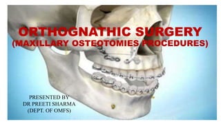
- 1. ORTHOGNATHIC SURGERY (MAXILLARY OSTEOTOMIES PROCEDURES) PRESENTED BY DR PREETI SHARMA (DEPT. OF OMFS) 1
- 2. CONTENTS • INTRODUCTION • HISTORY • SURGICAL ANATOMY • TYPES OF MAXILLARY OSTEOTOMY • COMPLICATIONS • REFERENCES 2
- 3. INTRODUCTION • Osteotomy- the surgical cutting or dividing of bone, usually to correct a deformity • Maxilla can be mobilized and repositioned and the healing continues as long as the mobilizes maxilla is pedicled on broad soft tissue base (soft palate, lateral pharyngeal wall, buccal mucosa). • Indicated in correction of malocclusion ,dentofacial deformities , craniofacial abnormalities etc. • Le Fort I osteotomy is a commonly performed maxillary procedure for the correction of dentofacial deformity. 3
- 4. HISTORY • Von Langenbeck (1859) - For the removal of nasopharyngeal polyps. • Cheever (1867) - For the treatment of complete nasal obstruction secondary to recurrent epistaxis for which a right hemi maxillary down fracture was used. • Wassmund (1927) - LeFort I osteotomy for the correction of the mid facial deformities and used orthopedic force post surgically • Axhausen (1934) - Described total mobilization of the maxilla with immediate repositioning for an open bite case • Schuchardt (1942) - First advocated the pterygomaxillary dysjunction. • Moore and Ward (1949) - Horizontal transaction of the pterygoid plates for advancement • Obwegeser (1965) - Complete mobilization of the maxilla so that repositioning could be accomplished without tension. 4
- 6. 6
- 8. Midface Osteotomies Segmental maxillary osteotomy Single tooth Anterior segmental Posterior segmental Horseshoe Total maxillary osteotomy LEFORT I SAME Classic Quadrangular LEFORT II Anterior LF II Pyramidal LF II Quadrangular LF II LEFORT III MIDFACE Zygomatic 8
- 9. SINGLE TOOTH OSTEOTOMY Indication: • Tooth mal position • Dental ankylosis • Closure of diastema Advantage: • Reduction in the treatment time. • Lower incidence of relapse Disadvantages: • Injury to teeth • Periodontal compromise • Devitalization of teeth 9
- 10. Procedure: *5mm and above the apices – adequate to maintain the vitality Anterior& posterior Subapical osteotomies -3mm is adequate(sheideman- joms.1985;43;408 ) *Yoshida-biologic responses of the pulp to single tooth osteotomy OOOO-1996;82 bell-revsclstion & bone healing after AMO. Jos1969;27;249,1978 10
- 11. ANTERIOR SEGMENTAL OSTEOTOMY Indications : • Correction of bi maxillary protrusion. • Marked protrusion of the maxillary teeth: • Anterior open bite where there is no vertical maxillary excess • To retract the anterior teeth when orthodontic treatment is not feaseable • When orthodontic tooth movement is inadvisable.( tooth ankylosis , root resorption) • To improve facial appearance in prognathic maxilla with incompetent lips 11
- 12. Wassmund 1927: • Both the labial and palatal blood supply are maintained. Indicated : • Closure of multiple interdental spaces and for anteroposterior repositioning of the premaxilla. • Maintain the best vascularity in comparison to all other ASMO methods. 12
- 13. Wunderer 1963 • Maintenance of the labial blood supply. • main advantage : Direct access for the palatal osteotomy , especially if posterior segments of the premaxilla must be removed • Indicated: -for setback of the anterior part of the maxilla -Blood flow studies have demonstrated that the transpalatal approach causes the greatest decrease in blood supply to the anterior maxilla 13
- 14. Cupar (1954) • Approach for down-fracture of the anterior maxilla • Commonly used technique • Minor version of total maxillary osteotomy • Indicated: for superior repositioning of the anterior maxilla in cases of vertical maxillary excess 14
- 15. Epker 1977 • Modified the Cupar technique for down-fracture of the anterior maxilla • Used only labial flaps and vertical tunnels labial to the teeth to be extracted, which were usually premolars on both sides • repositioning of the anterior maxilla superiorly, posteriorly and inferiorly Advantages : • preservation of the palatal pedicle, • ease of placement of internal fixation, • provide access to the nasal septal structures • a direct approach for removal of palatal bone. 15
- 16. 16 Vertical osteotomy Horizontal osteotomy Transpalatal osteotomy
- 19. (a) Down-fracture anterior segment of maxilla and maintaining palatal mucosa. (b) Midline or paramidline osteotomy for horizontal movement 19 CUPER METHOD
- 20. 20
- 21. (a) Alar cinch placement to control alar base, (b) V-Y closure of mucosa to maintain upper lip length 21
- 22. POSTERIOR SEGMENTAL OSTEOTOMY • Introduced by Schuchardt in 1954 as a two-staged procedure used in the surgical correction of open-bite deformity • Kufner (1971) - described a single buccal incision approach. • Perko (1967) - Bell technique 22
- 23. • Indications 1. Post maxillary alveolar hyperplasia 2. Total maxillary hyperplasia (when combined with AMO) 3. Distal repositioning of the post maxillary alveolar fragment to provide space for proper eruption of an impacted canine or bicuspid tooth 4. Spacing in the dentition that can be closed by ant repositioning of the posterior segment 5. Transverse excess or deficiency 6. Posterior open bite 7. Posterior cross bite 23
- 24. Schuchardt technique for posterior maxillary segmental osteotomies. (a) Limited buccal incision with combined horizontal and anterior vertical osteotomies (b) coronal sectional view indicating bone cuts (c) limited palatal incision located medial to planned lateral palatal osteotomy (d) coronal view of subnasal palatal alveolar and lateral sinus wall osteotomies 24
- 25. 25 Kufner technique for posterior maxillary segmental osteotomies. (a) Coronal sectional view of trans antral palatal osteotomy via chisel following lateral sinus wall osteotomy (b) Coronal view of trans antral palatal osteotomy trimming of bone segment
- 26. Perko–Bell technique for posterior maxillary osteotomies. (a) Limited buccal incision with combined horizontal and vertical osteotomies (b) limited palatal incision located medial to planned lateral palatal osteotomy (c) coronal sectional view indicating bone cuts with transantral medial nasal wall osteotomy 26
- 27. 27 Pterygomaxillary disjunction with a curved osteotome Down fracturing using digital pressure Fixation
- 28. Combination Anterior & Posterior Maxillary Osteotomy (Horseshoe osteotomy) • A combined form of anterior and posterior subapical osteotomies • Paul 1969 "total subapical maxillary osteotomy" were reported for midface hypoplasia. • This technique was further described by West & Epker 1972, Hall & Roddy 1975, Wolford & Epker 1975, West and McNeil 1975 and Hall & West 1976. Maloney (1982) reviewed this technique 28
- 29. Indication • Maxillary alveolar hyperplasia with or without an anterior open bite deformity • Transverse hypolplasia without a vertical component This procedure creates a three piece maxilla, with the central nasal portion left undisturbed, through the use of palatal parasagittal osteotomies The method has more or less been replaced by the traditional Le Fort I osteotomy 29
- 30. LEFORT 1 OSTEOTOMY Indications • To correct maxillary prognathism • Midface hypoplasia • Severe mandibular prognathism:reduce amount of mandibular setback • Superior repositioning of the maxilla, to correct vertical maxillary excess • Inferior repositioning of the maxilla, to correct vertical maxillary deficiency • Widening of the maxilla, to correct transverse discrepancies • Apertognathia, lefort I osteotomy should be given consideration because of the stability issues. • Often needed along with mandibular osteotomy to correct dentofacial asymmetries,asymmetric mandibular growth,condylar hyperplasia and hemifacial microsomia 30
- 31. SURGICAL TECHNIQUE • Step 1: Infiltration of the soft tissue with a vasoconstrictor • Step 2: Mucosal incision • Step 3: Completing the soft tissue incision through the periosteum • Leave at least 5 mm of nonkeratinized epithelium inferior to the incision • Incision should be increased posteriorly to approximately 10 mm at the buttress area. • A V-shaped incision at the labial frenum helps with alignment in later suturing The incision is angled superiorly so more soft tissue is left inferiorly for later ease of suturing and for sufficient blood supply support to the downfractued maxilla 31
- 32. • Step 4: Subperiosteal dissection • Step 5: Placement of reference marks • The subperiosteal dissection is carried superiorly and posteriorly to identify the piriform rim, infraorbital nerve, zygomatic buttress, and posterior maxilla • Vertical and horizontal reference marks are scored on the bone and the distances between them measured 32
- 33. • EXTERNAL REFRENCE MARKER 33
- 34. • Step 6: Anterior buccal osteotomy • Step 7: Posterior buccal osteotomy • A reciprocating saw is used to perform the osteotomy. • The osteotomy should be kept parallel to the occlusal plane. • The osteotomies can be kept parallel to the occlusal plane by placing a vertical step at the buttress area. • The maxilla can now be advanced parallel to the occlusal plane without any vertical changes. 34
- 35. • Step 8: Connecting the anterior and posterior osteotomies • Step 9: Placement of holes for interosseous wires • Connect the two horizontal osteotomies with a vertical osteotomy at the buttress using a 701 bur. • The holes should be placed in thick bone at the buttress areas and positioned in such a way that the vector of the positioning wire will support the repositioning of the maxilla 35
- 36. • Step 10: Separation of the tuberosity from the pterygoid plates • Step 11: Completion of the posterior osteotomy • The contents of the pterygoid maxillary fissure and soft palate are protected by placing the index finger at the hamulus palatally to feel the osteotome as it separates the tuberosity and the pterygoid plates. • The osteotome is directed medially and downward. • The osteotomy is completed using a small osteotome while the pterygoid osteotome is still in position, protecting the soft tissue behind the maxilla. 36
- 37. • Step 12: Osteotomy of the lateral nasal wall • Step 13: Repeating the procedure on the opposite side • Step 14: Completing the nasal spine subperiosteal dissection • The lateral nasal wall is separated. Note that the wall deviates posteriorly; therefore, care should be taken not to damage the descending palatine neurovascular bundle at the posterior end of the wall. • A small, wet sponge is placed on the completed side. • The osteotomies should be placed at the same level and angle on both sides according to the surgical treatment plan • Place a ramus retractor subperiosteally in the midline over the anterior nasal spine • (ANS). Dissect the rest of the periosteum from the ANS. 37
- 38. • Step 15: Completing the nasal mucosa dissection • Step 16: Osteotomy of the septal cartilage and vomer • Separate the septal cartilage from the ANS. Complete the dissection of the nasal mucosa from the nasal septum and floor. • The nasal mucosa is dissected from the ANS, nasal septum, and piriform rim. • The nasal cartilage and vomer are separated from the maxillary bone using a nasal septal osteotome. • Angle the osteotome toward the nasal floor to prevent tearing of the nasal mucosa. 38
- 39. • Step 17: Maxillary downfracture • Step 18: Redefining osteotomies when maxillary downfracture fails • The maxilla is downfractured with downward pressure on the anterior maxilla. • The maxilla should downfracture easily; if it does not, all the osteotomies, especially at the junction between the tuberosity and the pterygoid plates should be revised. • The maxilla should downfracture with ease. If it does not, following are the most probable reasons for the failure: -The pterygoid plates and tuberosity are not sufficiently separated. -The lateral nasal wall osteotomy is incomplete (too short). -Occasionally, the bone of the maxilla is very thick, making downfracture difficult. In such cases, tap the lateral nasal wall osteotomes into position (bilaterally), and carefully push down on the osteotomes to assist in the downfracture. 39
- 40. • Step 19: Mobilization of the maxilla • The right side of the maxilla is mobilized by pushing the maxilla anteriorly using a mobilizer (or pterygoid osteotome). • (r) The left side of the maxilla is mobilized; use a finger as a fulcrum, and protect the soft tissue at all times. 40
- 41. • Step 20: Reduction of the palatal aspect of the nasal septum • Step 21: Contouring the piriform rim • (y) The nasal septum and remaining part of the vomer is trimmed from the nasal floor with a bone nibbler. • (z) Adequate trimming of the vomer and even creation of a trough in the nasal floor is essential to accommodate the nasal septum after maxillary repositioning. This is especially important when the maxilla will be superiorly repositioned. • (aa) The piriform rim should be contoured with a vulcanite bur to accommodate the soft tissue of the base of the nose after maxillary repositioning. • (bb) A hole is drilled horizontally through the ANS. This hole will be used later for the placement of a cinch suture and for securing the nasal septum. 41
- 42. • Step 22: Reversing the hypotensive anesthesia and checking for hemorrhage • Step 23:Feeding the wire thru butress • Before final repositioning and fixation of the maxilla, it advised to reverse the hypotensive anesthesia. • While the patient is normotensive, undetected arterial hemorrhage can be discovered and addressed, which will help prevent postoperative hemorrhage. • 0.018-inch interosseous wires placed at the buttress area bilaterally. • When more rigidity is required (eg, maxillary expansion, maxillary downgrafting, multipiece Le Fort I procedures, and large movements [greater than 6 mm]), more plates are recommended. 42
- 43. • Step 24: Maxillomandibular wire fixation • Step 25: Maxillary repositioning • The first maxillomandibular wire around the four central incisors to achieve the planned interincisal relationship. • Place the wires around the orthodontic brackets • With the help of it the teeth could easily be “pulled into occlusion” or into the acrylic splint • Once the wires are removed, the teeth will return to their original position. • With maxillomandibular fixation in position, the maxillomandibular complex is rotated to achieve bone contact. • The vector of force on the condyle should be superior and slightly anterior.There should be no bony interferences during the rotation. 43
- 44. • Step 26: Turbinectomy • Step 27: Suturing the nasal mucosa • The hypertrophied turbinate is grasped with a tissue clamp and then removed using a diathermy knife. • Any tears in the nasal mucosa is identified, and repair them with a 4-0 chromic suture. 44
- 45. • Step 28: Checking the position of the nasal septum • Step 29: Tightening the buttress wires • The maxillomandibular complex closed to achieve the predetermined maxillary position is rotated. • The condyles are seated in the glenoid fossa is ensured, and the position of the nasal septum is also checked • The septum should lie freely, without any interference in the trough created in the nasal floor. • The nasal septum may be secured in position by placing a suture through it and through the horizontal hole in the base of the nasal spine after maxillary fixation. • Copious lavage of the maxillary sinuses and nasal floor with saline solution should be carried out before tightening the buttress wires because small bone fragments remaining in the sinus or nasal cavities will lead to postoperative infection. • The maxillomandibular complex then rotated superiorly to achieve bone apposition, and the interosseous buttress holding wires is tighten • Overtightening the wires is avoided in an attempt to achieve better bone apposition because the tension it would place on the bone would result in displacement and occlusal discrepancy once maxillomandibular fixation is removed. 45
- 46. • Step 30: Checking maxillary position using a caliper • Step 31: Replacing the maxillomandibular fixation • If there is any doubt about the condylar position at this stage, the maxillomandibular fixation should be removed and the occlusion checked by referring to intraoral bony reference marks or to an extraoral reference point (eg, a Kirschner wire in the frontal bone). • If the maxillary position and occlusion are satisfactory, replace the maxillomandibular fixation. 46
- 47. • Step 32: Placement of bone plates maxillomandibular fixation. • Semirigid fixation, which consists of bilateral titanium (1.5-mm) bone plates or resorbable (2.0- mm) plates placed anteriorly in the thick bone at the piriform areas and two interosseous (0.018-inch) wires placed at the zygomatic buttresses. • The plates should be adapted to fit passively. • Two screws should be placed on either side of the osteotomy, and care should be taken not to place screws into the roots of teeth, too close to the edge of the bone, or in thin bone. • If thin bone must be used, self-drilling screws are recommended. 47 Non – rigid Trans-osseous wiring IMF – 4-6 months Rigid/semi rigid Metal Plates Resorbable plates Mesh IMF- 4-6 weeks • For superior movement of the maxilla : two plates at the piriform rim with six screws per plate can be sufficient. • The most widely used are 2.0- and 1.5-mm screws with L plates or arched plates. • Usually for all osteotomies where bone contact buttressing is incomplete, additional plates are subsequently placed at the zygomatic buttress . • For downward movement of the maxilla interpositional grafts can be wedged beneath the miniplates
- 48. 48 Step 33: Removing the maxillomandibular fixation and checking the occlusion • Before the occlusion is checked, the mandible should be gently opened and closed as well as translated forward and from side to side to ensure that the articulating disc was not displaced while teeth were in maxillomandibular fixation. • Wait a few minutes after removing the maxillomandibular fixation before checking the occlusion. • Then, with slight pressure on the chin, close the mandible until the teeth occlude. If the planned occlusion has not been achieved, remove the rigid fixation and identify and correct the reason for failure.
- 49. • Step 34: Placement of nasal septum and cinch sutures • Secure the septum in the trough created in the nasal floor by a suture through the septum and the hole in the base of the ANS. (ii)The lateral alar soft tissue is grasped with a toothed forceps. (jj) The cinch suture is placed in a figure-eight fashion through the hole in the ANS. 49
- 50. 50 A, Classic alar cinch suture: Rauso et al. (2010)- involved anchoring the fibroareolar tissues directly under both alar and passing suture through the nasal spine. B, Classic alar cinch suture :Ritto et al. (2011)- involved anchoring the fibroareolar tissue intraorally under both alae separately. C, Classic alar cinch suture :Nirvikalpa et al. (2013)- involved using hypodermic needles under both alae to accurately identify the nasofacial skin fold; a thick suture bite was taken at this point. D, Modified alar cinch suture:Rauso (2010)- involved a reinsertion method by using hypodermic needles under both alae and passing the suture through the nasal spine. E, Modified alar cinch suture :Ritto et al. (2011)- involved a reinsertion method by using curved needles to anchor the fibroareolar tissue under both alae separately. F, Modified alar cinch suture Nirvikalpa et al. (2013)- involved using hypodermic needles under both alae to accurately identify the nasofacial skin fold; a thick suture bite was taken at this point, then the suture was passed through the nasal septum. Three studies with 146 participants undergoing LeFort I maxillary osteotomy were included in this review. The results showed that, compared with the classic method, both modified transseptal alar base suture and modified reinsertion sutures significantly decreased postoperative alar and alar base widening Vol. 117 No. 1 January 2014 Modified versus classic alar base sutures after LeFort I osteotomy: a systematic review Xianwen Liu, DDS, " Songsong Zhu, DDS, PhD, and Jing Hu, DDS, PhD° ab West China Hospital of Stomatology. Sichuan University. Chengdu, China: Harvard School of Dental Medicne, Boston, MA, USA
- 51. 51 Step 35: Submucosal suturing • The soft tissue incision is closed in layers—first the submucosal tissue and then the mucosa. • Starting posteriorly, and ending at the piriform rim on both sides by pulling the superior tissues slightly forward.
- 52. • Step 36: Mucosal suturing • Step 37: Placement of elastics or maxillomandibular fixation • Step 38: Applying a pressure dressing • a V-Y closure is performed and reapproximated the mucosal midline using 4-0 chromic sutures. • Two interrupted sutures is placed on both sides of the midline to reapproximate it • The mucosal suturing is done using a continuous suture, starting posteriorly and pulling the superior tissue slightly forward. • When lip lengthening is indicated, more horizontal sutures (or even bilateral V-Y suturing) may be used. • The period of maxillomandibular fixation may vary from a few days to 3 weeks. • Alternatively, for most patients, two to four inter occlusally placed elastics are sufficient to guide the teeth into the new occlusion. • The direction of the elastics should support the repositioning. • Long elastics should be avoided. • A pressure dressing is applied to help control swelling and/or prevent hematoma formation. 52
- 53. Quadrilateral (Quadrangular) Osteotomy • High-level osteotomy is a variant of the Le fort I osteotomy • extends up to the lower part of the zygoma, to a point just below the infraorbital nerve bilaterally Indications: • midface retrusion • excessive scleral exposure 53
- 54. Advantage: • improves the appearance of midfacial retrusion and flattening • improves zygomatic prominence and support for the lower eyelid. • This osteotomy has minimal surgical morbidity and has acceptable outcomes. • This may therefore be considered,especially in Asian patients, as a viable treatment alternative for midfacial advancement without augmentation of the malar region Lee H-J, Park H-S, Kyung H-M, Kwon T-G. Soft tissue changes and skeletal stability after modified quadrangular Le Fort I osteotomy. Int J Oral Maxillofac Surg. 2015;44:356–61. 54
- 55. • If the cuts are made from high to low, a significant inferior movement of the maxilla can be achieved which reduces the necessity for an interpositional bone graft and alloplastic onlay grafts for the zygomatic regions. • It is more stable than conventional inferior positioning of the maxilla as it produces less rotation of the nasal tip than the conventional Le Fort I osteotomy. 55
- 56. • The surgical technique is the same as for the conventional Le Fort I osteotomy, • but a significant sharp dissection of the masseter muscle from the zygoma is required to expose the prominence of the zygomatic bone. • The cuts are made just below the infraorbital nerves. • The bony cuts directed downward to the normal pterygomaxillary disjunction level. • The other steps :same as LeFort I • Fixation is with miniplates. (care must be taken to ensure the plates are not palpable in the infraorbital regions.) 56
- 57. 57 Only minor skeletal relapse (mean: 0.4 mm) was observed in the follow-up period (mean 14.2 months). Conclusion: The procedure should be considered whenever vertical maxilla relapse is of concern after anterocaudal displacement
- 58. F Osteotomy lines for variations of the Le Fort I osteotomy. (A) Conventional Le Fort I osteotomy (Axhausen 1934). (B) Modified quadrangular Le Fort I osteotomy (C) Quadrangular Le Fort I osteotomy (Keller and Sather, 1990). (D) Modified Le Fort I (maxillary–zygomatic) osteotomy (Abubaker and Sotereanos, 1991) . (E) Extended Le Fort I osteotomy (Nørholt et al., 1996). MODIFICATIONS 58
- 59. Laster ‘shark-fin’ osteotome was placed, with the fin within the cut, on the lateral wall of the maxillary sinus and slightly inclined down toward the occlusal plane The Laster ‘shark-fin’ osteotome with the double perpendicular cutting-edge. 59 Year 2002 complete or almost complete separation was obtained, whereas the use of the Obwegeser osteotome resulted in five sites with fractures of the maxillary tuberosity and three with high-level fractures of the pterygoid plates
- 60. The osteotome is driven from the nasal crest of the maxilla toward the pterygomaxillary junction. A narrow periosteal elevator (left) is used to protect the nasal mucosa Immediate downfracture of the maxilla is achieved by inwardly rotating the osteotome (arrow) Compared with classic pterygomaxillary dysjunction, the twist technique uses a frontal approach and a straight osteotome. This technical modification requires a substantially smaller incision, achieves an immediate effective separation of the maxilla, and enables adequate visualization J Oral Maxillofac Surg 71:389-392, 2013 60
- 61. 61
- 62. 62
- 63. SURGICAL ASSISTED MAXILLARY EXPANSION (SAME) • Brown first described SAME in 1938 - mid palatal split • A LeFort I type of osteotomy with a segmental split of the maxilla and the placement of a triangular unicortical iliac graft for correction of maxillary constriction was presented by Steinhauser in 1972. 63
- 64. Indications: • Increasing the maxillary arch perimeter so as to correct unilateral or bilateral posterior cross bite, with or without additional surgical procedures for other discrepancies. • Increasing the maxillary transverse width, especially when the transverse discrepancy is greater than 5 mm. • Alleviating dental crowding when bicuspid extractions are not indicated. • Reducing excessively prominent and visible buccal corridors when smiling. • When orthodontic maxillary expansion has failed. 64
- 65. Technique: • First the mandibular dentition should be decompensated • Expansion appliance should be placed preoperatively 65 HASS HYRAX
- 66. Steps: 66 Step 1: Incision Step 2: Buccal Osteotomy Step 3: Palatal incision A midpalatal incision (red line) with mucoperiosteal dissection (shaded areas) buccal osteotomy is made from the pterygomaxillary junction to the piriform rim anteriorly, using a reciprocating saw
- 67. 67 Step 4: Palatal Osteotomy Step 5: Midline Osteotomy
- 68. 68 Postoperative schedule : • After 5 days appliance is activated for first time. •1 week later the space that is being created between two front teeth is checked and the bite also is checked. •After another week the separation of teeth and the bite are checked one more time and activation is stopped. •Refered to the orthodontist for a check. •The appliance remains in place for 3 months without further activation. •Orthodontic treatment is then resumed
- 69. Modified SAME • Unilateral or asymmetric deformities • Osteotomy done on one side • On Non operated site buccal bone bending and dental tipping • Relapse occure non operated site • Mid palatal healing:6 months 69
- 70. Complications: a)Those due to inadequate surgery: • pain • dental tipping • periodontal breakdown • post orthodontic relapse b)Those due to expansion • lack of appliance expansion • deformation of the appliance due to processing errors • stripping or loosening of mid palatal screw 70
- 71. Segmented Le Fort I osteotomy • Required when open bite or a transversal expansion is required in the maxilla, a Le Fort I osteotomy approach can be combined with a multiple‐piece osteotomy to correct an unfavourable curve of Spee or a transverse discrepancy • maxilla can be sectioned into 2, 3, 4, 5, or 6 segments depending on the indications Segmentation of maxilla into 4 pieces. Segmentation of maxilla into 6 pieces 71
- 72. Hierarchy of predictability and stability for orthognathic surgical procedures. STABILITY OF LEFORT 1 Stability and predictability of orthognathic surgery Am J Orthod Dentofacial Orthop 2004;126:273-7 72
- 73. Soft tissue changes • Initially, swelling and bone irregularities contribute to the soft tissue changes observed: Approx. 1 year to dissipate. Soft tissue changes include: • forward movement of the base of the columella • nasal tip elevation, • flattening and shortening of the upper lip, • an increased nasolabial angle. • Alar cartilage width increases • the nares become more exposed. ( All soft tissue changes stabilize over time, except the increase in alar cartilage width, which remains close to immediate postoperative values) 73
- 74. Factors responsible for soft tissue changes: • preoperative soft tissue thickness • time lapsed since surgery • postoperative wound healing • surgical techniques 74
- 75. LEFORT II osteotomy • First presented by Henderson and Jackson in 1973. Indication • Relatively rare • Apert Syndrome(most common) • Address nose and maxilla • need for the correction of nasomaxillary hypoplasia 75
- 76. Variations: • Anterior Le fort II (Converse in 1971) • Pyramidal Le fort II(Henderson and Jackson in 1973) • Quadrangular Le fort II (Kufner in 1971) 76
- 77. INCISON: • Nasal root and medial orbital : coronal incision or paranasal incision • infraorbital rim and orbital floor: Conjunctival or sub ciliary incision • canine fossa and posterior maxilla: routine buccal vestibular incision 77
- 79. • Repositioning of maxilla Fixation Closure 79 bone grafts: preferably with fragments of autogenous, cancellous iliac bone.
- 80. Anterior "Le Fort II osteotomy” the osteotomy must be completed by cutting across the palate in the region of the first bicuspid 80
- 81. Rare Indication: -with nasomaxillary hypoplasia complicating other facial disharmonies Drawbacks: -strong tendency to relapse -retruded infraorbital rim can only be corrected in its medial portion 81 Pyramidal Le Fort II osteotomy
- 82. Quadrangular Le Fort II osteotomy • Defines the technique as "major maxillary osteotomy", which is a combination of the Le Fort I and Le Fort II classifications. • The main difference from the other Le Fort II type osteotomies is that there is no involvement of the nasal skeleton. • On the other hand the total infraorbital margin can be advanced by this approach. 82
- 83. • Medially the bone cut reaches the lacrimal fossa without changing the position of the lacrimal system. • Laterally the osteotomy goes across the zygomatic bone and continues from there to the pterygoid fossa and the pterygomaxillary junction. • With the separation from the nasal septum and the lateral nasal walls the mobilization of the mid facial segment is completed 83
- 84. • Advantage: -Lacrimal system is not within the operating field • Precaution: -damage the infraorbital nerve (recommend the use of small sharp chisels for the dissection of the orbital floor) -defects in the floor : filled with cancellous bone pieces • Disadvantage: -Relapse(MINOR DISADV. –overcorrection to be done) -Visible scar(to prevent –trans conjunctival incison - access limited./recommend a small incision in the lateral commissure of the eyelids (Lindorfand Steinhauser (1977). 84
- 85. LEFORT III Osteotomy • Sir Harold Gillies and colleagues : first time in 1951 • 1957, Longacre used autogenous rib grafts for improvement of total mid-face deficiency • 1967, Paul Tessier reported that mobilization and expansion of the entire midface by means of a sub-cranial le fort lll osteotomy was possible 85
- 86. Indication: • Underdevelopment of entire mid-face • Flat/sunken appearance of mid-face • Short nasal structure • Deficient nasal bridge • Exaggerated flatness of nasofrontal region • Reduced size of orbit • Exophthalmos • Retro position of zygoma 86
- 87. • Technique: Incision: Bicoronal incision Inferior eye lid incision Buccal vestibular incision 87
- 88. • Osteotomy 88
- 89. MIDFACE Osteotomy ZYGOMATIC/MALAR OSTEOTOMY Schematic representation of the malar osteotomy 89
- 91. COMPLICATIONS IN MAXILLARY OSTEOTOMIES • Preoperative • Intraoperative • Postoperative Dimitroulis 1998 J Adult Orthod Orthognath Surg 91
- 92. PREOPERATIVE • Lack of pre-treatment objectives: -Failure to recognize underlying skeletal abnormality -Unexpected adverse growth -Lack of patient co-operation • Orthodontics: -Insufficient decompensation -Inadequate transverse coordination -Uncorrected tooth size problems -Inadequate preoperative root divergence in segmental surgery -Improper Orthodontic appliances 92
- 93. • Preoperative planning error -In traditional treatment planning, the workup involves reproduction of the occlusal discrepancy on a semi-adjustable articulator through facebow transfer. -On this relation, desired mock surgical planning is done and splints are prepared -Errors and inaccuracies in this model surgery can contribute to compounding errors that are ultimately transferred to the operating room and patient -use of virtual surgical planning eliminates many of the uncertainties that go into preparing a case Virtual Surgical Planning in Orthognathic Surgery Farrell, Brian B. et al. Oral and Maxillofacial Surgery Clinics, Volume 26, Issue 4, 459 - 473 93
- 94. INTRA-OPERATIVE 1.VASCULAR • Serious intra operative bleeds are rare in orthognathic surgeries • Few reports from secondary haemorrhage has been reported • Intraoperative complications may occur secondary to maxillary or mandibular osteotomies • Kramer and colleagues found extensive bleeding in 1.1% of a prospective study of 1000 patients • Injury to the descending palatine artery during LeFort I osteotomy can be minimized by limiting the osteotomy to 30 mm posterior to the piriform rim in females and to 35 mm in males Kramer FJ, Baethage C, Swennen G, et al. Intra- and perioperative complications of the LeFort I osteotomy: A prospective evaluation of 1000 patients. J Craniofacial Surg 2004;15:971 Li, K.K., J.G. Meara, and A. Alexander, Jr., Location of the descending palatine artery in relation to the Le Fort I osteotomy. Journal of Oral and Maxillofacial Surgery, 1996.54(7): p. 822–825; discussion 826–827. 94
- 95. • Turvey and Fonseca reported that the main trunk of the maxillary artery was most vulnerable to the damage within the pterygopalatine fossa in the lateral position and they recommended angling the posterior lateral maxillary osteotomy downward to avoid damaging the artery • Osteotome used during pterygomaxillary dysjunction: 10-15mm(mean height of pterygomaxillary suture: 14.6mm) Turvey, T., and R. Fonseca, The anatomy of the internal maxillary artery in the pterygopalatine fossa: its relationship to maxillary surgery. Journal of Oral Surgery (American Dental Association: 1965), 1980. 38(2): p. 92. 95
- 96. • Greater palatine artery : -if reciprocating saw go deep within the posteromedial aspect of maxillary sinus -osteotome beyond 34mm to piriform rim along the lateral nasal wall -mobilization of maxilla without removing bony specules • Pterygoid venous plexus: -Maxillary venous bleeding most commonly involves the pterygoid venous plexus 96
- 97. Management of heamorrhage • Packing is suggested as the first attempt to tamponade the hemorrhage. • In delayed bleeding after LeFort I osteotomy, the surgeon should reopen surgical site and move the maxilla downward to find the bleeding source. • direct visualization of the bleeding source and cauterization of injured vessels stops the hemorrhage. • ligation of the external carotid artery and angiographic embolization. Felice O'Rayan, A.S., Complications with Orthognathic Surgery, in Oral and maxillofacial surgery, M. Fonseca, Turvey, Editor. 2009, Saunders Elsevier. p. 419–489 97
- 98. 2 NEURAL • The infraorbital nerve may be compressed, retracted or transected inadvertently during subperiosteal dissection. • resulted from incorrect separation during disimpaction of maxilla • 2.2% of individuals reported long term deficit to the upper lip and 9% to the teeth, palate, and gingiva • Usually temporary and recovery 2-8 weeks Robl et al . Complications in Orthognathic Surgery : Oral Maxillofacial Surg Clin N Am 26 (2014) 599–609 98
- 99. • TRIGEMINOCARDIAC REFLEX Surgery performed near the cranial nerves, especially the trigeminal nerve and its branches – greater palatine and posterior superior alveolar nerve may induce bradycardia by stimulation of the vagus nerve and finally activation of the parasympathetic system resulting in various types of dysrhythmia. • bradycardia < 60b/m, hypotension with a drop in the Management – • manipulation of the maxilla should be stopped immediately • Administration of anticholinergic medications such as atropine or glycopyrolate 99
- 100. 3.UNFAVOURABLE FRACTURE • MANDIBLE>MAXILLA • In maxilla unfavorable fractures may consist of pterygoid plate, sphenoid bone, and middle cranial fossa fractures. • Lanigan and Guest demonstrated pterygomaxillary dysjunction could cause disruption of the pterygopalatine fossa which could extend to the skull base. Lanigan, D.T., and P. Guest, Alternative approaches to pterygomaxillary separation. International Journal of Oral Maxillofacial Surgery, 1993. 22(3): p. 131–138. Undesirable split 1. fragmentation 2. comminution 3. higher fracture at the pterigoid level Renekye et al. reported the incidence of pterygoid plate fracture was 58% following LeFort I osteotomy using postoperative CT scans 100
- 101. 4 OROANTRAL COMMUNICATION • Palatal Bone is thickest in the midline where tissue is the thinnest. • Osteotomies in the midline are more likely to result in palatal tears and these may be less likely to heal than when a tear occurs laterally, in thicker tissues Management • <3 mm generally close spontaneous • >3 mm consider obturation • if >4-6 mm soft tissue flap Perciaccante V, Bays R. Maxillary orthognathic surgery.In: Miloro M, editor. Peterson’s principles of oral and maxillofacial surgery. 2nd edition. London: BC Decker; 2004. p. 1179–204 101
- 102. Neurologic complications • Brainstem infarct after Le Fort I osteotomy • CSF leak • AV fistula -The maxillary artery is at risk in midfacial advancement procedures because of its close proximity to the pterygomaxillary junction in the pterygopalatine fossa. -Transarterial embolisation is an effective treatment of the fistula when a surgical ligature cannot be done British Journal of Oral and Maxillofacial Surgery 55 (2017) 641–643 Int. J. Oral Maxillofac. Surg. 2010; 39: 292–307 Journal of Cranio-Maxillo-Facial Surgery (2010) 38, 251e254 POST OPERATIVE COMPLICATION 102
- 103. Nasal Abnormalities 1.Septal deviation :compression or displacement from inadequate bone removal of the nasal crest of the maxilla or inadequate trimming of the cartilagenous septum 2. Alar base widening: An alar base cinch suture is placed at the end of the procedure to control width, because soft tissue reflection leads to widening of the nose. 3. Tip over-rotation :Reduction of ANS prevents excessive rotation of the nasal tip 4. Dorsal deformities :A twisting dorsum and tip deviation may be related to inadequate septum reduction Felice O'Rayan, A.S., Complications with Orthognathic Surgery, in Oral and maxillofacial surgery, M. Fonseca, Turvey, Editor. 2009, Saunders Elsevier. p. 419–489 103
- 104. Aesthetic complications • common problem in superior repositioning is the bunching of the buccal tissues leading to “chubby cheeks” • 6 months time its settle down. 104
- 105. Loss of vascularity : aseptic necrosis • Anterior maxillary osteotomy • Transversal maxillary segmentations Cause: -Transection/kinking of vascular pedicle -Major anatomical irregularities -Poor flap design, Tearing of flaps • Consequences : -Loss of entire maxilla or segment, -Flattening of papilla, -Non vital teeth 105 • Prevention -Fewer Segmentation: avoid small segments -Avoid damage to pedicle
- 106. Nonunion/delayed union • Relatively high prior to the rigid fixation. • With rigid fixation most common cause is traumatic occlusion. • Traumatic occlusion causes unbalanced force on the maxilla, and if the bony healing has not progressed to the strength of resisting this forces , maxillary mobility occurs Management In cases of mobile maxilla • soft diet • discontinuation or decreased strength of elastic traction, • modified splint to balance occlusion, • local and systemic management of infection • elimination of parafunctional habits and • close observation. 106
- 107. Surgical management in a malunion or nonunion of the maxilla involves: • Recreation of the osteotomy with aggressive mobilization • Removal of all fibrous tissue • Passive repositioning of segments • Rigid fixation to resist segment displacement (consider auxiliary fixation, transpalatal support) • Grafting for continuity 107
- 108. Malocclusion •Immediate anterior open bite 1. Inadequate removal of posterior interferences with displacement of the condyles from the fossa during fixation •Late open bite development 1. Collapse of transverse expansion a. Lack of intraoperative methods to maintain expansion (grafting, splint placement) b. Lack of postoperative efforts by the orthodontist to maintain expansion (trans palatal arch) 2. Orthodontic relapse 108
- 109. • Relapse • Depends on : - Degree of surgical advancement - Degree of inferior repositioning of anterior maxilla - Use of bone grafts in large advancements • Other Causes : - Increased soft tissue stretching : results in drift of the screws during bone healing - Reduced area of bone contact at the lateral aspects of the maxilla - compromised union • Prevention: - Advance the maxilla at least 2mm more than the ideal overjet to compensate for relapse -Provision of a period of MMF (3—4 weeks) in addition to rigid fixation in large advancements Van Sickels BJOMS 1996;34:279—85. 109
- 110. Ophthalmic complications • Loss of function of the lacrimal gland - Damage to greater petrosal or vidian nerves interrupting parasympathetic supply to lacrimal gland • Oculomotor and abducens palsy – Ptosis & ophthalmoplegia – Superior orbital fissure fracture • Blindness -Blindness after orthognathic surgery is usually not from a direct injury to the optic nerve itself, but more commonly is the result of an ischaemic injury to the blood supply to the optic nerve, either directly from a fracture extending through the orbit to the optic canal or foramen, or indirectly from swelling and edema around the nerve in the optic canal disrupting its blood supply. • Can occur during pterygomaxillary dysjunction or maxillary downfracture •J. Oral Maxillofac. Surg. 2018; 47: 79–82 110
- 111. Loss of function of lacrimal gland 111
- 112. REFERENCES 112
- 113. 113