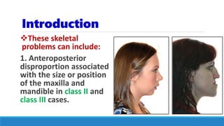
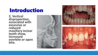
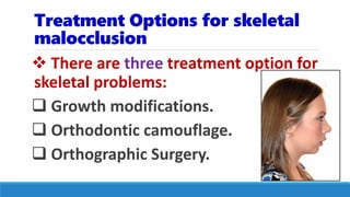
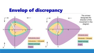
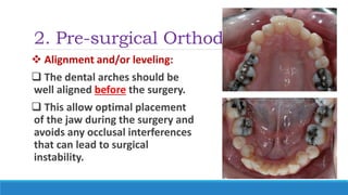
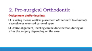
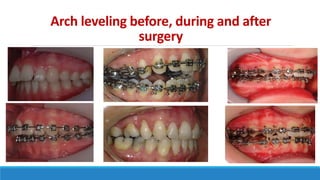
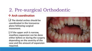
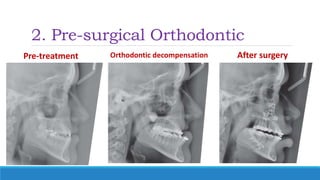
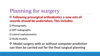
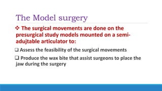
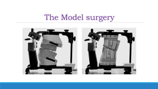
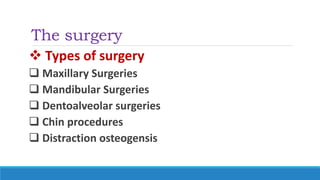
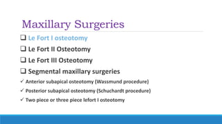
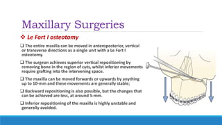
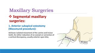
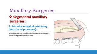
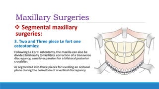
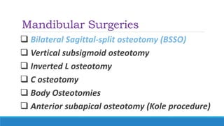
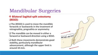
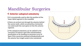
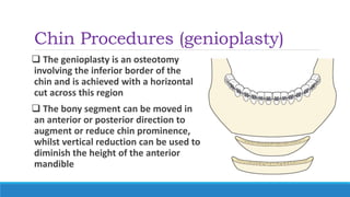
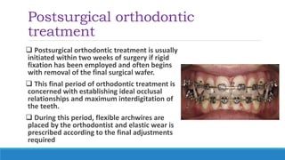
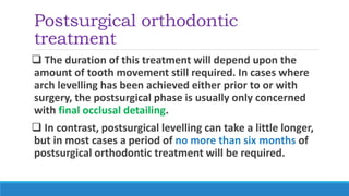
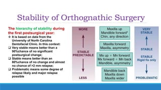
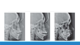
‫الرحيم‬‫الرحمن‬‫هللا‬‫بسم‬
Sudan International University
Faculty of Dentistry
Department of Orthodontics
Orthognathic Surgery
Mohanad Elsherif
BDS (U of K), MFD RCSI, MFDS RCPS(Glasg), MSc (Orthodontics), M.Orth. RCSEd
Introduction
 Malocclusion can be skeletal and/or dental
in nature.
 Dental malocclusion can be corrected with
orthodontic treatment alone using fixed or
removable appliances.
 Skeletal malocclusion means that there are
problems with the size and/or the position of
the jaws.
Introduction
These skeletal
problems can include:
1. Anteroposterior
disproportion associated
with the size or position
of the maxilla and
mandible in class II and
class III cases.
Introduction
2. Vertical
disproportion,
associated with
excessive or
reduced
maxillary incisor
tooth show,
increased
overbite or open
bite.
Introduction
3. Transverse anomalies
Introduction
4. symmetries of the face
and jaws.
Treatment Options for skeletal
malocclusion
 There are three treatment option for
skeletal problems:
 Growth modifications.
 Orthodontic camouflage.
 Orthographic Surgery.
Growth Modification Vs Camouflage VS
Surgery
 This will depend on:
 The patient age.
 The severity of skeletal discrepancy ( mild,
moderate or sever problem?).
 The pattern and direction of future growth.
 The degree of dentoalvelor compensation.
 The patient concerns ( if he/ She desire major
facial changes.
Envelop of discrepancy
The arrows
designate the
direction of the
movement in the
diagram
Steps of combined Orhodontic-
Surgical treatment
 Pre-treatment assessment
 Pre-surgical Orthodontics
 The Surgery
 Postsurgical Orthodontics
1. Pre-treatment assessment
 Patients should be assessed within the
environment of a joint clinic that involves both
surgeon and orthodontist.
 This allows a preliminary plan to be presented
and explained to the patient, affords them the
opportunity to ask any questions and come to an
informed decision with regard to undertaking such
treatment.
1. Pre-treatment assessment
 History
 Clinical examination
 Diagnostic records (Photos, DPT, lateral
Ceph, others)
 Psychological assessment
1. Pre-treatment assessment
 Psychological assessment:
 Ideally all patients should be assessed by a psychologist to establish
their motives and to determine whether their goals are realistic.
 A few patients have great difficulty in adapting to significant
changes in their facial appearance. This is more a problem in older
individuals.
 Also, a period of psychological adjustment following facial surgery
must be expected. In part, this is related to the use of steroids and
Steroid withdrawal, causes mood instability at 3 to 6 weeks post-
surgery.
Psychological assessment
1. Pre-treatment assessment
 Psychological assessment is a must in patient
with:
 A history of previous cosmetic surgery.
 Minimal facial deformity.
 Expectations that clearly exceed surgical feasibility.
 An obsessional concern with certain features.
2. Pre-surgical Orthodontic
The aim of the pre-surgical orthodontic is to prepare the
patient for the surgery.
 This involve one or more from the following steps:
 Alignment and/or leveling
 Arch coordination.
 Orthodontic decompensation.
 Provide space for osteotomy cuts (if required).
2. Pre-surgical Orthodontic
 Alignment and/or leveling:
 The dental arches should be
well aligned before the surgery.
 This allow optimal placement
of the jaw during the surgery and
avoids any occlusal interferences
that can lead to surgical
instability.
2. Pre-surgical Orthodontic
Alignment and/or leveling:
 Leveling means vertical placement of the teeth to eliminate
excessive or reversed curve of spee.
 Unlike alignment, leveling can be done before, during or
after the surgery depending on the case.
Arch leveling before, during and after
surgery
2. Pre-surgical Orthodontic
 Arch coordination
 The dental arches should be
coordinated in the transverse
plane following surgical
movement.
 if the upper arch is narrow,
maxillary expansion can be done
either before or during the surgery
depending on the severity of the
case and the amount of expansion
required.
2. Pre-surgical Orthodontic
 Orthodontic decompensation:
 Dental compensation means that natural movement of
the teeth to mask the severity of underline skeletal
discrepancy.
 This is will make the malocclusion and the overjet
appear less sever than its actually is.
 Compensation is more evident in class III than class II
cases.
Dental compensation
in class III:
proclination of the
upper incisors and
retroclination of the
lower incisors
Dental compensation
in class II:
retroclination of the
upper incisors and
proclination of the
lower incisors
2. Pre-surgical Orthodontic
 Orthodontic
decompensation:
 One of the major goals of
presurgical orthodontics is to
eliminate any dental
compensation.
 This allow maximum surgical
correction to be undertaken
during surgery.
2. Pre-surgical Orthodontic
Pre-treatment Orthodontic decompensation After surgery
2. Pre-surgical Orthodontic
Pre-treatment Orthodontic decompensation After surgery
Planning for surgery
 Following presurgical orthodontics a new sets of
records should be undertaken, This includes:
 Photographs.
 DPT radiographs.
 Lateral cephalometric.
 Study models.
 Model surgery with or without computer prediction
can then be carried out for the final surgical planning
The Model surgery
 The surgical movements are done on the
presurgical study models mounted on a semi-
adujtable articulator to:
 Assess the feasibility of the surgical movements
 Produce the wax bite that assist surgeons to place the
jaw during the surgery
The Model surgery
The Model surgery
The surgery
The surgery
 Types of surgery
 Maxillary Surgeries
 Mandibular Surgeries
 Dentoalveolar surgeries
 Chin procedures
 Distraction osteogensis
Maxillary Surgeries
 Le Fort I osteotomy
 Le Fort II Osteotomy
 Le Fort III Osteotomy
 Segmental maxillary surgeries
 Anterior subapical osteotomy (Wassmund procedure)
 Posterior subapical osteotomy (Schuchardt procedure)
 Two piece or three piece lefort I osteotomy
Maxillary Surgeries
 Le Fort I osteotomy
 The entire maxilla can be moved in anteroposterior, vertical
or transverse directions as a single unit with a Le Fort I
osteotomy.
 The surgeon achieves superior vertical repositioning by
removing bone in the region of cuts, whilst inferior movements
require grafting into the intervening space.
 The maxilla can be moved forwards or upwards by anything
up to 10-mm and these movements are generally stable;
 Backward repositioning is also possible, but the changes that
can be achieved are less, at around 5-mm.
 Inferior repositioning of the maxilla is highly unstable and
generally avoided.
Maxillary Surgeries
 Segmental maxillary
surgeries:
1. Anterior subapical osteotomy
(Wassmund procedure):
Achieves isolated movement of the canine and incisor
teeth, for either reduction of an overjet or correction of
a vertical discrepancy, usually anterior open bite;
Maxillary Surgeries
 Segmental maxillary
surgeries:
2. Posterior subapical osteotomy
(Wassmund procedure):
It is occasionally used for isolated correction of a
unilateral posterior crossbite
Maxillary Surgeries
 Segmental maxillary
surgeries:
3. Two and Three piece Le fort one
osteotomies:
Following Le Fort I osteotomy, the maxilla can also be
divided bilaterally to facilitate correction of a transverse
discrepancy, usually expansion for a bilateral posterior
crossbite;
or segmented into three pieces for levelling an occlusal
plane during the correction of a vertical discrepancy
Mandibular Surgeries
 Bilateral Sagittal-split osteotomy (BSSO)
 Vertical subsigmoid osteotomy
 Inverted L osteotomy
 C osteotomy
 Body Osteotomies
 Anterior subapical osteotomy (Kole procedure)
Mandibular Surgeries
 Bilateral Sagittal-split osteotomy
(BSSO):
 The (BSSO) is used to move the mandible
forwards or backwards in the treatment of
retrognathia, prognathia or asymmetry
 The mandible can be moved in either a
forward or backward direction using a BSSO.
 Both these movements demonstrate good
stability, particularly mandibular
advancement, although the upper limit is
around 10-mm.
Mandibular Surgeries
 Anterior subapical osteotomy
 It is occasionally used to alter the position of the
lower labial segment in the mandible
 Done via vertical cuts through the alveolus behind
the canine teeth, which are joined by a horizontal
cut underneath the root apices to free the anterior
segment.
 The subapical osteotomy can be utilized in the
correction of anterior open bite and bimaxillary
proclination or for levelling an excessive curve of
Spee if this cannot be achieved orthodontically and
the anterior face height needs to be maintained
Chin Procedures (genioplasty)
 The genioplasty is an osteotomy
involving the inferior border of the
chin and is achieved with a horizontal
cut across this region
 The bony segment can be moved in
an anterior or posterior direction to
augment or reduce chin prominence,
whilst vertical reduction can be used to
diminish the height of the anterior
mandible
Postsurgical orthodontic
treatment
 Postsurgical orthodontic treatment is usually
initiated within two weeks of surgery if rigid
fixation has been employed and often begins
with removal of the final surgical wafer.
 This final period of orthodontic treatment is
concerned with establishing ideal occlusal
relationships and maximum interdigitation of
the teeth.
 During this period, flexible archwires are
placed by the orthodontist and elastic wear is
prescribed according to the final adjustments
required
Postsurgical orthodontic
treatment
 The duration of this treatment will depend upon the
amount of tooth movement still required. In cases where
arch levelling has been achieved either prior to or with
surgery, the postsurgical phase is usually only concerned
with final occlusal detailing.
 In contrast, postsurgical levelling can take a little longer,
but in most cases a period of no more than six months of
postsurgical orthodontic treatment will be required.
Stability of Orthognathic Surgery
The hierarchy of stability during
the first postsurgical year:
 It is based on data from the
University of North Carolina
Dentofacial Clinic. In this context:
 Very stable means better than a
90%chance of no significant
postsurgical change;
 Stable means better than an
80%chance of no change and almost
no chance of >2 mm relapse;
 Problematic means some degree of
relapse likely and major relapse
possible
Case
Declaration
 The author wish to declare that; these presentations are his original work, all
materials and pictures collection, typing and slide design has been done by the
author.
 Most of these materials has been done for undergraduate students, although
postgraduate students may find some useful basic and advanced information.
 The universities title at the front page indicate where the lecture was first
presented. The author was working as a lecturer of orthodontics at Ibn Sina
University, Sudan International University, and as a Master student in Orthodontics at
University of Khartoum.
 The author declare that all materials and photos in these presentations has been
collected from different textbooks, papers and online websites. These pictures are
presented here for education and demonstration purposes only. The author are not
attempting to plagiarize or reproduced unauthorized material, and the intellectual
properties of these photos belong to their original authors.
Declaration
 As the authors reviews several textbooks, papers and other references during
preparation of these materials, it was impossible to cite every textbook and journal
article, the main textbooks that has been reviewed during preparation of these
presentations were:
Contemporary Orthodontics 5th edition; Proffit, William R, Henry W. Fields, and
David M. Sarver.
Handbook of Orthodontics. 1st edition; Cobourne, Martyn T, and Andrew T. DiBiase.
 Clinical cases in orthodontics. Martyn T. Cobourne, Padhraig S. Fleming, Andrew T.
DiBiase, Sofia Ahmad
Essentials of orthodontics: Diagnosis and Treatment; Robert N. Staley, Neil T. Reske
Orthodontics: Current Principles & Techniques 5th edition; Graber, Lee W, Robert L.
Vanarsdall, and Katherine W. L. Vig
Orthodontics: The Art and Science. 3rd Edition. Bhalajhi, S.I.
Declaration
 For the purposes of dissemination and sharing of knowledge, these
lectures were given to several colleagues and students. It were also
uploaded to SlideShare website by the author. Colleagues and students
may download, use, and modify these materials as they see fit for non-
profit purposes. The author retain the copyright of the original work.
 The author wish to thank his family, teachers, colleagues and students
for their love and support throughout his career. I also wish to express
my sincere gratitude to all orthodontic pillars for their tremendous
contribution to our specialty.
 Finally, the author welcome any advices and enquires through his
email address: Mohanad-07@hotmail.com
Thank You

Orthognathic surgery

  • 1.
    ‫الرحيم‬‫الرحمن‬‫هللا‬‫بسم‬ Sudan International University Facultyof Dentistry Department of Orthodontics Orthognathic Surgery Mohanad Elsherif BDS (U of K), MFD RCSI, MFDS RCPS(Glasg), MSc (Orthodontics), M.Orth. RCSEd
  • 2.
    Introduction  Malocclusion canbe skeletal and/or dental in nature.  Dental malocclusion can be corrected with orthodontic treatment alone using fixed or removable appliances.  Skeletal malocclusion means that there are problems with the size and/or the position of the jaws.
  • 3.
    Introduction These skeletal problems caninclude: 1. Anteroposterior disproportion associated with the size or position of the maxilla and mandible in class II and class III cases.
  • 4.
    Introduction 2. Vertical disproportion, associated with excessiveor reduced maxillary incisor tooth show, increased overbite or open bite.
  • 5.
  • 6.
  • 7.
    Treatment Options forskeletal malocclusion  There are three treatment option for skeletal problems:  Growth modifications.  Orthodontic camouflage.  Orthographic Surgery.
  • 8.
    Growth Modification VsCamouflage VS Surgery  This will depend on:  The patient age.  The severity of skeletal discrepancy ( mild, moderate or sever problem?).  The pattern and direction of future growth.  The degree of dentoalvelor compensation.  The patient concerns ( if he/ She desire major facial changes.
  • 9.
    Envelop of discrepancy Thearrows designate the direction of the movement in the diagram
  • 10.
    Steps of combinedOrhodontic- Surgical treatment  Pre-treatment assessment  Pre-surgical Orthodontics  The Surgery  Postsurgical Orthodontics
  • 11.
    1. Pre-treatment assessment Patients should be assessed within the environment of a joint clinic that involves both surgeon and orthodontist.  This allows a preliminary plan to be presented and explained to the patient, affords them the opportunity to ask any questions and come to an informed decision with regard to undertaking such treatment.
  • 12.
    1. Pre-treatment assessment History  Clinical examination  Diagnostic records (Photos, DPT, lateral Ceph, others)  Psychological assessment
  • 13.
    1. Pre-treatment assessment Psychological assessment:  Ideally all patients should be assessed by a psychologist to establish their motives and to determine whether their goals are realistic.  A few patients have great difficulty in adapting to significant changes in their facial appearance. This is more a problem in older individuals.  Also, a period of psychological adjustment following facial surgery must be expected. In part, this is related to the use of steroids and Steroid withdrawal, causes mood instability at 3 to 6 weeks post- surgery.
  • 14.
  • 15.
    1. Pre-treatment assessment Psychological assessment is a must in patient with:  A history of previous cosmetic surgery.  Minimal facial deformity.  Expectations that clearly exceed surgical feasibility.  An obsessional concern with certain features.
  • 16.
    2. Pre-surgical Orthodontic Theaim of the pre-surgical orthodontic is to prepare the patient for the surgery.  This involve one or more from the following steps:  Alignment and/or leveling  Arch coordination.  Orthodontic decompensation.  Provide space for osteotomy cuts (if required).
  • 17.
    2. Pre-surgical Orthodontic Alignment and/or leveling:  The dental arches should be well aligned before the surgery.  This allow optimal placement of the jaw during the surgery and avoids any occlusal interferences that can lead to surgical instability.
  • 18.
    2. Pre-surgical Orthodontic Alignmentand/or leveling:  Leveling means vertical placement of the teeth to eliminate excessive or reversed curve of spee.  Unlike alignment, leveling can be done before, during or after the surgery depending on the case.
  • 19.
    Arch leveling before,during and after surgery
  • 20.
    2. Pre-surgical Orthodontic Arch coordination  The dental arches should be coordinated in the transverse plane following surgical movement.  if the upper arch is narrow, maxillary expansion can be done either before or during the surgery depending on the severity of the case and the amount of expansion required.
  • 21.
    2. Pre-surgical Orthodontic Orthodontic decompensation:  Dental compensation means that natural movement of the teeth to mask the severity of underline skeletal discrepancy.  This is will make the malocclusion and the overjet appear less sever than its actually is.  Compensation is more evident in class III than class II cases.
  • 22.
    Dental compensation in classIII: proclination of the upper incisors and retroclination of the lower incisors Dental compensation in class II: retroclination of the upper incisors and proclination of the lower incisors
  • 23.
    2. Pre-surgical Orthodontic Orthodontic decompensation:  One of the major goals of presurgical orthodontics is to eliminate any dental compensation.  This allow maximum surgical correction to be undertaken during surgery.
  • 24.
    2. Pre-surgical Orthodontic Pre-treatmentOrthodontic decompensation After surgery
  • 25.
    2. Pre-surgical Orthodontic Pre-treatmentOrthodontic decompensation After surgery
  • 26.
    Planning for surgery Following presurgical orthodontics a new sets of records should be undertaken, This includes:  Photographs.  DPT radiographs.  Lateral cephalometric.  Study models.  Model surgery with or without computer prediction can then be carried out for the final surgical planning
  • 27.
    The Model surgery The surgical movements are done on the presurgical study models mounted on a semi- adujtable articulator to:  Assess the feasibility of the surgical movements  Produce the wax bite that assist surgeons to place the jaw during the surgery
  • 28.
  • 29.
  • 30.
  • 31.
    The surgery  Typesof surgery  Maxillary Surgeries  Mandibular Surgeries  Dentoalveolar surgeries  Chin procedures  Distraction osteogensis
  • 32.
    Maxillary Surgeries  LeFort I osteotomy  Le Fort II Osteotomy  Le Fort III Osteotomy  Segmental maxillary surgeries  Anterior subapical osteotomy (Wassmund procedure)  Posterior subapical osteotomy (Schuchardt procedure)  Two piece or three piece lefort I osteotomy
  • 33.
    Maxillary Surgeries  LeFort I osteotomy  The entire maxilla can be moved in anteroposterior, vertical or transverse directions as a single unit with a Le Fort I osteotomy.  The surgeon achieves superior vertical repositioning by removing bone in the region of cuts, whilst inferior movements require grafting into the intervening space.  The maxilla can be moved forwards or upwards by anything up to 10-mm and these movements are generally stable;  Backward repositioning is also possible, but the changes that can be achieved are less, at around 5-mm.  Inferior repositioning of the maxilla is highly unstable and generally avoided.
  • 34.
    Maxillary Surgeries  Segmentalmaxillary surgeries: 1. Anterior subapical osteotomy (Wassmund procedure): Achieves isolated movement of the canine and incisor teeth, for either reduction of an overjet or correction of a vertical discrepancy, usually anterior open bite;
  • 35.
    Maxillary Surgeries  Segmentalmaxillary surgeries: 2. Posterior subapical osteotomy (Wassmund procedure): It is occasionally used for isolated correction of a unilateral posterior crossbite
  • 36.
    Maxillary Surgeries  Segmentalmaxillary surgeries: 3. Two and Three piece Le fort one osteotomies: Following Le Fort I osteotomy, the maxilla can also be divided bilaterally to facilitate correction of a transverse discrepancy, usually expansion for a bilateral posterior crossbite; or segmented into three pieces for levelling an occlusal plane during the correction of a vertical discrepancy
  • 37.
    Mandibular Surgeries  BilateralSagittal-split osteotomy (BSSO)  Vertical subsigmoid osteotomy  Inverted L osteotomy  C osteotomy  Body Osteotomies  Anterior subapical osteotomy (Kole procedure)
  • 38.
    Mandibular Surgeries  BilateralSagittal-split osteotomy (BSSO):  The (BSSO) is used to move the mandible forwards or backwards in the treatment of retrognathia, prognathia or asymmetry  The mandible can be moved in either a forward or backward direction using a BSSO.  Both these movements demonstrate good stability, particularly mandibular advancement, although the upper limit is around 10-mm.
  • 39.
    Mandibular Surgeries  Anteriorsubapical osteotomy  It is occasionally used to alter the position of the lower labial segment in the mandible  Done via vertical cuts through the alveolus behind the canine teeth, which are joined by a horizontal cut underneath the root apices to free the anterior segment.  The subapical osteotomy can be utilized in the correction of anterior open bite and bimaxillary proclination or for levelling an excessive curve of Spee if this cannot be achieved orthodontically and the anterior face height needs to be maintained
  • 40.
    Chin Procedures (genioplasty) The genioplasty is an osteotomy involving the inferior border of the chin and is achieved with a horizontal cut across this region  The bony segment can be moved in an anterior or posterior direction to augment or reduce chin prominence, whilst vertical reduction can be used to diminish the height of the anterior mandible
  • 41.
    Postsurgical orthodontic treatment  Postsurgicalorthodontic treatment is usually initiated within two weeks of surgery if rigid fixation has been employed and often begins with removal of the final surgical wafer.  This final period of orthodontic treatment is concerned with establishing ideal occlusal relationships and maximum interdigitation of the teeth.  During this period, flexible archwires are placed by the orthodontist and elastic wear is prescribed according to the final adjustments required
  • 42.
    Postsurgical orthodontic treatment  Theduration of this treatment will depend upon the amount of tooth movement still required. In cases where arch levelling has been achieved either prior to or with surgery, the postsurgical phase is usually only concerned with final occlusal detailing.  In contrast, postsurgical levelling can take a little longer, but in most cases a period of no more than six months of postsurgical orthodontic treatment will be required.
  • 43.
    Stability of OrthognathicSurgery The hierarchy of stability during the first postsurgical year:  It is based on data from the University of North Carolina Dentofacial Clinic. In this context:  Very stable means better than a 90%chance of no significant postsurgical change;  Stable means better than an 80%chance of no change and almost no chance of >2 mm relapse;  Problematic means some degree of relapse likely and major relapse possible
  • 44.
  • 48.
    Declaration  The authorwish to declare that; these presentations are his original work, all materials and pictures collection, typing and slide design has been done by the author.  Most of these materials has been done for undergraduate students, although postgraduate students may find some useful basic and advanced information.  The universities title at the front page indicate where the lecture was first presented. The author was working as a lecturer of orthodontics at Ibn Sina University, Sudan International University, and as a Master student in Orthodontics at University of Khartoum.  The author declare that all materials and photos in these presentations has been collected from different textbooks, papers and online websites. These pictures are presented here for education and demonstration purposes only. The author are not attempting to plagiarize or reproduced unauthorized material, and the intellectual properties of these photos belong to their original authors.
  • 49.
    Declaration  As theauthors reviews several textbooks, papers and other references during preparation of these materials, it was impossible to cite every textbook and journal article, the main textbooks that has been reviewed during preparation of these presentations were: Contemporary Orthodontics 5th edition; Proffit, William R, Henry W. Fields, and David M. Sarver. Handbook of Orthodontics. 1st edition; Cobourne, Martyn T, and Andrew T. DiBiase.  Clinical cases in orthodontics. Martyn T. Cobourne, Padhraig S. Fleming, Andrew T. DiBiase, Sofia Ahmad Essentials of orthodontics: Diagnosis and Treatment; Robert N. Staley, Neil T. Reske Orthodontics: Current Principles & Techniques 5th edition; Graber, Lee W, Robert L. Vanarsdall, and Katherine W. L. Vig Orthodontics: The Art and Science. 3rd Edition. Bhalajhi, S.I.
  • 50.
    Declaration  For thepurposes of dissemination and sharing of knowledge, these lectures were given to several colleagues and students. It were also uploaded to SlideShare website by the author. Colleagues and students may download, use, and modify these materials as they see fit for non- profit purposes. The author retain the copyright of the original work.  The author wish to thank his family, teachers, colleagues and students for their love and support throughout his career. I also wish to express my sincere gratitude to all orthodontic pillars for their tremendous contribution to our specialty.  Finally, the author welcome any advices and enquires through his email address: Mohanad-07@hotmail.com
  • 51.