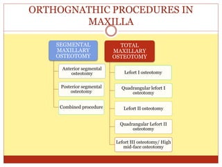
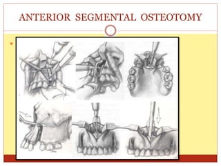
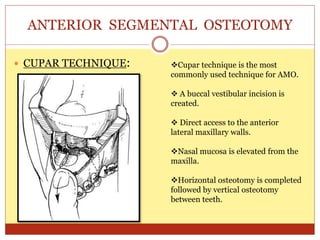
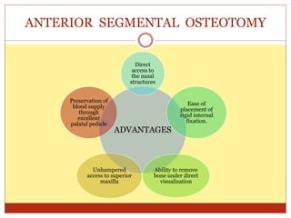
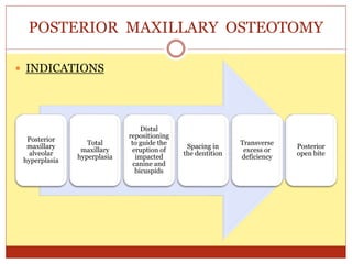
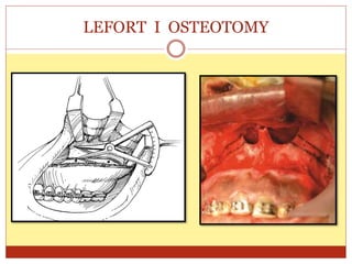
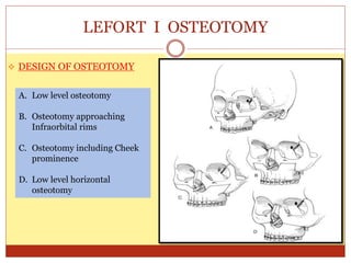
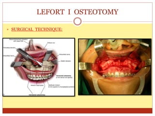
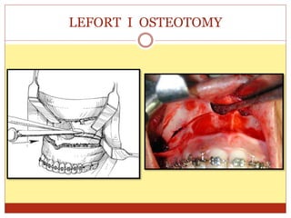
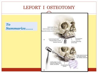
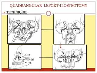
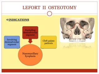
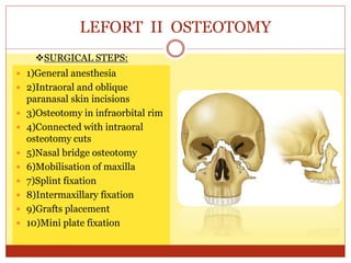
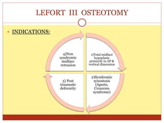
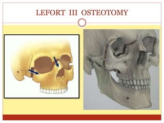
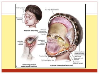
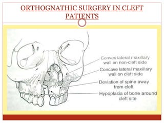
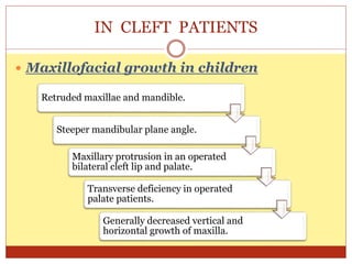
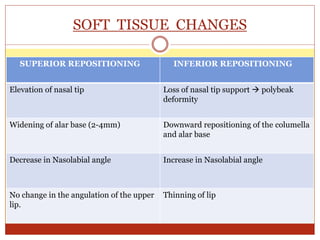
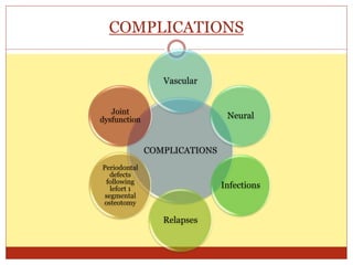
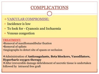
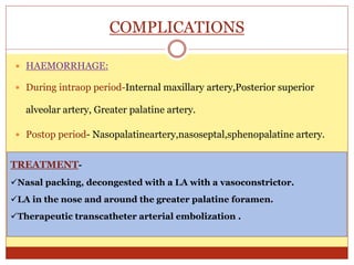
PRESENTED BY- DR. SAHANA M.S
MAXILLARY ORTHOGNATHIC
PROCEDURES AND SOFT
TISSUE CHANGES
CONTENTS
 INTRODUCTION
 AIMS & OBJECTIVES
 HISTORY
 ANATOMICAL CONSIDERATIONS
 ANTERIOR AND POSTERIOR MAXILLARY SEGMENTAL
OSTEOTOMY
 SARME
 LEFORT -I OSTEOTOMY
 MAXILLARY QUADRANGULAR LEFORT I AND QUADRANGULAR
LEFORT II OSTEOTOMY
 HIGH LEVEL MIDFACE OSTEOTOMY
 CLEFT OSTEOTOMIES
 COMPLICATIONS
 CONCLUSION
 REFERENCES
INTRODUCTION
 Dentofacial deformities affect 20%of the population.
 The incidence of Transverse Maxillary Deficiency is estimated to be
between 8% & 18%
 Patients with dentofacial deformities may demonstrate various degrees
of functional and aesthetic compromise.
 Such deformities may be limited to either one jaw or both the jaws.
 Tremendous advancement in field of orthognathic surgery have been
made since 1970s.
AIMS & OBJECTIVES
1
• Prioritize the problem list of the patient.
2
• Review of the risk-benefit considerations.
3
• Consideration of the patient’s expectations
& values.
Fonseca vol 2- Orthognathic surgery
HISTORY
 1859: Von-Langenback did the first orthognathic
procedure.
 1921:Cohn-Stock introduced the anterior maxillary
osteotomy.
 1927:Wassmund reported the first total maxillary
osteotomy.
 Bells research- biologic basis for orthognathic surgery.
GOALS
Correct Masticatory / Swallowing
abnormalities
Establish Functional occlusion
Correct inability to open and close the
jaws
Correct TMJ dysfunction, pathosis /
pain & Myofacial pain
Correct Structural abnormalities and
speech problems
ANATOMICAL CONSIDERATIONS
 VASCULAR ANATOMY:
All the vessels are branches of
the ECA.
Anastomosed ascending
palatine and ascending
pharyngeal artery joins the
lesser palatine artery.
Sphenopalatine, posterior
superior alveolar and
infraorbital arteries are the
others
ORTHOGNATHIC PROCEDURES IN
MAXILLA
SEGMENTAL
MAXILLARY
OSTEOTOMY
Anterior segmental
osteotomy
Posterior segmental
osteotomy
Combined procedure
TOTAL
MAXILLARY
OSTEOTOMY
Lefort I osteotomy
Quadrangular lefort I
osteotomy
Lefort II osteotomy
Quadrangular Lefort II
osteotomy
Lefort III osteotomy/ High
mid-face osteotomy
ANTERIOR SEGMENTAL OSTEOTOMY
 INDICATIONS
Excess vertical or
antero-posterior
dimension of maxillary
alveolar process
To close an anterior
open bite
To retract anterior
maxillary teeth
When orthodontic teeth
movement cannot be
done
To correct marked
protrusion of maxillary
teeth with normal
incisor inclination
between teeth and bone
ANTERIOR SEGMENTAL OSTEOTOMY
 TECHNIQUES:
WUNDERER TECHNIQUE
WASSMUND TECHNIQUE
CUPAR TECHNIQUE
 WUNDERER TECHNIQUE:
ANTERIOR SEGMENTAL OSTEOTOMY
 WASSMUND TECHNIQUE:
ANTERIOR SEGMENTAL OSTEOTOMY
 CUPAR TECHNIQUE:
ANTERIOR SEGMENTAL OSTEOTOMY
Cupar technique is the most
commonly used technique for AMO.
 A buccal vestibular incision is
created.
 Direct access to the anterior
lateral maxillary walls.
Nasal mucosa is elevated from the
maxilla.
Horizontal osteotomy is completed
followed by vertical osteotomy
between teeth.
ADVANTAGES
Direct
access to
the nasal
structures
Ease of
placement of
rigid internal
fixation.
Ability to remove
bone under direct
visualization
Unhampered
access to superior
maxilla
Preservation of
blood supply
through
excellent
palatal pedicle
ANTERIOR SEGMENTAL OSTEOTOMY
POSTERIOR MAXILLARY OSTEOTOMY
 INDICATIONS
Posterior
maxillary
alveolar
hyperplasia
Total
maxillary
hyperplasia
Distal
repositioning
to guide the
eruption of
impacted
canine and
bicuspids
Spacing in
the dentition
Transverse
excess or
deficiency
Posterior
open bite
POSTERIOR MAXILLARY OSTEOTOMY
CONTRAINDICATIONS:
Damage to the vital structures.
When repositioning of the entire
maxilla is recommended.
COMBINED ANTERIOR AND POSTERIOR
MAXILLARY OSTEOTOMY
Also called as
Horse shoe
osteotomy
Used for patient
with maxillary
alveolar
hyperplasia
with or without
anterior open
bite deformity
Transverse
hypoplasia
without vertical
component
Abandoned
procedure
SURGICALLY ASSISTED MAXILLARY
EXPANSION
 Brown-1938- midpalatal split
 Timms – major resistance to expansion is midpalatal suture.
 Kennedy –lateral maxillary osteotomy with midpalatal split
 Shetty-all bony buttress contribute resistance for expansion but
midpalatal suture followed by pterygomaxillary
articulations were the primary areas of resistance.
SURGICALLY ASSISTED MAXILLARY
EXPANSION
 INDICATIONS
Skeletal maxillomandibular transverse
discrepancy >5mm
Significant transverse maxillary deficiency
associated with wide mandible
Failed orthodontic expansion
To avoid segmental osteotomies
Significant nasal stenosis
SURGICALLY ASSISTED MAXILLARY
EXPANSION
SURGICALLY ASSISTED MAXILLARY
EXPANSION
 TECHNIQUE
 TECHNIQUE
SURGICALLY ASSISTED MAXILLARY
EXPANSION
Maxilla should remain stationary – 5 days then 0.5mm /day
0.5mm-1mm/day –Expansion > this causes gingival recession
LEFORT I OSTEOTOMY
 INDICATIONS
• Deficient
vertical
maxilla
• Convex
profile (class
II~ horizontal
excess)
• Vertical
maxillary
excess (
gummy
smile)
• Post trauma
• Cleft lip & palate
• Nasomaxillary
hypoplasia
• Severe mandibular
prognathism (class
III)
Anterior Superior
Inferior
Posterior
LEFORT I OSTEOTOMY
LEFORT I OSTEOTOMY
LEFORT I OSTEOTOMY
 DESIGN OF OSTEOTOMY
A. Low level osteotomy
B. Osteotomy approaching
Infraorbital rims
C. Osteotomy including Cheek
prominence
D. Low level horizontal
osteotomy
LEFORT I OSTEOTOMY
 SURGICAL TECHNIQUE:
LEFORT I OSTEOTOMY
LEFORT I OSTEOTOMY
LEFORT I OSTEOTOMY
LEFORT I OSTEOTOMY
LEFORT I OSTEOTOMY
LEFORT I OSTEOTOMY
To
Summarize.......
SURGICAL COMPLICATIONS OF SEGMENTAL LEFORT I
OSTEOTOMY
M.W.Ho, M.A.Boyle, J.C.Cooper, M.D.Dodd, D.Richardson
CONCLUSION:
The overall complication rate was 27%. There was no segmental loss of
bone or teeth.
Our results show that complications in this cohort were relatively low, and
that segmental maxillary surgery is safe as an adjunct in carefully selected
cases.
British Journal Of Oral & Maxillofacial Surgery, 23 Sep 2010
QUADRANGULAR LEFORT-I OSTEOTOMY
 INDICATIONS
1) Maxillary –
zygomatic
horizontal
deficiency
2) Class 111
skeletal
malocclusion
3) Maxillary
vertical excess or
deficiency
4) Maxillary
transverse
deficiency
QUADRANGULAR LEFORT-I OSTEOTOMY
QUADRANGULAR LEFORT-II OSTEOTOMY
INDICATIONS
1)Maxillary – zygomatic
horizontal deficiency
2)Class 111 skeletal
malocclusion
3)Maxillary vertical excess
or deficiency
4)Maxillary transverse
deficiency
CONTRAINDICATIONS
1)Vertical excess or
deficiency of more
tham 5mm
2)Patients with
retruded nasal
projection
3)Patients with
anterior open bite
QUADRANGULAR LEFORT-II OSTEOTOMY
 TECHNIQUE:
LEFORT II OSTEOTOMY
Nasomaxillary
hyoplasia
Involving
dentoalveolar
segment
Excluding
dentoalveolar
segment
Cleft palate
patients
INDICATIONS
LEFORT II OSTEOTOMY
 1)General anesthesia
 2)Intraoral and oblique
paranasal skin incisions
 3)Osteotomy in infraorbital rim
 4)Connected with intraoral
osteotomy cuts
 5)Nasal bridge osteotomy
 6)Mobilisation of maxilla
 7)Splint fixation
 8)Intermaxillary fixation
 9)Grafts placement
 10)Mini plate fixation
SURGICAL STEPS:
LEFORT III OSTEOTOMY
 INDICATIONS:
1)Total midface
hypoplasia
primarily in AP &
vertical dimension
2)Syndromic
synostosis
(Aperts,
Crouzons
syndrome)
3) Post
traumatic
deformity
4)Non
syndromic
midface
retrusion
LEFORT III OSTEOTOMY
ORTHOGNATHIC SURGERY IN CLEFT
PATIENTS
 Most patients with cleft express some amount of mid face
deficiency.
 Problems due to cleft:-
 Nasal breathing
 Speech, hearing and olfaction.
 Sometimes exorbitism and eyelid in adequacy.
IN CLEFT PATIENTS
 Maxillofacial growth in children
Retruded maxillae and mandible.
Steeper mandibular plane angle.
Maxillary protrusion in an operated
bilateral cleft lip and palate.
Transverse deficiency in operated
palate patients.
Generally decreased vertical and
horizontal growth of maxilla.
ORTHOGNATHIC IN CLEFTS
OSTEOTOMY CUTS
INCISIONS
ORTHOGNATHIC IN CLEFTS
LATERAL VIEW
ORTHOGNATHIC IN CLEFTS
DOWNFRACTURING THE MAXILLA
CLOSURE
ORTHOGNATHIC IN CLEFTS
BONE GRAFTING
SOFT TISSUE CHANGES
 Most important aspect at end of the surgery –achievement
of an aesthetically pleasing facial soft tissue envelope .
 With the refinement of surgical procedures and the advent
of rigid fixation techniques the surgeon can predict the final
outcome of osseous and soft tissue changes .
SOFT TISSUE CHANGES
 NASAL CHANGES
Affects lower aspect of
nasal dorsum
Widening of alar base
Shortening of columellar
height, Alar height and
Nasal tip projection.
Changes in Nasolabial
angle.
SOFT TISSUE CHANGES
Direction
Maxillary
Movement
Alar Bases Nasal Tip Nasolabial
Angle
Superior Increase Increase Decrease
Anterior Increase Increase Decrease
Inferior Inferior Decrease Increase
Posterior None Decrease Increase
SOFT TISSUE CHANGES
 LABIAL CHANGES
ANTERIOR SEGMENTAL
REPOSITIONING- SETBACK
ANTERIOR SEGMENTAL
REPOSITIONING- ADVANCEMENT
Increase in nasolabial angle Decrease in nasolabial angle
Lengthning of upper lip Slight shortening and thinning of
lip(2mm)
Decrease in interlabial gap Alar base widening
Uncurling and retraction of lower lip Advancement of upper lip,subnasale and
nose
SOFT TISSUE CHANGES
SUPERIOR REPOSITIONING INFERIOR REPOSITIONING
Elevation of nasal tip Loss of nasal tip support  polybeak
deformity
Widening of alar base (2-4mm) Downward repositioning of the columella
and alar base
Decrease in Nasolabial angle Increase in Nasolabial angle
No change in the angulation of the upper
lip.
Thinning of lip
COMPLICATIONS
COMPLICATIONS
Vascular
Neural
Infections
Relapses
Periodontal
defects
following
lefort 1
segmental
osteotomy
Joint
dysfunction
COMPLICATIONS
 VASCULAR COMPROMISE:
 Incidence is low
 To look for - Cyanosis and Ischaemia
 Venous congestion
TREATMENT:
•Removal of maxillomandibular fixation
•Removal of splints
•Angiographs to detect site of spasm or occlusion
Administration of Anticoagulants, Beta blockers, Vasodilators,
Hyperbaric oxygen therapy
After irreversible damage debridement of necrotic tissue is undertaken
followed by intraoral free graft
COMPLICATIONS
 HAEMORRHAGE:
 During intraop period-Internal maxillary artery,Posterior superior
alveolar artery, Greater palatine artery.
 Postop period- Nasopalatineartery,nasoseptal,sphenopalatine artery.
TREATMENT-
Nasal packing, decongested with a LA with a vasoconstrictor.
LA in the nose and around the greater palatine foramen.
Therapeutic transcatheter arterial embolization .
COMPLICATIONS
 NEURAL:-
 Injury to the nerve is more common in mandibular procedures
after BSSO than in cases of total down fracture of maxilla
 Study conducted by Kari Panula et al states that incidence of
infraorbital nerve injuries is about 6% on 12 months follow up
when tested objectively.
 During Le Fort osteotomy it is recognized that the nasopalatine
and posterior, middle, and anterior superior alveolar nerves are
completely severed as an intrinsic part of the surgical procedure.
 Despite ligation and division of the neurovascular bundle,
sensory recovery does occur and is most likely to represent
collateral axonal sprouting from adjacent nerves.
COMPLICATIONS
 INFECTIONS:-
 Overall, the incidence of infection is reported to be between
0% and 18% with either a perioperative or a combined
perioperative and postoperative antibiotic course.
 Rates of infection between 0% and 53% have been reported
without antibiotics.
Oral Maxillofacial Surg Clin N Am 15 (2003) 229–242 231
COMPLICATIONS
 RELAPSE:-
 Relapse is usually three dimensional, with vertical, horizontal,
and sagittal components that may occur concurrently.
 On comparison mandibular procedures>maxillary procedures
 Relapse tends to be proportionately greater with greater
advancements.
 Bone grafting large advancements (>8 mm) may help to
reduce relapse.
COMPLICATIONS
 TEMPOROMANDIBULAR DISORDERS:-
 Researchers believe that malocclusion plays some role in TMD.
 Numerous studies support the notion that orthognathic
surgery decreases the overall prevalence of TMD signs and symptoms.
 With respect to orthognathic surgery patients with preexisting TMD,
subjective improvement ranges from 0% to 75%, with a mean of 18%
 Objective improvement ranges from 7% to 72%, with a mean of 48%.
 R.A. Bays, G.F. Bouloux / Oral Maxillofacial Surg Clin N Am 15 (2003) 229–242
COMPLICATIONS- RARE
 Permanent Blindness
 Nasolacrimal obstruction or injury
 Orbital compartment syndrome
 Avascular necrosis (most of which were segmental)
 False aneurysm of sphenopalatine artery
 False aneurysm of maxillary artery
 Carotid-cavernous sinus fistula
 Vomerosphenoidal dysarticulation
 Cranial nerve III palsy
 Keratitis
SEQUENCING
 MANDIBLE FIRST:-
Timothy Turvey journal of oral and maxillofacial surgery 2011 august 69(8):2217-2224
Downgrafting the maxilla
If uncertain about the interocclusal
registration accuracy
When intraoperative MMF wires are to
be used
When fixation of the maxilla may not
be rigid
When performing concomitant TMJ
surgery
CONCLUSION
 Esthetic appearance is critical and of utmost importance in our
society
 Dentofacial and craniofacial deformities not only have a significant
psychological effect on individuals but also on general health status
of same.
 Correction of these deformities cannot be done always by same
protocol and requires indepth knowledge of the subject and can
involve other specialities also.
REFERENCES
1. Oral & maxillofacial surgery-Fonseca vol 2
2. Oral & maxillofacial trauma-Rowe & Williams vol 2
3. Principles of Oral & maxillofacial surgery-Peterson vol 2
4. Killeys- 5th Edition
5. Maxillofacial trauma & facial reconstruction-Peter Ward Booth
vol 1
6. Essentials of Orthognathic surgery- Reyneke
MAXILLARY ORTHOGNATHIC PROCEDURES AND SOFT TISSUE CHANGES-2.pptx

MAXILLARY ORTHOGNATHIC PROCEDURES AND SOFT TISSUE CHANGES-2.pptx

  • 1.
    PRESENTED BY- DR.SAHANA M.S MAXILLARY ORTHOGNATHIC PROCEDURES AND SOFT TISSUE CHANGES
  • 2.
    CONTENTS  INTRODUCTION  AIMS& OBJECTIVES  HISTORY  ANATOMICAL CONSIDERATIONS  ANTERIOR AND POSTERIOR MAXILLARY SEGMENTAL OSTEOTOMY  SARME  LEFORT -I OSTEOTOMY  MAXILLARY QUADRANGULAR LEFORT I AND QUADRANGULAR LEFORT II OSTEOTOMY  HIGH LEVEL MIDFACE OSTEOTOMY  CLEFT OSTEOTOMIES  COMPLICATIONS  CONCLUSION  REFERENCES
  • 3.
    INTRODUCTION  Dentofacial deformitiesaffect 20%of the population.  The incidence of Transverse Maxillary Deficiency is estimated to be between 8% & 18%  Patients with dentofacial deformities may demonstrate various degrees of functional and aesthetic compromise.  Such deformities may be limited to either one jaw or both the jaws.  Tremendous advancement in field of orthognathic surgery have been made since 1970s.
  • 4.
    AIMS & OBJECTIVES 1 •Prioritize the problem list of the patient. 2 • Review of the risk-benefit considerations. 3 • Consideration of the patient’s expectations & values. Fonseca vol 2- Orthognathic surgery
  • 5.
    HISTORY  1859: Von-Langenbackdid the first orthognathic procedure.  1921:Cohn-Stock introduced the anterior maxillary osteotomy.  1927:Wassmund reported the first total maxillary osteotomy.  Bells research- biologic basis for orthognathic surgery.
  • 6.
    GOALS Correct Masticatory /Swallowing abnormalities Establish Functional occlusion Correct inability to open and close the jaws Correct TMJ dysfunction, pathosis / pain & Myofacial pain Correct Structural abnormalities and speech problems
  • 7.
  • 8.
    ANATOMICAL CONSIDERATIONS  VASCULARANATOMY: All the vessels are branches of the ECA. Anastomosed ascending palatine and ascending pharyngeal artery joins the lesser palatine artery. Sphenopalatine, posterior superior alveolar and infraorbital arteries are the others
  • 9.
    ORTHOGNATHIC PROCEDURES IN MAXILLA SEGMENTAL MAXILLARY OSTEOTOMY Anteriorsegmental osteotomy Posterior segmental osteotomy Combined procedure TOTAL MAXILLARY OSTEOTOMY Lefort I osteotomy Quadrangular lefort I osteotomy Lefort II osteotomy Quadrangular Lefort II osteotomy Lefort III osteotomy/ High mid-face osteotomy
  • 10.
    ANTERIOR SEGMENTAL OSTEOTOMY INDICATIONS Excess vertical or antero-posterior dimension of maxillary alveolar process To close an anterior open bite To retract anterior maxillary teeth When orthodontic teeth movement cannot be done To correct marked protrusion of maxillary teeth with normal incisor inclination between teeth and bone
  • 11.
    ANTERIOR SEGMENTAL OSTEOTOMY TECHNIQUES: WUNDERER TECHNIQUE WASSMUND TECHNIQUE CUPAR TECHNIQUE
  • 12.
  • 13.
  • 14.
     CUPAR TECHNIQUE: ANTERIORSEGMENTAL OSTEOTOMY Cupar technique is the most commonly used technique for AMO.  A buccal vestibular incision is created.  Direct access to the anterior lateral maxillary walls. Nasal mucosa is elevated from the maxilla. Horizontal osteotomy is completed followed by vertical osteotomy between teeth.
  • 15.
    ADVANTAGES Direct access to the nasal structures Easeof placement of rigid internal fixation. Ability to remove bone under direct visualization Unhampered access to superior maxilla Preservation of blood supply through excellent palatal pedicle ANTERIOR SEGMENTAL OSTEOTOMY
  • 16.
    POSTERIOR MAXILLARY OSTEOTOMY INDICATIONS Posterior maxillary alveolar hyperplasia Total maxillary hyperplasia Distal repositioning to guide the eruption of impacted canine and bicuspids Spacing in the dentition Transverse excess or deficiency Posterior open bite
  • 17.
    POSTERIOR MAXILLARY OSTEOTOMY CONTRAINDICATIONS: Damageto the vital structures. When repositioning of the entire maxilla is recommended.
  • 18.
    COMBINED ANTERIOR ANDPOSTERIOR MAXILLARY OSTEOTOMY Also called as Horse shoe osteotomy Used for patient with maxillary alveolar hyperplasia with or without anterior open bite deformity Transverse hypoplasia without vertical component Abandoned procedure
  • 19.
    SURGICALLY ASSISTED MAXILLARY EXPANSION Brown-1938- midpalatal split  Timms – major resistance to expansion is midpalatal suture.  Kennedy –lateral maxillary osteotomy with midpalatal split  Shetty-all bony buttress contribute resistance for expansion but midpalatal suture followed by pterygomaxillary articulations were the primary areas of resistance.
  • 20.
    SURGICALLY ASSISTED MAXILLARY EXPANSION INDICATIONS Skeletal maxillomandibular transverse discrepancy >5mm Significant transverse maxillary deficiency associated with wide mandible Failed orthodontic expansion To avoid segmental osteotomies Significant nasal stenosis
  • 21.
  • 22.
  • 23.
     TECHNIQUE SURGICALLY ASSISTEDMAXILLARY EXPANSION Maxilla should remain stationary – 5 days then 0.5mm /day 0.5mm-1mm/day –Expansion > this causes gingival recession
  • 24.
    LEFORT I OSTEOTOMY INDICATIONS • Deficient vertical maxilla • Convex profile (class II~ horizontal excess) • Vertical maxillary excess ( gummy smile) • Post trauma • Cleft lip & palate • Nasomaxillary hypoplasia • Severe mandibular prognathism (class III) Anterior Superior Inferior Posterior
  • 25.
  • 26.
  • 27.
    LEFORT I OSTEOTOMY DESIGN OF OSTEOTOMY A. Low level osteotomy B. Osteotomy approaching Infraorbital rims C. Osteotomy including Cheek prominence D. Low level horizontal osteotomy
  • 28.
    LEFORT I OSTEOTOMY SURGICAL TECHNIQUE:
  • 29.
  • 30.
  • 31.
  • 32.
  • 33.
  • 34.
  • 37.
    SURGICAL COMPLICATIONS OFSEGMENTAL LEFORT I OSTEOTOMY M.W.Ho, M.A.Boyle, J.C.Cooper, M.D.Dodd, D.Richardson CONCLUSION: The overall complication rate was 27%. There was no segmental loss of bone or teeth. Our results show that complications in this cohort were relatively low, and that segmental maxillary surgery is safe as an adjunct in carefully selected cases. British Journal Of Oral & Maxillofacial Surgery, 23 Sep 2010
  • 38.
    QUADRANGULAR LEFORT-I OSTEOTOMY INDICATIONS 1) Maxillary – zygomatic horizontal deficiency 2) Class 111 skeletal malocclusion 3) Maxillary vertical excess or deficiency 4) Maxillary transverse deficiency
  • 39.
  • 41.
    QUADRANGULAR LEFORT-II OSTEOTOMY INDICATIONS 1)Maxillary– zygomatic horizontal deficiency 2)Class 111 skeletal malocclusion 3)Maxillary vertical excess or deficiency 4)Maxillary transverse deficiency CONTRAINDICATIONS 1)Vertical excess or deficiency of more tham 5mm 2)Patients with retruded nasal projection 3)Patients with anterior open bite
  • 42.
  • 43.
  • 44.
    LEFORT II OSTEOTOMY 1)General anesthesia  2)Intraoral and oblique paranasal skin incisions  3)Osteotomy in infraorbital rim  4)Connected with intraoral osteotomy cuts  5)Nasal bridge osteotomy  6)Mobilisation of maxilla  7)Splint fixation  8)Intermaxillary fixation  9)Grafts placement  10)Mini plate fixation SURGICAL STEPS:
  • 45.
    LEFORT III OSTEOTOMY INDICATIONS: 1)Total midface hypoplasia primarily in AP & vertical dimension 2)Syndromic synostosis (Aperts, Crouzons syndrome) 3) Post traumatic deformity 4)Non syndromic midface retrusion
  • 46.
  • 48.
    ORTHOGNATHIC SURGERY INCLEFT PATIENTS  Most patients with cleft express some amount of mid face deficiency.  Problems due to cleft:-  Nasal breathing  Speech, hearing and olfaction.  Sometimes exorbitism and eyelid in adequacy.
  • 49.
    IN CLEFT PATIENTS Maxillofacial growth in children Retruded maxillae and mandible. Steeper mandibular plane angle. Maxillary protrusion in an operated bilateral cleft lip and palate. Transverse deficiency in operated palate patients. Generally decreased vertical and horizontal growth of maxilla.
  • 50.
  • 51.
  • 52.
  • 53.
  • 54.
    SOFT TISSUE CHANGES Most important aspect at end of the surgery –achievement of an aesthetically pleasing facial soft tissue envelope .  With the refinement of surgical procedures and the advent of rigid fixation techniques the surgeon can predict the final outcome of osseous and soft tissue changes .
  • 55.
    SOFT TISSUE CHANGES NASAL CHANGES Affects lower aspect of nasal dorsum Widening of alar base Shortening of columellar height, Alar height and Nasal tip projection. Changes in Nasolabial angle.
  • 56.
    SOFT TISSUE CHANGES Direction Maxillary Movement AlarBases Nasal Tip Nasolabial Angle Superior Increase Increase Decrease Anterior Increase Increase Decrease Inferior Inferior Decrease Increase Posterior None Decrease Increase
  • 57.
    SOFT TISSUE CHANGES LABIAL CHANGES ANTERIOR SEGMENTAL REPOSITIONING- SETBACK ANTERIOR SEGMENTAL REPOSITIONING- ADVANCEMENT Increase in nasolabial angle Decrease in nasolabial angle Lengthning of upper lip Slight shortening and thinning of lip(2mm) Decrease in interlabial gap Alar base widening Uncurling and retraction of lower lip Advancement of upper lip,subnasale and nose
  • 58.
    SOFT TISSUE CHANGES SUPERIORREPOSITIONING INFERIOR REPOSITIONING Elevation of nasal tip Loss of nasal tip support  polybeak deformity Widening of alar base (2-4mm) Downward repositioning of the columella and alar base Decrease in Nasolabial angle Increase in Nasolabial angle No change in the angulation of the upper lip. Thinning of lip
  • 59.
  • 60.
    COMPLICATIONS  VASCULAR COMPROMISE: Incidence is low  To look for - Cyanosis and Ischaemia  Venous congestion TREATMENT: •Removal of maxillomandibular fixation •Removal of splints •Angiographs to detect site of spasm or occlusion Administration of Anticoagulants, Beta blockers, Vasodilators, Hyperbaric oxygen therapy After irreversible damage debridement of necrotic tissue is undertaken followed by intraoral free graft
  • 61.
    COMPLICATIONS  HAEMORRHAGE:  Duringintraop period-Internal maxillary artery,Posterior superior alveolar artery, Greater palatine artery.  Postop period- Nasopalatineartery,nasoseptal,sphenopalatine artery. TREATMENT- Nasal packing, decongested with a LA with a vasoconstrictor. LA in the nose and around the greater palatine foramen. Therapeutic transcatheter arterial embolization .
  • 63.
    COMPLICATIONS  NEURAL:-  Injuryto the nerve is more common in mandibular procedures after BSSO than in cases of total down fracture of maxilla  Study conducted by Kari Panula et al states that incidence of infraorbital nerve injuries is about 6% on 12 months follow up when tested objectively.  During Le Fort osteotomy it is recognized that the nasopalatine and posterior, middle, and anterior superior alveolar nerves are completely severed as an intrinsic part of the surgical procedure.  Despite ligation and division of the neurovascular bundle, sensory recovery does occur and is most likely to represent collateral axonal sprouting from adjacent nerves.
  • 64.
    COMPLICATIONS  INFECTIONS:-  Overall,the incidence of infection is reported to be between 0% and 18% with either a perioperative or a combined perioperative and postoperative antibiotic course.  Rates of infection between 0% and 53% have been reported without antibiotics. Oral Maxillofacial Surg Clin N Am 15 (2003) 229–242 231
  • 65.
    COMPLICATIONS  RELAPSE:-  Relapseis usually three dimensional, with vertical, horizontal, and sagittal components that may occur concurrently.  On comparison mandibular procedures>maxillary procedures  Relapse tends to be proportionately greater with greater advancements.  Bone grafting large advancements (>8 mm) may help to reduce relapse.
  • 66.
    COMPLICATIONS  TEMPOROMANDIBULAR DISORDERS:- Researchers believe that malocclusion plays some role in TMD.  Numerous studies support the notion that orthognathic surgery decreases the overall prevalence of TMD signs and symptoms.  With respect to orthognathic surgery patients with preexisting TMD, subjective improvement ranges from 0% to 75%, with a mean of 18%  Objective improvement ranges from 7% to 72%, with a mean of 48%.  R.A. Bays, G.F. Bouloux / Oral Maxillofacial Surg Clin N Am 15 (2003) 229–242
  • 67.
    COMPLICATIONS- RARE  PermanentBlindness  Nasolacrimal obstruction or injury  Orbital compartment syndrome  Avascular necrosis (most of which were segmental)  False aneurysm of sphenopalatine artery  False aneurysm of maxillary artery  Carotid-cavernous sinus fistula  Vomerosphenoidal dysarticulation  Cranial nerve III palsy  Keratitis
  • 68.
    SEQUENCING  MANDIBLE FIRST:- TimothyTurvey journal of oral and maxillofacial surgery 2011 august 69(8):2217-2224 Downgrafting the maxilla If uncertain about the interocclusal registration accuracy When intraoperative MMF wires are to be used When fixation of the maxilla may not be rigid When performing concomitant TMJ surgery
  • 69.
    CONCLUSION  Esthetic appearanceis critical and of utmost importance in our society  Dentofacial and craniofacial deformities not only have a significant psychological effect on individuals but also on general health status of same.  Correction of these deformities cannot be done always by same protocol and requires indepth knowledge of the subject and can involve other specialities also.
  • 70.
    REFERENCES 1. Oral &maxillofacial surgery-Fonseca vol 2 2. Oral & maxillofacial trauma-Rowe & Williams vol 2 3. Principles of Oral & maxillofacial surgery-Peterson vol 2 4. Killeys- 5th Edition 5. Maxillofacial trauma & facial reconstruction-Peter Ward Booth vol 1 6. Essentials of Orthognathic surgery- Reyneke