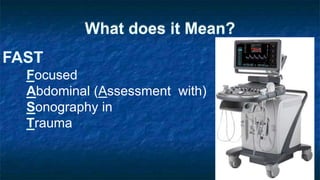
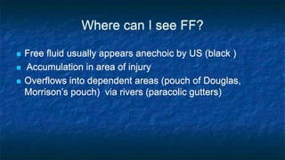
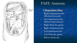
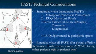
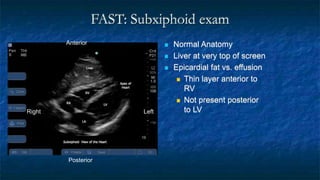
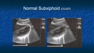
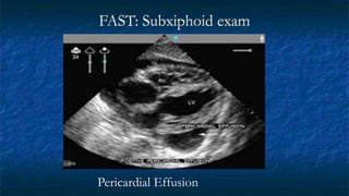
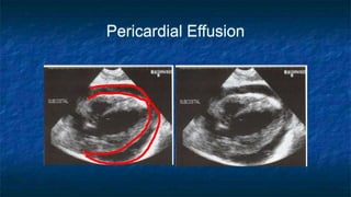
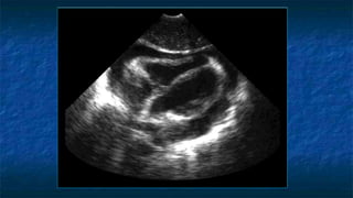
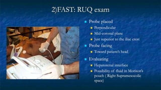
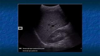
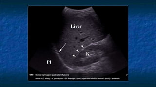
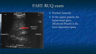
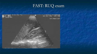
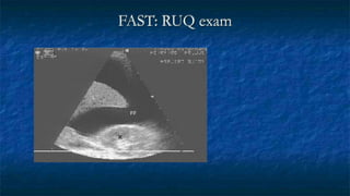
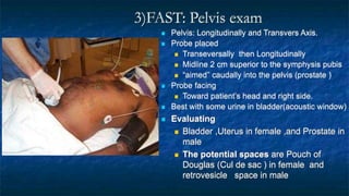
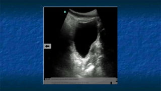
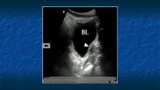
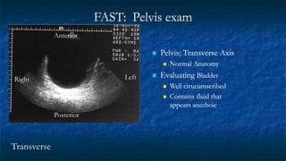
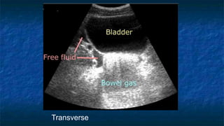
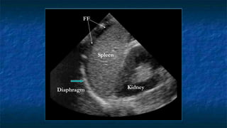
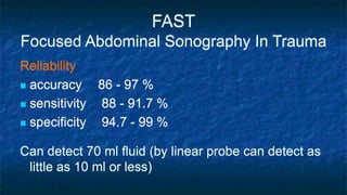
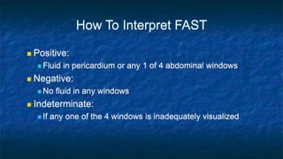
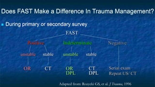
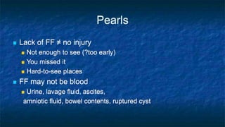
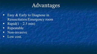
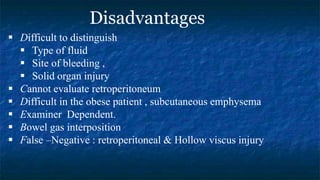
.
Dr. Ashraf Hussein Ismail
Emergency Medicine Consultant
What does it Mean?
FAST
Focused
Abdominal (Assessment with)
Sonography in
Trauma
INTRODUCTION
 1980s- US for trauma in Japan, Germany
 1990s- US for trauma in North America
 The term FAST introduced in 1996
Where can I see FF?
 Free fluid usually appears anechoic by US (black )
 Accumulation in area of injury
 Overflows into dependent areas (pouch of Douglas,
Morrison’s pouch) via rivers (paracolic gutters)
FAST: Anatomy
7 Dependent Sites
1. Right Supramesocolic
(Morison’s pouch)
2. Left Supramesocolic
(Splenorenal rescess)
3. Right Pericolic gutter
4. Right Inframesocolic
5. Left Inframesocolic
6. Left Pericolic gutter
7. Pelvic cul-de-sac
FAST: Technical Considerations
• Standerded views (standerded FAST ):
1- Subxiphoid/Subcostal: Pericardium
2- RUQ: Morrison’s Pouch
3-Pelvis: Pelvic Cul-de-sac (Douglas )
Transverse
Longitudinal
4- LUQ: Splenorenal & perisplenic spaces
• Extended views (E-FAST) :For pleural effusion
Remember: Probe marker almost ALWAYS facing
either patient’s right or patient’s head
Supine patient
1
4
2
3
1) Subxiphoid exam
 Probe placed
 Transversally
 Midline plane
 Just below subxiphoid region
 Probe facing towards patient’s right
FAST: Subxiphoid exam
 Normal Anatomy
 Liver at very top of screen
 Epicardial fat vs. effusion
 Thin layer anterior to
RV
 Not present posterior
to LV
Anterior
Posterior
Left
Right
Normal Subxiphoid exam
FAST: Subxiphoid exam
Pericardial Effusion
Types of pericardial effusions, subxiphoid cardiac view.
Left image: typical effusion, middle image: clotted effusion , right image : with cardiac tamponade
.
2)FAST: RUQ exam
 Probe placed
 Perpendicular
 Mid-coronal plane
 Just superior to the iliac crest
 Probe facing
 Toward patient’s head
 Evaluating
 Hepatorenal interface
 Possibility of fluid in Morison’s
pouch ( Right Supramesocolic
space)
FAST: RUQ exam
 Normal Anatomy
 In the supine patient, the
hepatorenal space
(Morison’s Pouch) is the
most dependent space
Anterior
Posterior
Inferior
Superior
Morison’s
Pouch
FAST: RUQ exam
L
K
FF
RS
D
FAST: RUQ exam
L
K
FF
3)FAST: Pelvis exam
 Pelvis: Longitudinally and Transvers Axis.
 Probe placed
 Transeversally then Longitudinally
 Midline 2 cm superior to the symphysis pubis
 “aimed” caudally into the pelvis (prostate )
 Probe facing
 Toward patient’s head and right side.
 Best with some urine in bladder(acoustic window)
 Evaluating
 Bladder ,Uterus in female ,and Prostate in
male
 The potential spaces are Pouch of
Douglas (Cul de sac ) in female and
retrovesicle space in male
FAST: Pelvis exam
 Pelvis: Transverse Axis
 Normal Anatomy
 Evaluating Bladder
 Well cirucumscribed
 Contains fluid that
appears anechoic
Transverse
Anterior
Right Left
Posterior
Transverse
FAST: Pelvis exam - Pathology
Transverse
Bladder
FF
Transverse
4)FAST: LUQ exam
 Probe placed
 Perpendicular
 Mid - coronal plane
 Just superior to the iliac crest
 Probe facing
 Towards patient’s head
 Evaluating
 Spleno-renal interface
 Possibility of fluid in splenorenal recess
and presplenic /subphrenic space( most
common space for fluid collection in
LUQ)
FF
Kidney
Spleen
FF
Diaphragm
FAST Demo
FAST
Focused Abdominal Sonography In Trauma
Reliability
 accuracy 86 - 97 %
 sensitivity 88 - 91.7 %
 specificity 94.7 - 99 %
Can detect 70 ml fluid (by linear probe can detect as
little as 10 ml or less)
How To Interpret FAST
 Positive:
 Fluid in pericardium or any 1 of 4 abdominal windows
 Negative:
 No fluid in any windows
 Indeterminate:
 If any one of the 4 windows is inadequately visualized
Does FAST Make a Difference In Trauma Management?
 During primary or secondary survey
FAST
Positive Negative
Indeterminate
unstable stable
OR CT
unstable stable
OR
DPL
CT
DPL
Serial exam
Repeat US/ CT
Adapted from: Rozycki GS, et al. J Trauma, 1996
Pearls
 Lack of FF ≠ no injury
 Not enough to see (?too early)
 You missed it
 Hard-to-see places
 FF may not be blood
 Urine, lavage fluid, ascites,
amniotic fluid, bowel contents, ruptured cyst
Advantages
 Easy & Early to Diagnose in
Resuscitation/Emergency room
 Rapid(1 – 2.5 min)
 Repeatable
 Non-invasive
 Low cost.
 Difficult to distinguish
 Type of fluid
 Site of bleeding ,
 Solid organ injury
 Cannot evaluate retroperitoneum
 Difficult in the obese patient , subcutaneous emphysema
 Examiner Dependent.
 Bowel gas interposition
 False –Negative : retroperitoneal & Hollow viscus injury
Disadvantages
Pitfalls and limits
• -Pre-exesiting fluid collection ( Ascites , dialysis )
• -Pelvic fluid collection (female ) .
• -Fluid filled bowel loops .
• -Contained injury (hollow viscus, bowel wall
contusion, pancreatic trauma and renal pedicle injury)
• -Echogenic clot.
The scan should be repeated during the secondary survey and
also if the patient demonstrates clinical deterioration, since free
fluid may have accumulated in the intervening time .
The quality of images obtained may also be a limiting factor with
patient obesity , gas in the bowel leading to degradation in image
quality , subcutaneous emphysema , non-mobile patient and
pnetrating injury.
Does FAST replace CT?
 Unstable patient, (+) FAST  OR
 Stable patient, low force injury, (-) FAST  consider observing patient.
CT is far more sensitive than FAST for detecting and characterizing abdominal injury in
trauma. The gold standard for characterizing intraparenchymal injury.
“Death begins with a CT.” Never send an unstable patient to CT. FAST, however, can be
performed during resuscitation. FAST
Positive Negative
Indeterminate
unstable stable
OR CT
unstable stable
OR
DPL
CT
DPL
Serial exam Repeat US/
CT
Extended FAST (E-FAST)
RUQ, LUQ views:
 Check above diaphragm for hemothorax
 CXR = US in detection of hemothorax
Ma and Mateer. Ann Emerg Med, 1997
 50-175cc vs. 20cc or less
 US does not replace CXR
Suprapubic view:
 Check uterus for pregnancy
Hemothorax
KD
SP
FF
D
Pleural Fluid
Lung Scanning for Pneumothorax
Comet tails sign
and sliding lung
Loss of comet tail and lung
sliding movement
Questions?
6- Abdominal Trauma and FAST Scan.pptx

6- Abdominal Trauma and FAST Scan.pptx

  • 1.
    . Dr. Ashraf HusseinIsmail Emergency Medicine Consultant
  • 2.
    What does itMean? FAST Focused Abdominal (Assessment with) Sonography in Trauma
  • 3.
    INTRODUCTION  1980s- USfor trauma in Japan, Germany  1990s- US for trauma in North America  The term FAST introduced in 1996
  • 4.
    Where can Isee FF?  Free fluid usually appears anechoic by US (black )  Accumulation in area of injury  Overflows into dependent areas (pouch of Douglas, Morrison’s pouch) via rivers (paracolic gutters)
  • 5.
    FAST: Anatomy 7 DependentSites 1. Right Supramesocolic (Morison’s pouch) 2. Left Supramesocolic (Splenorenal rescess) 3. Right Pericolic gutter 4. Right Inframesocolic 5. Left Inframesocolic 6. Left Pericolic gutter 7. Pelvic cul-de-sac
  • 6.
    FAST: Technical Considerations •Standerded views (standerded FAST ): 1- Subxiphoid/Subcostal: Pericardium 2- RUQ: Morrison’s Pouch 3-Pelvis: Pelvic Cul-de-sac (Douglas ) Transverse Longitudinal 4- LUQ: Splenorenal & perisplenic spaces • Extended views (E-FAST) :For pleural effusion Remember: Probe marker almost ALWAYS facing either patient’s right or patient’s head Supine patient 1 4 2 3
  • 8.
    1) Subxiphoid exam Probe placed  Transversally  Midline plane  Just below subxiphoid region  Probe facing towards patient’s right
  • 11.
    FAST: Subxiphoid exam Normal Anatomy  Liver at very top of screen  Epicardial fat vs. effusion  Thin layer anterior to RV  Not present posterior to LV Anterior Posterior Left Right
  • 12.
  • 13.
  • 14.
  • 16.
    Types of pericardialeffusions, subxiphoid cardiac view. Left image: typical effusion, middle image: clotted effusion , right image : with cardiac tamponade .
  • 17.
    2)FAST: RUQ exam Probe placed  Perpendicular  Mid-coronal plane  Just superior to the iliac crest  Probe facing  Toward patient’s head  Evaluating  Hepatorenal interface  Possibility of fluid in Morison’s pouch ( Right Supramesocolic space)
  • 20.
    FAST: RUQ exam Normal Anatomy  In the supine patient, the hepatorenal space (Morison’s Pouch) is the most dependent space Anterior Posterior Inferior Superior Morison’s Pouch
  • 21.
  • 22.
  • 23.
    3)FAST: Pelvis exam Pelvis: Longitudinally and Transvers Axis.  Probe placed  Transeversally then Longitudinally  Midline 2 cm superior to the symphysis pubis  “aimed” caudally into the pelvis (prostate )  Probe facing  Toward patient’s head and right side.  Best with some urine in bladder(acoustic window)  Evaluating  Bladder ,Uterus in female ,and Prostate in male  The potential spaces are Pouch of Douglas (Cul de sac ) in female and retrovesicle space in male
  • 30.
    FAST: Pelvis exam Pelvis: Transverse Axis  Normal Anatomy  Evaluating Bladder  Well cirucumscribed  Contains fluid that appears anechoic Transverse Anterior Right Left Posterior
  • 31.
  • 32.
    FAST: Pelvis exam- Pathology Transverse
  • 33.
  • 34.
    4)FAST: LUQ exam Probe placed  Perpendicular  Mid - coronal plane  Just superior to the iliac crest  Probe facing  Towards patient’s head  Evaluating  Spleno-renal interface  Possibility of fluid in splenorenal recess and presplenic /subphrenic space( most common space for fluid collection in LUQ)
  • 39.
  • 40.
  • 42.
  • 43.
    FAST Focused Abdominal SonographyIn Trauma Reliability  accuracy 86 - 97 %  sensitivity 88 - 91.7 %  specificity 94.7 - 99 % Can detect 70 ml fluid (by linear probe can detect as little as 10 ml or less)
  • 44.
    How To InterpretFAST  Positive:  Fluid in pericardium or any 1 of 4 abdominal windows  Negative:  No fluid in any windows  Indeterminate:  If any one of the 4 windows is inadequately visualized
  • 45.
    Does FAST Makea Difference In Trauma Management?  During primary or secondary survey FAST Positive Negative Indeterminate unstable stable OR CT unstable stable OR DPL CT DPL Serial exam Repeat US/ CT Adapted from: Rozycki GS, et al. J Trauma, 1996
  • 46.
    Pearls  Lack ofFF ≠ no injury  Not enough to see (?too early)  You missed it  Hard-to-see places  FF may not be blood  Urine, lavage fluid, ascites, amniotic fluid, bowel contents, ruptured cyst
  • 47.
    Advantages  Easy &Early to Diagnose in Resuscitation/Emergency room  Rapid(1 – 2.5 min)  Repeatable  Non-invasive  Low cost.
  • 48.
     Difficult todistinguish  Type of fluid  Site of bleeding ,  Solid organ injury  Cannot evaluate retroperitoneum  Difficult in the obese patient , subcutaneous emphysema  Examiner Dependent.  Bowel gas interposition  False –Negative : retroperitoneal & Hollow viscus injury Disadvantages
  • 49.
    Pitfalls and limits •-Pre-exesiting fluid collection ( Ascites , dialysis ) • -Pelvic fluid collection (female ) . • -Fluid filled bowel loops . • -Contained injury (hollow viscus, bowel wall contusion, pancreatic trauma and renal pedicle injury) • -Echogenic clot. The scan should be repeated during the secondary survey and also if the patient demonstrates clinical deterioration, since free fluid may have accumulated in the intervening time . The quality of images obtained may also be a limiting factor with patient obesity , gas in the bowel leading to degradation in image quality , subcutaneous emphysema , non-mobile patient and pnetrating injury.
  • 50.
  • 51.
    Does FAST replaceCT?  Unstable patient, (+) FAST  OR  Stable patient, low force injury, (-) FAST  consider observing patient. CT is far more sensitive than FAST for detecting and characterizing abdominal injury in trauma. The gold standard for characterizing intraparenchymal injury. “Death begins with a CT.” Never send an unstable patient to CT. FAST, however, can be performed during resuscitation. FAST Positive Negative Indeterminate unstable stable OR CT unstable stable OR DPL CT DPL Serial exam Repeat US/ CT
  • 53.
    Extended FAST (E-FAST) RUQ,LUQ views:  Check above diaphragm for hemothorax  CXR = US in detection of hemothorax Ma and Mateer. Ann Emerg Med, 1997  50-175cc vs. 20cc or less  US does not replace CXR Suprapubic view:  Check uterus for pregnancy
  • 54.
  • 55.
  • 56.
    Lung Scanning forPneumothorax Comet tails sign and sliding lung
  • 58.
    Loss of comettail and lung sliding movement
  • 62.