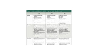
Evaluación por Ecografía Focalizada en Trauma
(FAST)
• Focused Abdominal Sonography
• for Trauma (FAST)
• FAST is performed as part of the secondary survey in victims of
• torso trauma (see Chapter 16 ). It is a temporally (2–5 minutes)
• and anatomically limited real-time sonographic examination
• whose core components include a direct sonographic search for
• free fluid in the pericardial sac, both upper abdominal quadrants,
• and the intraperitoneal recesses adjacent to the urinary
• bladder. Scanning is optimally performed in two orthogonal
• planes (e.g., longitudinal and transverse), and intraparenchymal
• and retroperitoneal injuries are generally not sought but are
• sometimes seen. FAST may be extended to detect a hemothorax
• and pneumothorax. Commercially available, portable handheld
• real-time imaging devices are technically adequate to
• perform FAST.
• FAST is uniformly accurate for the detection of intraperitoneal
• fluid with moderately large volumes 400 cm 3 (at smaller
• volumes, accuracy varies with user experience). 3 – 5 Unfortunately,
• isolated hepatosplenic injuries with minimal or no hemoperitoneum
• represent as many as one third of solid organ injuries.
• 6 , 7 Fortunately, small isolated intraparenchymal lesions
• with less than 250 mL of intraperitoneal blood rarely require
• endovascular or surgical intervention (liver 1%, spleen
• 5%). 8 False-positive interpretation of FAST images can
• result from improper machine settings (gain), sonolucent perinephric
• fat (which is rarely sonolucent in both axial and coronal
• scanning planes), fluid-filled loops of bowel, bladder,
• various types of fluid-filled intra-abdominal cysts, and physiological
• SURGEON-PERFORMED
• ULTRASOUND IN TRAUMA
• ■ FAST
• Developed for the evaluation of injured patients, the F ocused
• A ssessment for the S onographic Examination of the T rauma
• Patient (FAST) is a rapid diagnostic examination to assess
• patients with potential injuries to the torso. The test sequentially
• surveys for the presence or absence of fluid in the pericardial
• sac and in the dependent abdominal regions, including Morison’s pouch region in the right upper quadrant (RUQ),
• the left upper quadrant (LUQ) behind the spleen and between
• the spleen and kidney, and the pelvis posterior to the bladder.
• Surgeons can perform the FAST during the primary
• or secondary survey of the American College of Surgeons
• Advanced Trauma Life Support 19 algorithm and, although
• minimal patient preparation is needed, a full urinary bladder is
• ideal to provide an acoustic window for visualization of blood
• in the pelvis.
• Blood, as any fluid, will accumulate in dependent regions of
FAST_110421.pptx
FAST_110421.pptx
FAST_110421.pptx
FAST_110421.pptx

FAST_110421.pptx

  • 2.
    Evaluación por EcografíaFocalizada en Trauma (FAST)
  • 3.
    • Focused AbdominalSonography • for Trauma (FAST) • FAST is performed as part of the secondary survey in victims of • torso trauma (see Chapter 16 ). It is a temporally (2–5 minutes) • and anatomically limited real-time sonographic examination • whose core components include a direct sonographic search for • free fluid in the pericardial sac, both upper abdominal quadrants, • and the intraperitoneal recesses adjacent to the urinary • bladder. Scanning is optimally performed in two orthogonal • planes (e.g., longitudinal and transverse), and intraparenchymal • and retroperitoneal injuries are generally not sought but are • sometimes seen. FAST may be extended to detect a hemothorax • and pneumothorax. Commercially available, portable handheld • real-time imaging devices are technically adequate to • perform FAST. • FAST is uniformly accurate for the detection of intraperitoneal • fluid with moderately large volumes 400 cm 3 (at smaller • volumes, accuracy varies with user experience). 3 – 5 Unfortunately, • isolated hepatosplenic injuries with minimal or no hemoperitoneum • represent as many as one third of solid organ injuries. • 6 , 7 Fortunately, small isolated intraparenchymal lesions • with less than 250 mL of intraperitoneal blood rarely require • endovascular or surgical intervention (liver 1%, spleen • 5%). 8 False-positive interpretation of FAST images can • result from improper machine settings (gain), sonolucent perinephric • fat (which is rarely sonolucent in both axial and coronal • scanning planes), fluid-filled loops of bowel, bladder, • various types of fluid-filled intra-abdominal cysts, and physiological
  • 5.
    • SURGEON-PERFORMED • ULTRASOUNDIN TRAUMA • ■ FAST • Developed for the evaluation of injured patients, the F ocused • A ssessment for the S onographic Examination of the T rauma • Patient (FAST) is a rapid diagnostic examination to assess • patients with potential injuries to the torso. The test sequentially • surveys for the presence or absence of fluid in the pericardial • sac and in the dependent abdominal regions, including Morison’s pouch region in the right upper quadrant (RUQ), • the left upper quadrant (LUQ) behind the spleen and between • the spleen and kidney, and the pelvis posterior to the bladder. • Surgeons can perform the FAST during the primary • or secondary survey of the American College of Surgeons • Advanced Trauma Life Support 19 algorithm and, although • minimal patient preparation is needed, a full urinary bladder is • ideal to provide an acoustic window for visualization of blood • in the pelvis. • Blood, as any fluid, will accumulate in dependent regions of