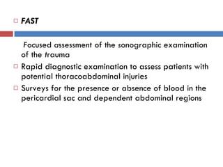
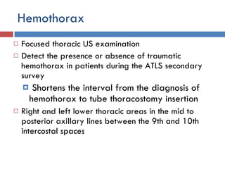
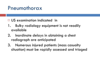
FAST  Dr. vinayak lokare JMMC & RI
Blunt abdominal trauma
Is there fluid within the peritoneal cavity?  Is there fluid in the pericardial sac?
FAST F ocused  a ssessment of the  s onographic examination of the  t rauma  Rapid diagnostic examination to assess patients with potential thoracoabdominal injuries Surveys for the presence or absence of blood in the pericardial sac and dependent abdominal regions
Four areas examined  sequentially  in FAST –  pericardial sac  right upper quadrant (RUQ), left upper quadrant (LUQ), pelvis
 
Pericardial sac  First to be examined  To set the gain – anechoic  3.5-MHz convex transducer positioned in the subxiphoid region
 
Difficulties severe chest wall injury, a very narrow subcostal area,  subcutaneous emphysema,  morbid obesity  parasternal US
 
False-positive and false-negative pericardial US examinations  Massive hemothorax or  Mediastinal blood Repeating FAST after insertion of a tube thoracostomy
Right upper quadrant  The transducer is placed in the right midaxillary line between the 11th and 12th ribs Liver, kidney, and diaphragm are viewed in the sagittal section  Morison's pouch and in the subphrenic space
 
Left upper quadrant Transducer positioned in the left posterior axillary line between the 10th and 11th ribs Spleen and kidney and the subphrenic space are visualized
 
Pelvis  Transducer is directed for a  transverse  view and placed about 4 cm cephalad to the symphysis pubis It is swept inferiorly to obtain a coronal view of the full bladder and the pelvis to examine for the presence or absence of blood
 
Advantages  Portable, Rapid,  Hemodynamically unstable patient Inexpensive,  Accurate Early diagnosis of hemopericardium before the patients underwent physiologic deterioration Hypotensive  patients
noninvasive,  repeatable
Disadvantages  Operator variability  Morbidly obese patients Those with large amounts of subcutaneous air False-negative FAST at least300 ml of fluid must be present before it can be reliably detected by FAST.
Comparison with CT  FAST does not readily identify intraparenchymal or retroperitoneal injuries
Indications of CT in false-negative FAST Fractures of the pelvis  Fractures of thoracolumbar spine Major thoracic trauma (pulmonary contusion, lower rib fractures) Hematuria
 
Hemothorax Focused thoracic US examination Detect the presence or absence of traumatic hemothorax in patients during the ATLS secondary survey Shortens the interval from the diagnosis of hemothorax to tube thoracostomy insertion  Right and left lower thoracic areas in the mid to posterior axillary lines between the 9th and 10th intercostal spaces
 
Normal  Hemothorax
Pneumothorax US examination indicated  in  1.    Bulky radiology equipment is not readily available    2.    Inordinate delays in obtaining a chest radiograph are anticipated     3.    Numerous injured patients (mass casualty situation) must be rapidly assessed and triaged
5.0- to 7.5-MHz linear-array transducer Third to fourth intercostal space in the midclavicular line Presumed unaffected thoracic cavity is examined first Normal  examination rib (seen as black on the US ) pleural sliding comet tail artifact
Comet tail appearance
Sternal Fracture Visualized on a lateral x-ray view of the chest which is difficult to obtain in a multisystem-injured patient 5.0- or 8.0-MHz linear-array transducer Beginning at the suprasternal notch transducer is slowly advanced caudally


Fast

  • 1.
    FAST Dr.vinayak lokare JMMC & RI
  • 2.
  • 3.
    Is there fluidwithin the peritoneal cavity? Is there fluid in the pericardial sac?
  • 4.
    FAST F ocused a ssessment of the s onographic examination of the t rauma Rapid diagnostic examination to assess patients with potential thoracoabdominal injuries Surveys for the presence or absence of blood in the pericardial sac and dependent abdominal regions
  • 5.
    Four areas examined sequentially in FAST – pericardial sac right upper quadrant (RUQ), left upper quadrant (LUQ), pelvis
  • 6.
  • 7.
    Pericardial sac First to be examined To set the gain – anechoic 3.5-MHz convex transducer positioned in the subxiphoid region
  • 8.
  • 9.
    Difficulties severe chestwall injury, a very narrow subcostal area, subcutaneous emphysema, morbid obesity parasternal US
  • 10.
  • 11.
    False-positive and false-negativepericardial US examinations Massive hemothorax or Mediastinal blood Repeating FAST after insertion of a tube thoracostomy
  • 12.
    Right upper quadrant The transducer is placed in the right midaxillary line between the 11th and 12th ribs Liver, kidney, and diaphragm are viewed in the sagittal section Morison's pouch and in the subphrenic space
  • 13.
  • 14.
    Left upper quadrantTransducer positioned in the left posterior axillary line between the 10th and 11th ribs Spleen and kidney and the subphrenic space are visualized
  • 15.
  • 16.
    Pelvis Transduceris directed for a transverse view and placed about 4 cm cephalad to the symphysis pubis It is swept inferiorly to obtain a coronal view of the full bladder and the pelvis to examine for the presence or absence of blood
  • 17.
  • 18.
    Advantages Portable,Rapid, Hemodynamically unstable patient Inexpensive, Accurate Early diagnosis of hemopericardium before the patients underwent physiologic deterioration Hypotensive patients
  • 19.
  • 20.
    Disadvantages Operatorvariability Morbidly obese patients Those with large amounts of subcutaneous air False-negative FAST at least300 ml of fluid must be present before it can be reliably detected by FAST.
  • 21.
    Comparison with CT FAST does not readily identify intraparenchymal or retroperitoneal injuries
  • 22.
    Indications of CTin false-negative FAST Fractures of the pelvis Fractures of thoracolumbar spine Major thoracic trauma (pulmonary contusion, lower rib fractures) Hematuria
  • 23.
  • 24.
    Hemothorax Focused thoracicUS examination Detect the presence or absence of traumatic hemothorax in patients during the ATLS secondary survey Shortens the interval from the diagnosis of hemothorax to tube thoracostomy insertion Right and left lower thoracic areas in the mid to posterior axillary lines between the 9th and 10th intercostal spaces
  • 25.
  • 26.
  • 27.
    Pneumothorax US examinationindicated in 1.    Bulky radiology equipment is not readily available    2.    Inordinate delays in obtaining a chest radiograph are anticipated    3.    Numerous injured patients (mass casualty situation) must be rapidly assessed and triaged
  • 28.
    5.0- to 7.5-MHzlinear-array transducer Third to fourth intercostal space in the midclavicular line Presumed unaffected thoracic cavity is examined first Normal examination rib (seen as black on the US ) pleural sliding comet tail artifact
  • 29.
  • 30.
    Sternal Fracture Visualizedon a lateral x-ray view of the chest which is difficult to obtain in a multisystem-injured patient 5.0- or 8.0-MHz linear-array transducer Beginning at the suprasternal notch transducer is slowly advanced caudally
  • 31.
  • 32.
  • 33.