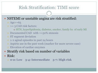
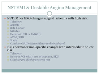
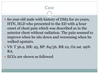
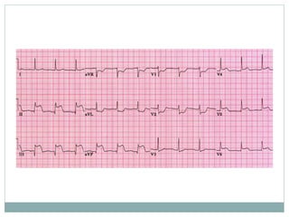
Acute Coronary Syndrome
MINI LECTURE
KELVIN NGUYEN
OBJECTIVES
 Definition of ACS
 UA, NSTEMI, and STEMI
 Risk stratification in NSTEMI
 Management
Acute Coronary Syndrome
Definition: a constellation of symptoms related to
obstruction of coronary arteries with chest pain being the
most common symptom in addition to nausea, vomiting,
diaphoresis etc.
Chest pain concerned for ACS is often radiating to the left
arm or angle of the jaw, pressure-like in character, and
associated with nausea and sweating. Chest pain is often
categorized into typical and atypical angina.
Acute coronary syndrome
 Based on ECG and cardiac enzymes, ACS is classified
into:
 STEMI: ST elevation, elevated cardiac enzymes
 NSTEMI: ST depression, T-wave inversion, elevated cardiac
enzymes
 Unstable Angina: Non specific EKG changes, normal
cardiac enzymes
Unstable Angina
 Occurs at rest and prolonged, usually lasting >20
minutes
 New onset angina that limits activity
 Increasing angina: Pain that occurs more frequently,
lasts longer periods or is increasingly limiting the
patients activity
EKG
 STEMI:
 Q waves , ST elevations, hyper acute T waves; followed by T wave
inversions.
 Clinically significant ST segment elevations:
 > than 1 mm (0.1 mV) in at least two anatomical contiguous leads
 or 2 mm (0.2 mV) in two contiguous precordial leads (V2 and V3)
 Note: LBBB and pacemakers can interfere with diagnosis of MI on
EKG
EKG
 NSTEMI:
 ST depressions (0.5 mm at least) or T wave inversions ( 1.0
mm at least) without Q waves in 2 contiguous leads with
prominent R wave or R/S ratio >1.
 Isolated T wave inversions:
 can correlate with increased risk for MI
 may represent Wellen’s syndrome:
 critical LAD stenosis
 >2mm inversions in anterior precordial leads
 Unstable Angina:
 May present with nonspecific or transient ST segment
depressions or elevations
Cardiac Enzymes
 Troponin is primarily used for diagnosing MI
because it has good sensitivity and specificity.
 CK-MB is more useful in certain situations such as post
reperfusion MI or if troponin test is not available
 Other conditions can cause elevation in troponin
such as renal failure or heart failure
 The increasing troponin trend is the important thing
to look for in diagnosing MI. Order Troponin
together with ECG when doing serial testing to rule
out ACS.
Risk Stratification: TIMI score
 NSTEMI or unstable angina are risk stratified:
 Age>=65
 >= 3 CAD risk factors:
 HTN, hyperlipidemia, diabetes, smoker, family hx of early MI
 Documented CAD with >=50% stenosis
 ST segment deviation
 ≥ 2 aginal episodes in past 24 hours
 Aspirin use in the past week (marker for more severe case)
 Elevation of cardiac enzymes
 Stratify risk based on number of variables
 Risk:
 0-2: Low 3-4: Intermediate 5-7: High risk
NSTEMI & Unstable Angina Management
 NSTEMI or EKG changes suggest ischemia with high risk:
 Telemetry
 Aspirin
 Beta blocker
 Nitrates
 Heparin (UFH or LMWH)
 ACE-I/ARB
 Statin
 Consider GP IIb/IIIa inhibitor and clopidogrel
 EKG normal or non-specific changes with intermediate or low
risk:
 Telemetry
 Rule out ACS with 3 sets of troponin, EKG
 Consider pre-discharge stress test
STEMI Management
 STEMI patients usually go straight to the cath lab from
the ED. Goal: door to balloon 90 minutes.
 Initial management for STEMI:
 Cardiac monitor
 Supplemental O2
 Nitrates*
 Beta blocker
 Morphine
 Clopidogrel
 Aspirin
 Good IV access
 Call cardiology fellow!
Case
 60 year old male with history of DM2 for 20 years,
HTN, HLD who presented to the ED with 4 hour
onset of chest pain which was described as in the
anterior chest without radiation. The pain seemed to
improve when he sits down and worsening when he
walked upstairs.
 VS: T 36.9, HR: 95, BP: 84/56, RR 22, O2 sat. 99%
RA.
 ECGs are shown as followed
 What will you do?
 What’s your diagnosis?
 What should be done now?
References
 2005 American Heart Association Guidelines for Cardiopulmonary Resuscitation and Emergency
Cardiovascular Care. Circulation 2005;112:IV-89-IV-110
 2013 ACCF/AHA Guideline for the Management of ST-Elevation Myocardial Infarction : A Report of
the American College of Cardiology Foundation/American Heart Association Task Force on Practice
Guidelines. Circulation 2013, epublished April 29th 2013 and print published june 4th 2013.
 Herman LK, et al. Comparison of frequency of inducible myocardial ischemia in patients presenting to
emergency department with typical versus atypical or nonanginal chest pain. Am J Cardiol. 2010
105:1561-4.
 www.uptodate.com:
 Overview of the acute management of unstable angina and acute non-ST elevation myocardial infarction
 Initial evaluation and management of suspected acute coronary syndrome in the emergency department
 Criteria for the diagnosis of acute myocardial infarction

acs.ppt

  • 1.
    Acute Coronary Syndrome MINILECTURE KELVIN NGUYEN
  • 2.
    OBJECTIVES  Definition ofACS  UA, NSTEMI, and STEMI  Risk stratification in NSTEMI  Management
  • 3.
    Acute Coronary Syndrome Definition:a constellation of symptoms related to obstruction of coronary arteries with chest pain being the most common symptom in addition to nausea, vomiting, diaphoresis etc. Chest pain concerned for ACS is often radiating to the left arm or angle of the jaw, pressure-like in character, and associated with nausea and sweating. Chest pain is often categorized into typical and atypical angina.
  • 4.
    Acute coronary syndrome Based on ECG and cardiac enzymes, ACS is classified into:  STEMI: ST elevation, elevated cardiac enzymes  NSTEMI: ST depression, T-wave inversion, elevated cardiac enzymes  Unstable Angina: Non specific EKG changes, normal cardiac enzymes
  • 5.
    Unstable Angina  Occursat rest and prolonged, usually lasting >20 minutes  New onset angina that limits activity  Increasing angina: Pain that occurs more frequently, lasts longer periods or is increasingly limiting the patients activity
  • 6.
    EKG  STEMI:  Qwaves , ST elevations, hyper acute T waves; followed by T wave inversions.  Clinically significant ST segment elevations:  > than 1 mm (0.1 mV) in at least two anatomical contiguous leads  or 2 mm (0.2 mV) in two contiguous precordial leads (V2 and V3)  Note: LBBB and pacemakers can interfere with diagnosis of MI on EKG
  • 7.
    EKG  NSTEMI:  STdepressions (0.5 mm at least) or T wave inversions ( 1.0 mm at least) without Q waves in 2 contiguous leads with prominent R wave or R/S ratio >1.  Isolated T wave inversions:  can correlate with increased risk for MI  may represent Wellen’s syndrome:  critical LAD stenosis  >2mm inversions in anterior precordial leads  Unstable Angina:  May present with nonspecific or transient ST segment depressions or elevations
  • 8.
    Cardiac Enzymes  Troponinis primarily used for diagnosing MI because it has good sensitivity and specificity.  CK-MB is more useful in certain situations such as post reperfusion MI or if troponin test is not available  Other conditions can cause elevation in troponin such as renal failure or heart failure  The increasing troponin trend is the important thing to look for in diagnosing MI. Order Troponin together with ECG when doing serial testing to rule out ACS.
  • 9.
    Risk Stratification: TIMIscore  NSTEMI or unstable angina are risk stratified:  Age>=65  >= 3 CAD risk factors:  HTN, hyperlipidemia, diabetes, smoker, family hx of early MI  Documented CAD with >=50% stenosis  ST segment deviation  ≥ 2 aginal episodes in past 24 hours  Aspirin use in the past week (marker for more severe case)  Elevation of cardiac enzymes  Stratify risk based on number of variables  Risk:  0-2: Low 3-4: Intermediate 5-7: High risk
  • 10.
    NSTEMI & UnstableAngina Management  NSTEMI or EKG changes suggest ischemia with high risk:  Telemetry  Aspirin  Beta blocker  Nitrates  Heparin (UFH or LMWH)  ACE-I/ARB  Statin  Consider GP IIb/IIIa inhibitor and clopidogrel  EKG normal or non-specific changes with intermediate or low risk:  Telemetry  Rule out ACS with 3 sets of troponin, EKG  Consider pre-discharge stress test
  • 11.
    STEMI Management  STEMIpatients usually go straight to the cath lab from the ED. Goal: door to balloon 90 minutes.  Initial management for STEMI:  Cardiac monitor  Supplemental O2  Nitrates*  Beta blocker  Morphine  Clopidogrel  Aspirin  Good IV access  Call cardiology fellow!
  • 12.
    Case  60 yearold male with history of DM2 for 20 years, HTN, HLD who presented to the ED with 4 hour onset of chest pain which was described as in the anterior chest without radiation. The pain seemed to improve when he sits down and worsening when he walked upstairs.  VS: T 36.9, HR: 95, BP: 84/56, RR 22, O2 sat. 99% RA.  ECGs are shown as followed
  • 15.
     What willyou do?  What’s your diagnosis?  What should be done now?
  • 16.
    References  2005 AmericanHeart Association Guidelines for Cardiopulmonary Resuscitation and Emergency Cardiovascular Care. Circulation 2005;112:IV-89-IV-110  2013 ACCF/AHA Guideline for the Management of ST-Elevation Myocardial Infarction : A Report of the American College of Cardiology Foundation/American Heart Association Task Force on Practice Guidelines. Circulation 2013, epublished April 29th 2013 and print published june 4th 2013.  Herman LK, et al. Comparison of frequency of inducible myocardial ischemia in patients presenting to emergency department with typical versus atypical or nonanginal chest pain. Am J Cardiol. 2010 105:1561-4.  www.uptodate.com:  Overview of the acute management of unstable angina and acute non-ST elevation myocardial infarction  Initial evaluation and management of suspected acute coronary syndrome in the emergency department  Criteria for the diagnosis of acute myocardial infarction

Editor's Notes

  • #4 Typical chest pain: met 3/3 criteria v.s. atypical chest pain, only met 2/3 criteria. 3 criteria are: 1. the presence of substernal chest pain or (2) discomfort that was provoked by exertion or emotional stress and (3) was relieved by rest and/or nitroglycerin.
  • #5 There is a subset of Q-wave v.s. non Q-wave MI (can fall under either NSTEMI or STEMI). Patients with nonQwave MI seem to have a better prognosis.
  • #7 Remember 50% of patients with history of LBBB do not present with chest pain in addition to difficult ECG interpretation  patient with LBBB is difficult to diagnose and manage and should have low threshold for acute MI in these patients.
  • #10 If intermediate risk or high risk, consider to be aggressive with treatments ,i.e. start heparin gtt early, admitting the patient to the CCU and contact cardiology ASAP.
  • #12 What will be contraindicated if the patient shows ST elevation in II, III, avF? Answer: not to give nitrate as there is a concern for right ventricular infarction and this situation is pre-load dependent. What happens if cath is not available at your hospital?
  • #14 Leads II, III and aVF reflect electrocardiogram changes associated with acute infarction of the inferior aspect of the heart. ST elevation, developing Q waves and T wave inversion may all be present depending on the timing of the ECG relative to the onset of myocardial infarction. Most frequently, inferior MI results from occlusion of the right coronary artery. Conduction abnormalities which may alert the physician to patients at risk include second degree AV block and complete heart block together with junctional escape beats. Note that the patient is also suffering from a concurrent posterior wall infarction as eveidenced by ST depression in leads V1 and V2.
  • #15 What is this ECG? Answer: right sided ECG. Electrocardiogram shows Q waves and prominent doming ST segment elevation in II, III, and aVF, findings which are characteristic of an acute inferior myocardial infarction. ST elevation in the right precordial leads - V4R, V5R, and V6R - indicates right ventricular involvement as well (arrows). The ST depressions in leads I and aVL represent reciprocal changes.
  • #16 ACITVATE STEMI code, call cardiology for emergent cath and ask which medications given to the patient before cath, draw troponin, IV fluid, O2, morphine, Aspirin 325 mg x1, and plavix 1 dose of 300 mg PO but NO nitrate!