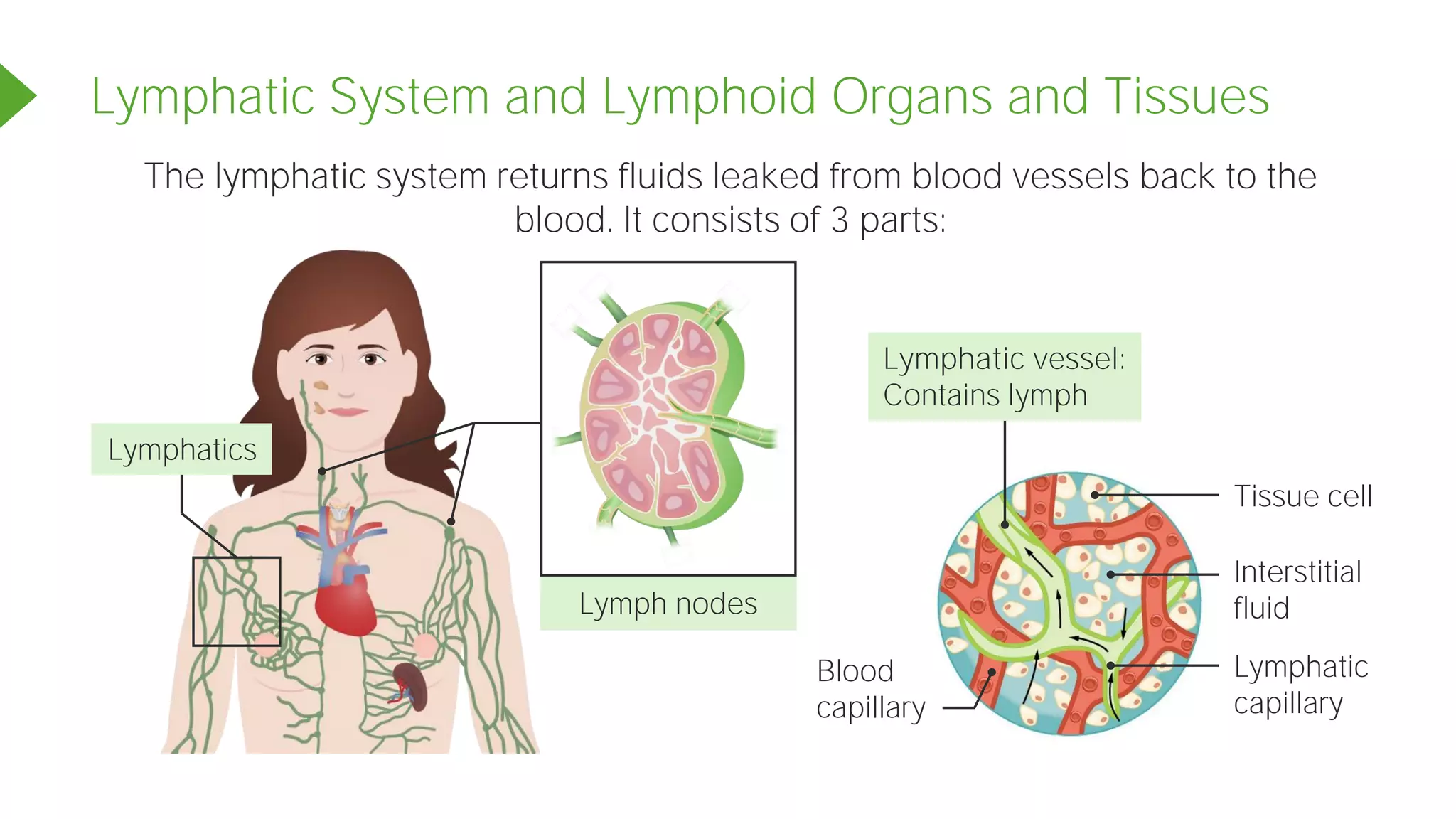
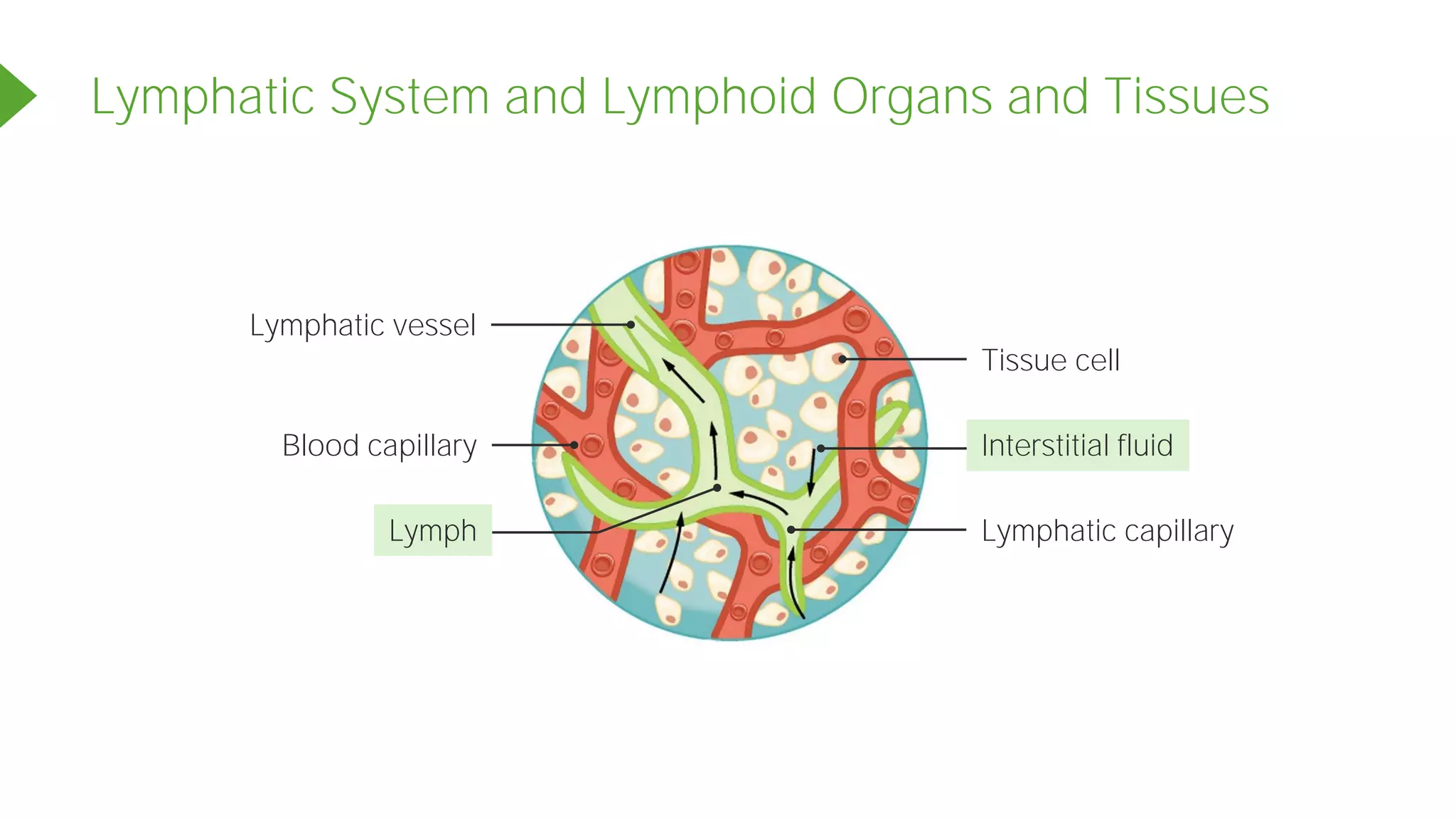
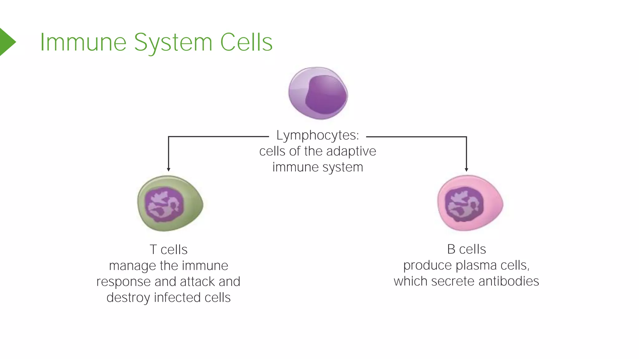
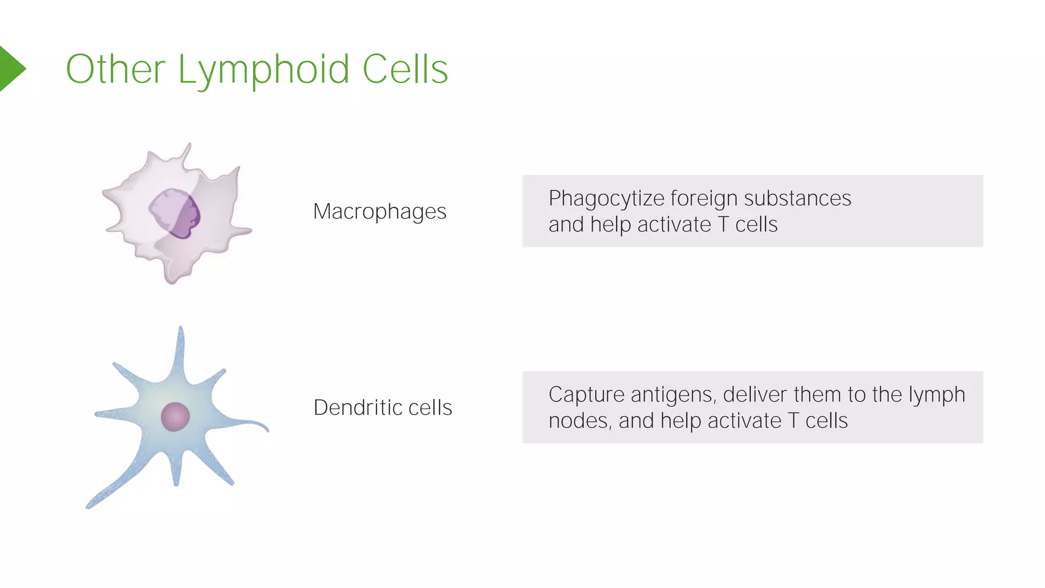
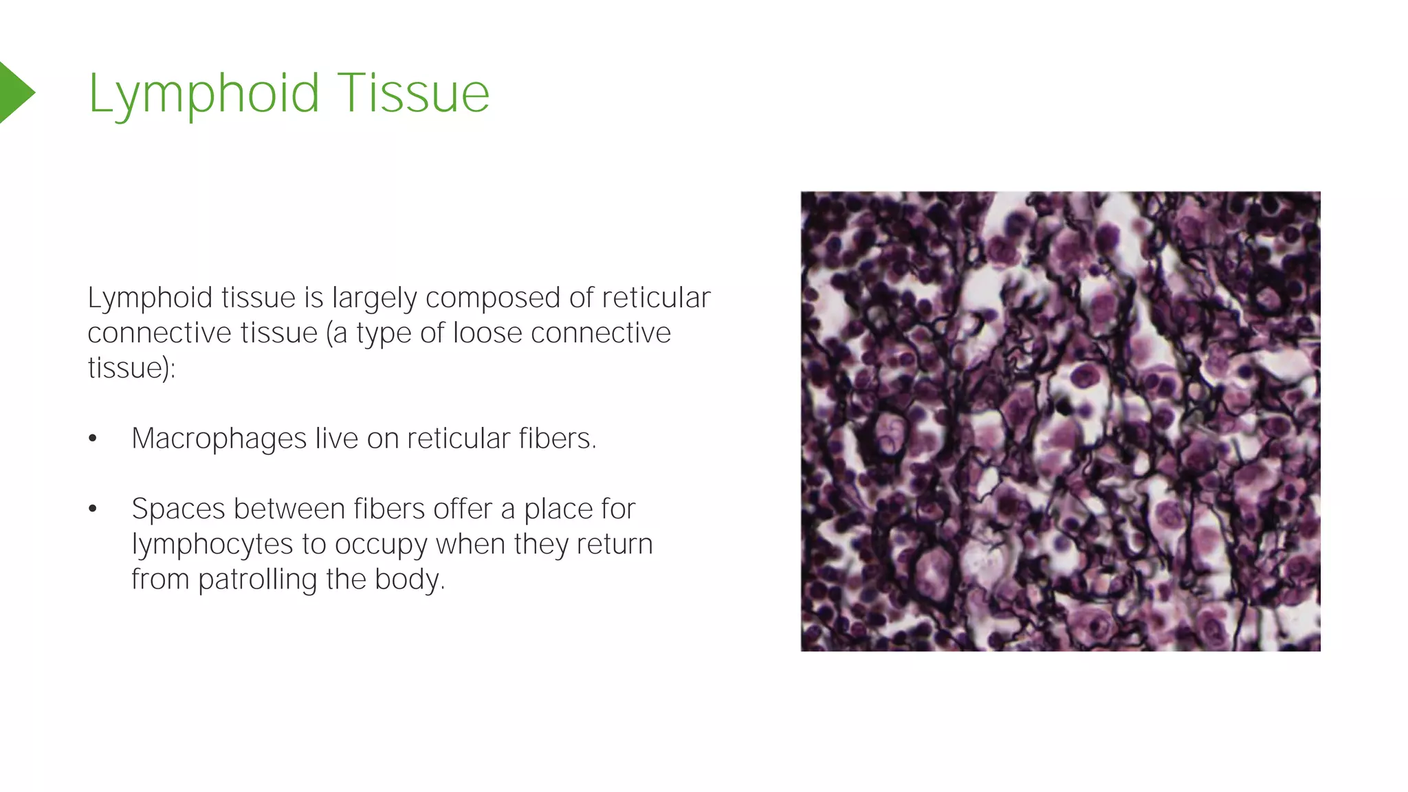
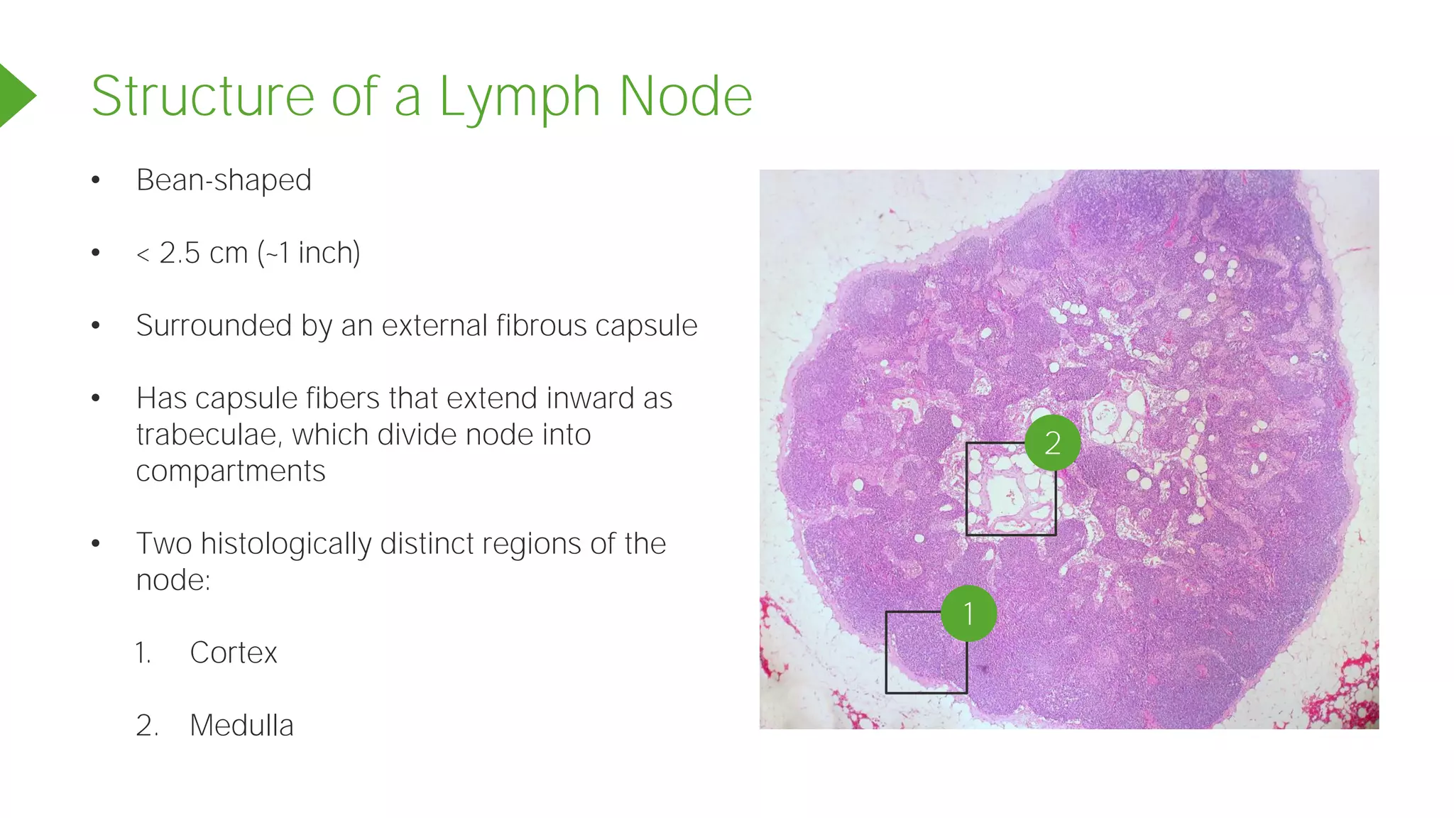
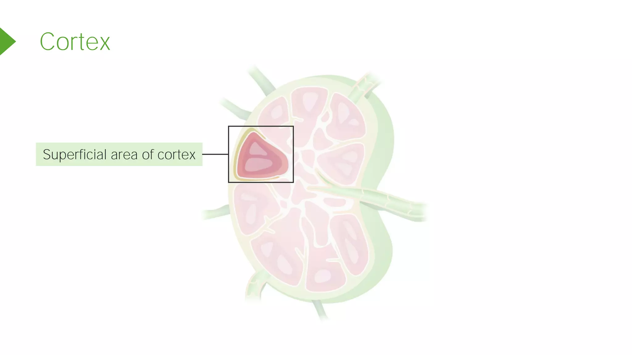
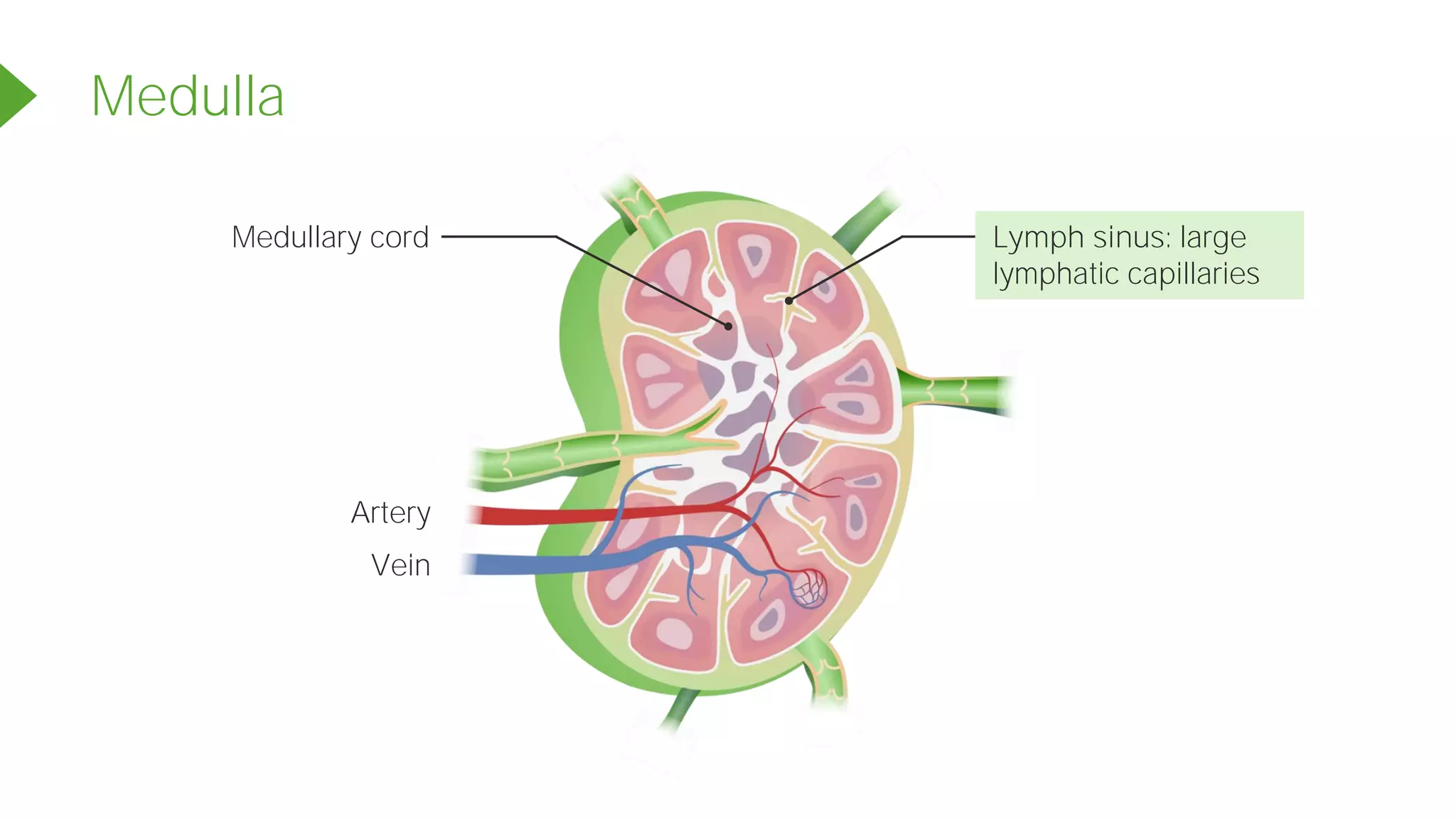
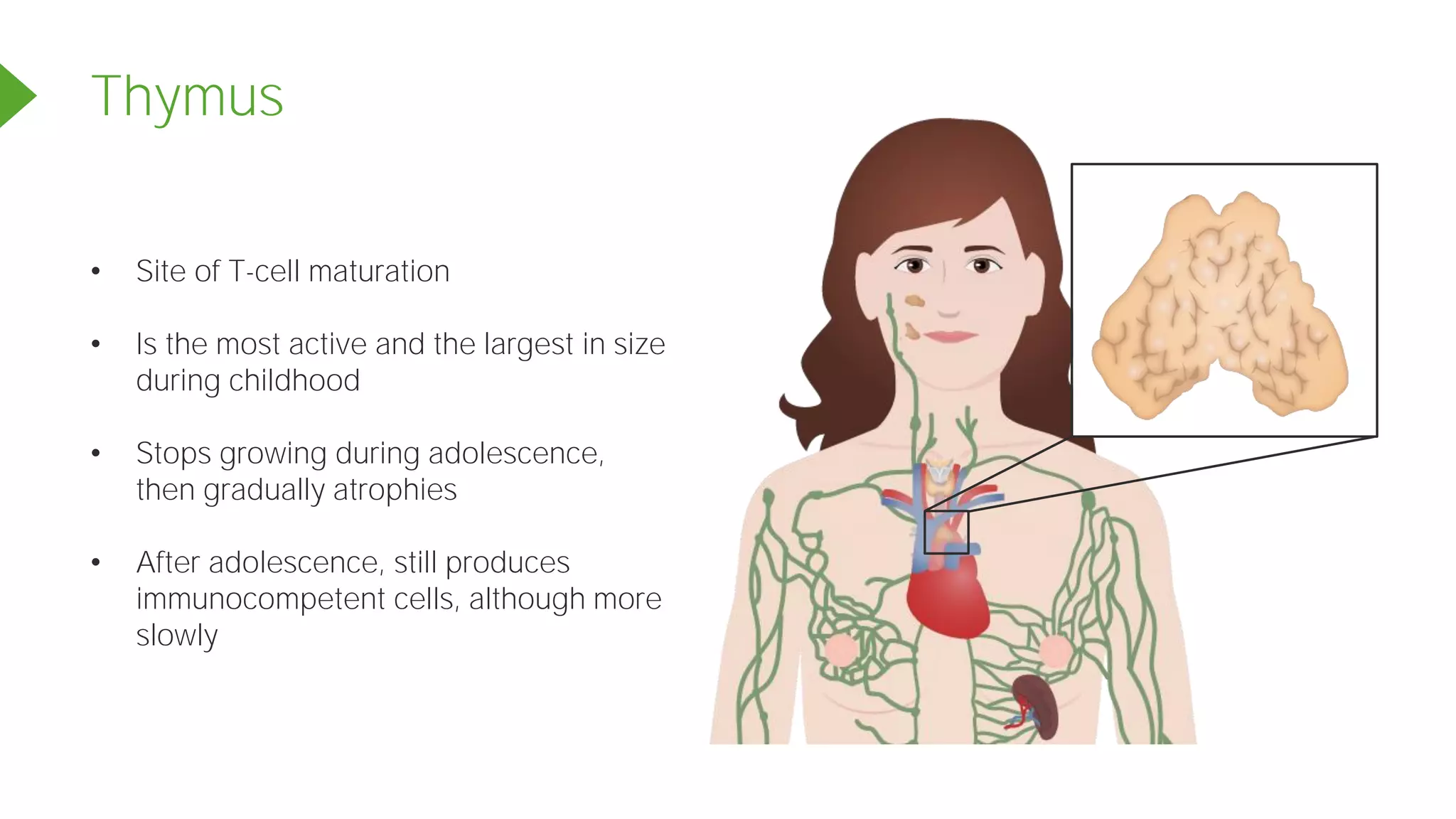
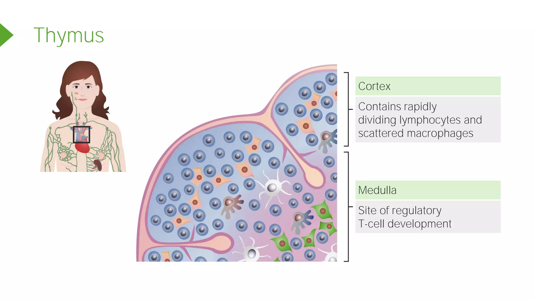
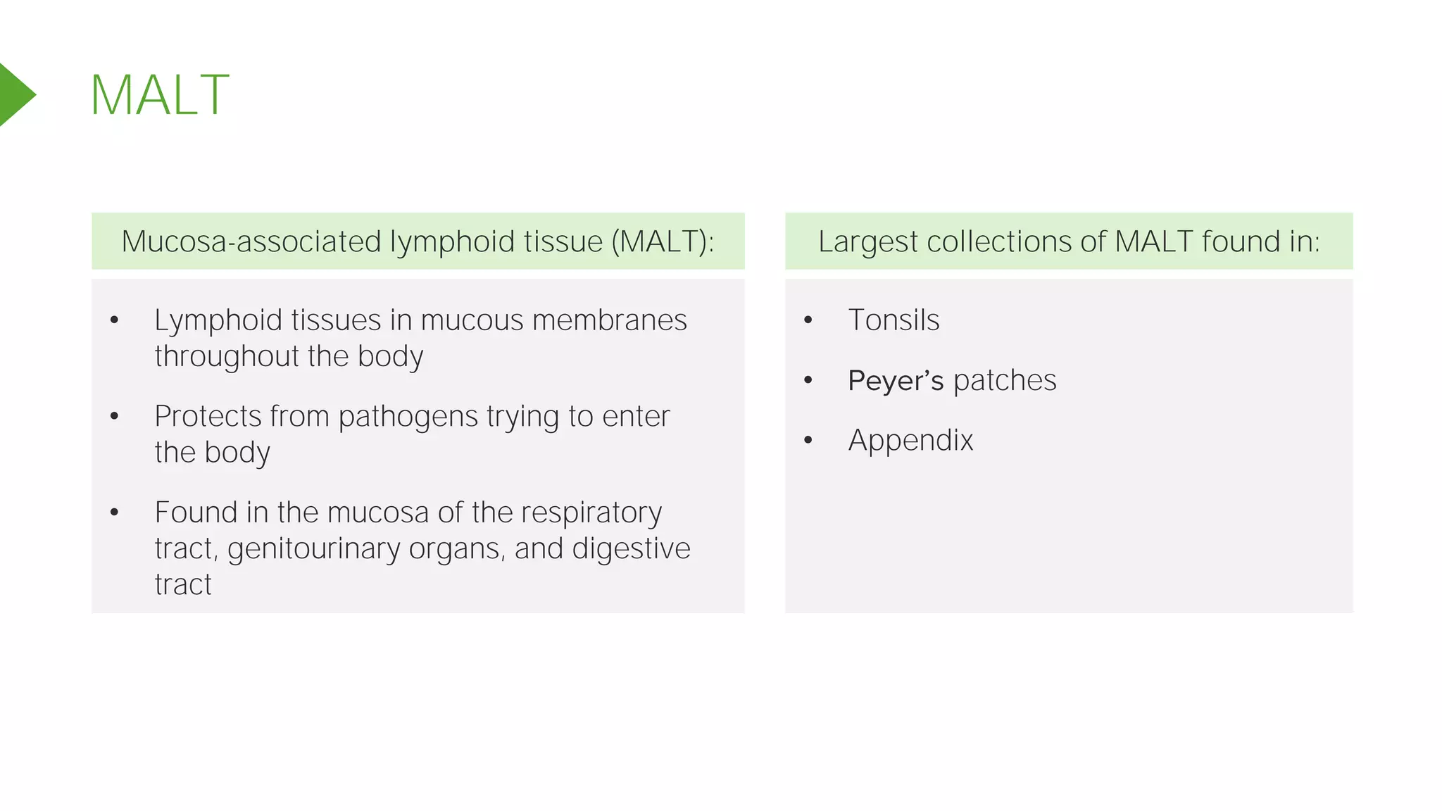
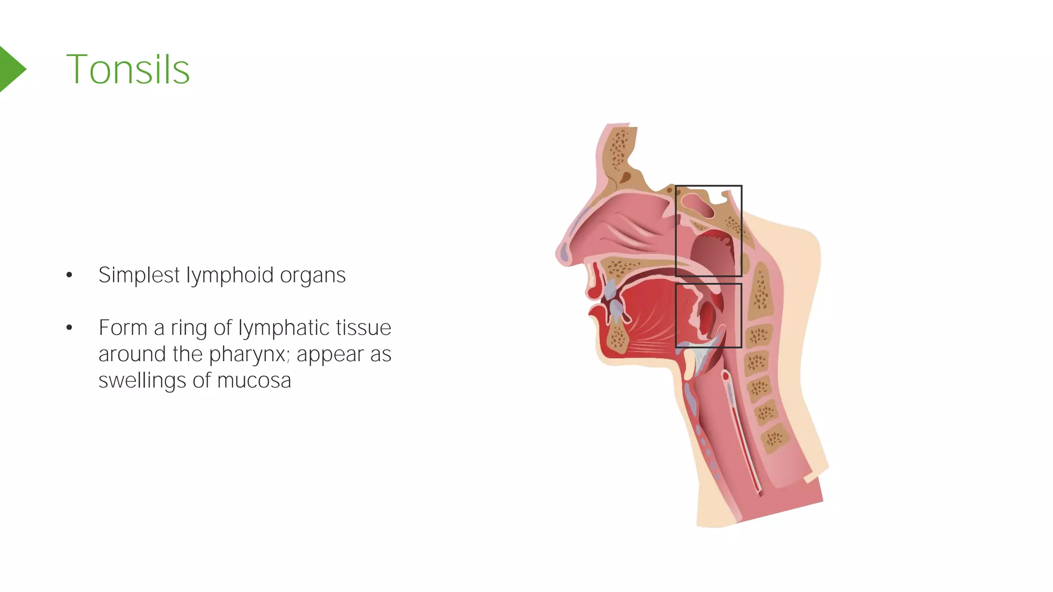
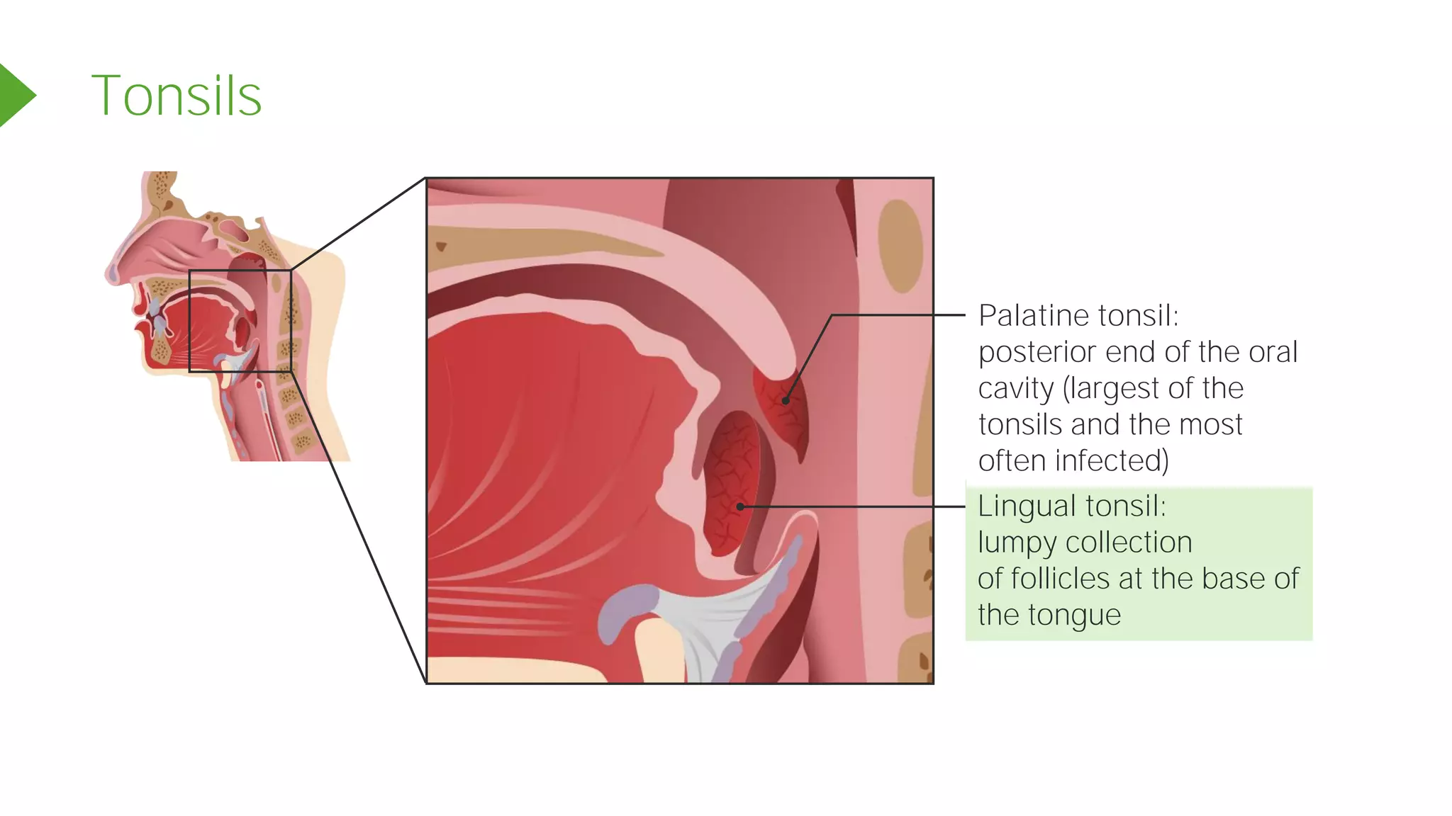
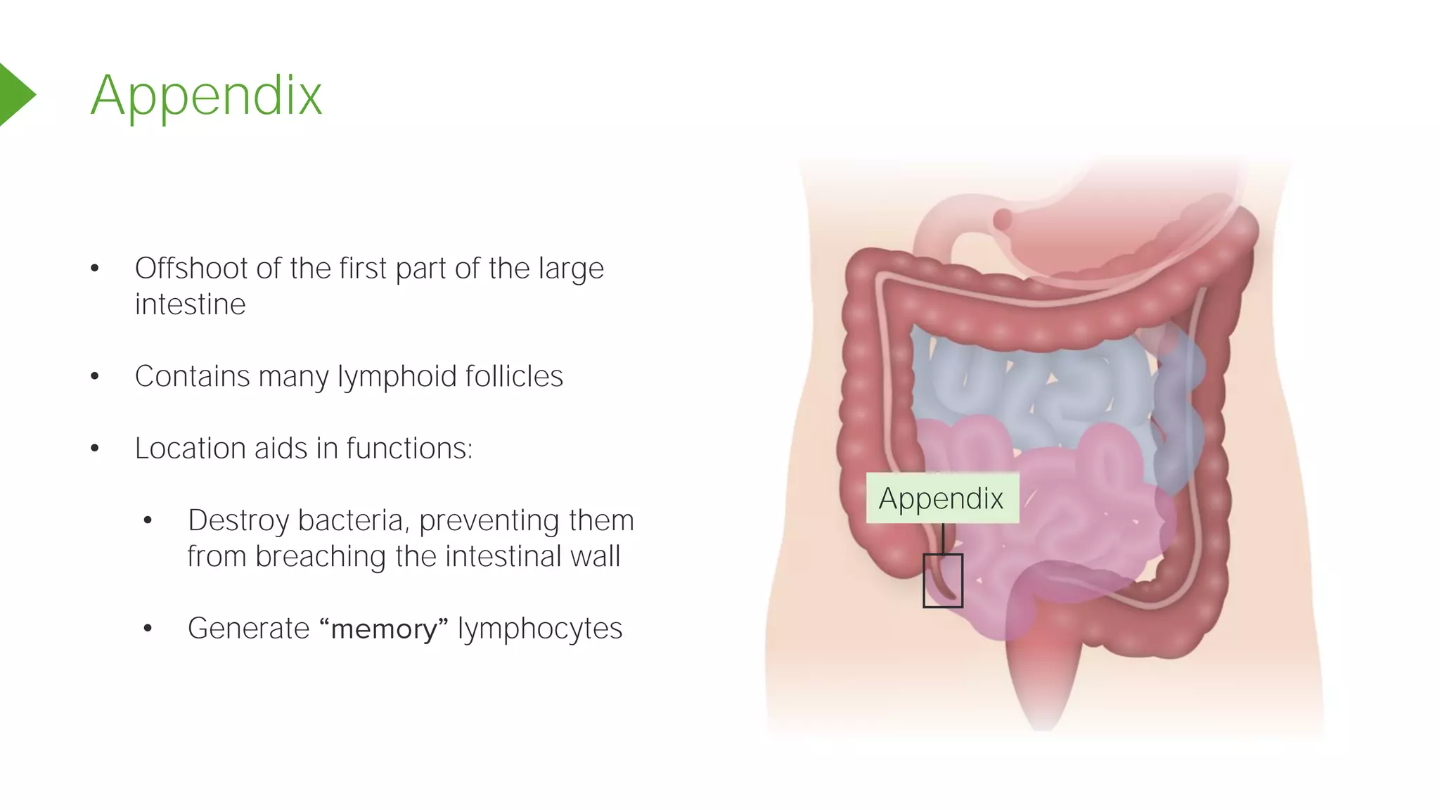
Lymphatic System
Principles of Anatomy and Physiology
The lymphatic system returns fluids leaked from blood vessels back to the
blood. It consists of 3 parts:
Lymphatics
Lymphatic vessel:
Contains lymph
Blood
capillary
Tissue cell
Interstitial
fluid
Lymphatic
capillary
Lymph nodes
Lymphatic System and Lymphoid Organs and Tissues
Blood capillary
Tissue cell
Interstitial fluid
Lymphatic capillary
Lymphatic vessel
Lymph
Lymphatic System and Lymphoid Organs and Tissues
Distribution and Structure of Lymphatic Vessels
Lymph capillary
Larger lymph
vessel
• Absent from bones, teeth, bone marrow,
and the CNS
• More permeable than blood capillaries:
• Can take up larger molecules and
particles that blood capillaries cannot
• Can act as a route for pathogens or
cancer cells to travel throughout the
body
Distribution and Special Features
Intimate association between blood capillaries and lymph capillaries
Blood capillary
Lymph capillary
Distribution and Structure of Lymphatic Vessels
Lymph vessel endothelial cells
Lymphatic capillary
• Anchored to the matrix
by collagen filaments
• Increase in extracellular
fluid (ECF) causes mini
valves to open
• Decrease in ECF causes
mini valves to close
Backflow prevention valve
Lymphatic Vessels in the GI Tract
Lacteal:
absorb digested fat
and deliver fatty lymph
(chyle) to the blood
• The larger lymphatic vessels have
structures and tunics similar
to those of the veins,
• walls, with more
internal valves.
• more
frequently.
• Collecting vessels in the skin
travel with the superficial veins,
but deep vessels travel with
arteries.
Larger Lymphatic Vessels
Lymphatic
collecting
vessels
Lymph node
Lymph duct
Lymph
trunk
Lymphatic Trunks
Formed by union of the largest collecting vessels to drain large areas of the body
Right lumbar trunk Left lumbar trunk
Right bronchomediastinal
trunk
Left bronchomediastinal
trunk
Right subclavian trunk Left subclavian trunk
Right jugular trunk Left jugular trunk
Intestinal trunk
Larger Lymphatic Vessels
Right lymphatic
duct
Drained by thoracic duct
Drained by right lymphatic duct
Thoracic duct
Cysterna chyli
of thoracic duct
Right subclavian
vein
Left subclavian
vein
Right internal jugular vein Left internal jugular vein
The lymph system is a low-pressure system like the venous system.
Lymph Transport
Lymph Transport
Milking action of skeletal muscle
Pressure changes in the thorax during breathing
Valves to prevent backflow
Pulsations of nearby arteries
Contractions of smooth muscles in walls of the lymphatics
Mechanisms
Physical activity increases the flow of lymph.
Immobilization of the area keeps needed
inflammatory material in the area for faster
healing.
Lymph Transport
Lymphatic System and Lymphoid Organs and Tissues
Lymphoid organs and tissues provide the structural basis of the immune
system by housing phagocytic cells and lymphocytes.
Tonsils
Thymus
Spleen
Lymph nodes
Lymphoid cells consist of immune system cells
found in lymphoid tissue and supporting cells
that form lymphoid tissue structures.
Lymphoid Cells, Tissues, and Organs
Protect against antigens
Examples: bacteria, toxins, viruses,
mismatched RBCs, cancer cells
Immune System Cells
Lymphocytes:
cells of the adaptive
immune system
T cells B cells
Immune System Cells
Lymphocytes:
cells of the adaptive
immune system
B cells
produce plasma cells,
which secrete antibodies
T cells
manage the immune
response and attack and
destroy infected cells
Other Lymphoid Cells
Macrophages
Phagocytize foreign substances
and help activate T cells
Dendritic cells
Capture antigens, deliver them to the lymph
nodes, and help activate T cells
Supporting Lymphoid Cells
Reticular
fibers
Reticular
cells
Stroma: network-like support that acts as scaffolding for
immune cells
Lymphoid Tissue
Houses and provides proliferation
sites for lymphocytes
Offers surveillance vantage
points for lymphocytes
and macrophages as they
filter through the lymph
Main functions of the lymphoid tissue
Lymphoid Tissue
Lymphoid tissue is largely composed of reticular
connective tissue (a type of loose connective
tissue):
• Macrophages live on reticular fibers.
• Spaces between fibers offer a place for
lymphocytes to occupy when they return
from patrolling the body.
2 Types of Tissue: Diffuse Lymphoid Tissue
• Loose arrangement of lymphoid
cells and some reticular fibers
• Found in virtually every organ of
the body
• Larger collections found in the
lamina propria of the mucous
membranes
2 Types of Tissue: Lymphoid Follicles (Nodules)
• Solid, spherical bodies consisting
of tightly packed lymphoid cells
and reticular fibers
• Isolated
patches and in the appendix
Germinal centers of
proliferating B cells
Nodules
Lymphoid Organs 2 Functional Categories
Primary lymphoid organs
• Areas where T and B cells
mature red bone marrow
and thymus
• T and B cells: originate in the bone
marrow, but only B cells mature
there; T cells mature in the thymus
Secondary lymphoid organs
• Areas where mature lymphocytes
first encounter their antigen and
become activated
• Nodes, spleen, mucosa-associated
lymphoid tissue (MALT), and
diffuse lymphoid tissues
Thymus
Tonsils
Spleen
Peyer s patches
Appendix
Lymph nodes
Lymph Nodes 2 Main Functions
Primary lymphoid organs
Secondary lymphoid organs
Lymph Nodes
• Lymph nodes are the principal secondary
lymphoid organs of the body.
• Most nodes are embedded deep in the
connective tissue.
• Some are closer to the body surface
(inguinal, axillary, and cervical regions).
Here, collecting vessels converge into
trunks.
Lymph Nodes 2 Main Functions
Cleansing the lymph
• Lymph nodes act as lymph
• Macrophages remove and destroy
microorganisms and debris that
enter the lymph.
• Lymph nodes prevent unwanted
substances from being delivered to
the blood.
Immune system activation
• Lymph nodes offer a place for
lymphocytes to become activated.
• They mount an attack against
antigens.
• Bean-shaped
• < 2.5 cm (~1 inch)
• Surrounded by an external fibrous capsule
• Has capsule fibers that extend inward as
trabeculae, which divide node into
compartments
• Two histologically distinct regions of the
node:
1. Cortex
2. Medulla
Structure of a Lymph Node
2
1
Cortex
Superficial area of cortex
Cortex
T cells
Cortex
Lymphoid nodule
Germinal centers:
Contain dividing B cells
Dendritic cells (DC):
Associated with T- and
B-cell activation
DC present the
antigen to the
B cell
Plasma cell
Memory cell
Cortex
T cell proliferates
DC present the antigen to the T cell
Medulla
Medullary cord: contains
macrophages and antibody-
producing plasma cells
Artery
Vein
Medulla
Lymph sinus: large
lymphatic capillaries
Medullary cord
Artery
Vein
Medullary cord
Artery
Vein
Circulation in the Lymph Nodes
Afferent lymphatic vessel
Subcapsular sinus
Efferent lymphatic vessel
Lymph sinus
Medullary sinus
Hilum
Artery
Vein
Circulation in the Lymph Nodes
Afferent lymphatic vessel
Efferent lymphatic vessel
Afferent lymphatic vessel Afferent lymphatic vessel
Hilum
Spleen
Splenic
vein
Splenic
artery
Hilum
Spleen
• Blood-rich organ
about the size of
a fist
• Is the largest
lymphoid organ
• Site of lymphocyte proliferation and immune
surveillance and response
• Cleanses the blood of aged blood cells and
platelets
• Macrophages: remove debris
Spleen Functions
Spleen
Spleen
Liver
Spleen 3 Additional Functions
Stores breakdown products
of RBCs (e.g., iron) for
later reuse
1
Stores blood platelets and
monocytes for release into
the blood when needed
2
May be the site of fetal
erythrocyte production
3
Spleen
Red pulp
Trabecula
Venule
Arteriole
White pulp
Spleen
White pulp
• Site where immune function occurs
• Contains mostly lymphocytes
on reticular fibers
• Clusters around the
central arteries
Red pulp
• Site where old blood cells and
bloodborne pathogens are destroyed
• Rich in RBCs and macrophages
that engulf them
• Composed of splenic cords
(reticular tissue) that separate blood-
filled splenic sinusoids (venous sinuses)
Thymus
• Bilobed lymphoid organ
found in the inferior neck
• Extends into the mediastinum
• Partially overlies the heart
Thymus:
• Site of T-cell maturation
• Is the most active and the largest in size
during childhood
• Stops growing during adolescence,
then gradually atrophies
• After adolescence, still produces
immunocompetent cells, although more
slowly
Thymus
Thymus
Contains rapidly
dividing lymphocytes and
scattered macrophages
Cortex
Site of regulatory
T-cell development
Medulla
Thymus Differences from Other Lymphoid Organs
Has no lymphoid follicles
1
Stroma:
3
• Made up of epithelial cells
• Provide an environment in which T lymphocytes become immunocompetent
Does not directly fight antigens
2
• Functions strictly in T-lymphocyte maturation
• Contains the blood-thymus barrier keeps immature T lymphocytes isolated
from any antigens to prevent premature activation
Lymphoid Nodules
MALT Tonsils
Appendix
• Lymphoid tissues in mucous membranes
throughout the body
• Protects from pathogens trying to enter
the body
• Found in the mucosa of the respiratory
tract, genitourinary organs, and digestive
tract
• Tonsils
• patches
• Appendix
Mucosa-associated lymphoid tissue (MALT): Largest collections of MALT found in:
MALT
• Simplest lymphoid organs
• Form a ring of lymphatic tissue
around the pharynx; appear as
swellings of mucosa
Tonsils
Tonsils
Lingual tonsil:
lumpy collection
of follicles at the base of
the tongue
Palatine tonsil:
posterior end of the oral
cavity (largest of the
tonsils and the most
often infected)
Tonsils
Tubal tonsil:
posterior end of
the oral cavity
Pharyngeal tonsil:
also called adenoids
• Tonsils' function: gather and
remove pathogens in food or air
• Contain follicles with germinal
centers and scattered lymphocytes
• Not fully encapsulated
Tonsils
Germinal
centers
Stratified
squamous
epithelium
Crypt
• The overlying epithelium
invaginates, forming tonsillar crypts.
• Bacteria or particulate matter enters
crypts, where they are trapped and
destroyed.
Tonsils
Germinal
centers
Stratified
squamous
epithelium
Crypt
Peyer s
patches
• Clusters of lymphoid follicles in the wall
of the distal portion of the small
intestine
• Also called aggregated lymphoid
nodules
• Structurally similar to tonsils
• Functions:
• Destroy bacteria
• Generate memory lymphocytes
Appendix
Appendix
• Offshoot of the first part of the large
intestine
• Contains many lymphoid follicles
• Location aids in functions:
• Destroy bacteria, preventing them
from breaching the intestinal wall
• Generate lymphocytes
In a Nutshell
The lymphatic system comprises the
lymphatic vessels, lymph, and lymph tissues
and organs, as well as immune cells.
Its functions are to collect, filter, and clean
interstitial fluid, and then return it to the
blood.
Like the veins of the circulatory system, the
lymphatic vessels are a low-pressure system.
The lymph organs play an integral role in the
body s immune response to antigens.

14.0 The Lymphatic System.pdf

  • 1.
    Lymphatic System Principles ofAnatomy and Physiology
  • 2.
    The lymphatic systemreturns fluids leaked from blood vessels back to the blood. It consists of 3 parts: Lymphatics Lymphatic vessel: Contains lymph Blood capillary Tissue cell Interstitial fluid Lymphatic capillary Lymph nodes Lymphatic System and Lymphoid Organs and Tissues
  • 3.
    Blood capillary Tissue cell Interstitialfluid Lymphatic capillary Lymphatic vessel Lymph Lymphatic System and Lymphoid Organs and Tissues
  • 4.
    Distribution and Structureof Lymphatic Vessels Lymph capillary Larger lymph vessel • Absent from bones, teeth, bone marrow, and the CNS • More permeable than blood capillaries: • Can take up larger molecules and particles that blood capillaries cannot • Can act as a route for pathogens or cancer cells to travel throughout the body
  • 5.
    Distribution and SpecialFeatures Intimate association between blood capillaries and lymph capillaries Blood capillary Lymph capillary
  • 6.
    Distribution and Structureof Lymphatic Vessels Lymph vessel endothelial cells Lymphatic capillary • Anchored to the matrix by collagen filaments • Increase in extracellular fluid (ECF) causes mini valves to open • Decrease in ECF causes mini valves to close Backflow prevention valve
  • 7.
    Lymphatic Vessels inthe GI Tract Lacteal: absorb digested fat and deliver fatty lymph (chyle) to the blood
  • 8.
    • The largerlymphatic vessels have structures and tunics similar to those of the veins, • walls, with more internal valves. • more frequently. • Collecting vessels in the skin travel with the superficial veins, but deep vessels travel with arteries. Larger Lymphatic Vessels Lymphatic collecting vessels Lymph node Lymph duct Lymph trunk
  • 9.
    Lymphatic Trunks Formed byunion of the largest collecting vessels to drain large areas of the body Right lumbar trunk Left lumbar trunk Right bronchomediastinal trunk Left bronchomediastinal trunk Right subclavian trunk Left subclavian trunk Right jugular trunk Left jugular trunk Intestinal trunk
  • 10.
    Larger Lymphatic Vessels Rightlymphatic duct Drained by thoracic duct Drained by right lymphatic duct Thoracic duct Cysterna chyli of thoracic duct Right subclavian vein Left subclavian vein Right internal jugular vein Left internal jugular vein
  • 11.
    The lymph systemis a low-pressure system like the venous system. Lymph Transport
  • 12.
    Lymph Transport Milking actionof skeletal muscle Pressure changes in the thorax during breathing Valves to prevent backflow Pulsations of nearby arteries Contractions of smooth muscles in walls of the lymphatics Mechanisms
  • 13.
    Physical activity increasesthe flow of lymph. Immobilization of the area keeps needed inflammatory material in the area for faster healing. Lymph Transport
  • 14.
    Lymphatic System andLymphoid Organs and Tissues Lymphoid organs and tissues provide the structural basis of the immune system by housing phagocytic cells and lymphocytes. Tonsils Thymus Spleen Lymph nodes
  • 15.
    Lymphoid cells consistof immune system cells found in lymphoid tissue and supporting cells that form lymphoid tissue structures. Lymphoid Cells, Tissues, and Organs
  • 16.
    Protect against antigens Examples:bacteria, toxins, viruses, mismatched RBCs, cancer cells Immune System Cells Lymphocytes: cells of the adaptive immune system T cells B cells
  • 17.
    Immune System Cells Lymphocytes: cellsof the adaptive immune system B cells produce plasma cells, which secrete antibodies T cells manage the immune response and attack and destroy infected cells
  • 18.
    Other Lymphoid Cells Macrophages Phagocytizeforeign substances and help activate T cells Dendritic cells Capture antigens, deliver them to the lymph nodes, and help activate T cells
  • 19.
    Supporting Lymphoid Cells Reticular fibers Reticular cells Stroma:network-like support that acts as scaffolding for immune cells
  • 20.
    Lymphoid Tissue Houses andprovides proliferation sites for lymphocytes Offers surveillance vantage points for lymphocytes and macrophages as they filter through the lymph Main functions of the lymphoid tissue
  • 21.
    Lymphoid Tissue Lymphoid tissueis largely composed of reticular connective tissue (a type of loose connective tissue): • Macrophages live on reticular fibers. • Spaces between fibers offer a place for lymphocytes to occupy when they return from patrolling the body.
  • 22.
    2 Types ofTissue: Diffuse Lymphoid Tissue • Loose arrangement of lymphoid cells and some reticular fibers • Found in virtually every organ of the body • Larger collections found in the lamina propria of the mucous membranes
  • 23.
    2 Types ofTissue: Lymphoid Follicles (Nodules) • Solid, spherical bodies consisting of tightly packed lymphoid cells and reticular fibers • Isolated patches and in the appendix Germinal centers of proliferating B cells Nodules
  • 24.
    Lymphoid Organs 2Functional Categories Primary lymphoid organs • Areas where T and B cells mature red bone marrow and thymus • T and B cells: originate in the bone marrow, but only B cells mature there; T cells mature in the thymus Secondary lymphoid organs • Areas where mature lymphocytes first encounter their antigen and become activated • Nodes, spleen, mucosa-associated lymphoid tissue (MALT), and diffuse lymphoid tissues
  • 25.
    Thymus Tonsils Spleen Peyer s patches Appendix Lymphnodes Lymph Nodes 2 Main Functions Primary lymphoid organs Secondary lymphoid organs
  • 26.
    Lymph Nodes • Lymphnodes are the principal secondary lymphoid organs of the body. • Most nodes are embedded deep in the connective tissue. • Some are closer to the body surface (inguinal, axillary, and cervical regions). Here, collecting vessels converge into trunks.
  • 27.
    Lymph Nodes 2Main Functions Cleansing the lymph • Lymph nodes act as lymph • Macrophages remove and destroy microorganisms and debris that enter the lymph. • Lymph nodes prevent unwanted substances from being delivered to the blood. Immune system activation • Lymph nodes offer a place for lymphocytes to become activated. • They mount an attack against antigens.
  • 28.
    • Bean-shaped • <2.5 cm (~1 inch) • Surrounded by an external fibrous capsule • Has capsule fibers that extend inward as trabeculae, which divide node into compartments • Two histologically distinct regions of the node: 1. Cortex 2. Medulla Structure of a Lymph Node 2 1
  • 29.
  • 30.
  • 31.
    Cortex Lymphoid nodule Germinal centers: Containdividing B cells Dendritic cells (DC): Associated with T- and B-cell activation DC present the antigen to the B cell Plasma cell Memory cell
  • 32.
    Cortex T cell proliferates DCpresent the antigen to the T cell
  • 33.
    Medulla Medullary cord: contains macrophagesand antibody- producing plasma cells Artery Vein
  • 34.
    Medulla Lymph sinus: large lymphaticcapillaries Medullary cord Artery Vein
  • 35.
    Medullary cord Artery Vein Circulation inthe Lymph Nodes Afferent lymphatic vessel Subcapsular sinus Efferent lymphatic vessel Lymph sinus Medullary sinus Hilum
  • 36.
    Artery Vein Circulation in theLymph Nodes Afferent lymphatic vessel Efferent lymphatic vessel Afferent lymphatic vessel Afferent lymphatic vessel Afferent lymphatic vessel Afferent lymphatic vessel Hilum
  • 37.
    Spleen Splenic vein Splenic artery Hilum Spleen • Blood-rich organ aboutthe size of a fist • Is the largest lymphoid organ
  • 38.
    • Site oflymphocyte proliferation and immune surveillance and response • Cleanses the blood of aged blood cells and platelets • Macrophages: remove debris Spleen Functions
  • 39.
  • 40.
    Spleen 3 AdditionalFunctions Stores breakdown products of RBCs (e.g., iron) for later reuse 1 Stores blood platelets and monocytes for release into the blood when needed 2 May be the site of fetal erythrocyte production 3
  • 41.
  • 42.
    Spleen White pulp • Sitewhere immune function occurs • Contains mostly lymphocytes on reticular fibers • Clusters around the central arteries Red pulp • Site where old blood cells and bloodborne pathogens are destroyed • Rich in RBCs and macrophages that engulf them • Composed of splenic cords (reticular tissue) that separate blood- filled splenic sinusoids (venous sinuses)
  • 43.
    Thymus • Bilobed lymphoidorgan found in the inferior neck • Extends into the mediastinum • Partially overlies the heart Thymus:
  • 44.
    • Site ofT-cell maturation • Is the most active and the largest in size during childhood • Stops growing during adolescence, then gradually atrophies • After adolescence, still produces immunocompetent cells, although more slowly Thymus
  • 45.
    Thymus Contains rapidly dividing lymphocytesand scattered macrophages Cortex Site of regulatory T-cell development Medulla
  • 46.
    Thymus Differences fromOther Lymphoid Organs Has no lymphoid follicles 1 Stroma: 3 • Made up of epithelial cells • Provide an environment in which T lymphocytes become immunocompetent Does not directly fight antigens 2 • Functions strictly in T-lymphocyte maturation • Contains the blood-thymus barrier keeps immature T lymphocytes isolated from any antigens to prevent premature activation
  • 47.
  • 48.
    • Lymphoid tissuesin mucous membranes throughout the body • Protects from pathogens trying to enter the body • Found in the mucosa of the respiratory tract, genitourinary organs, and digestive tract • Tonsils • patches • Appendix Mucosa-associated lymphoid tissue (MALT): Largest collections of MALT found in: MALT
  • 49.
    • Simplest lymphoidorgans • Form a ring of lymphatic tissue around the pharynx; appear as swellings of mucosa Tonsils
  • 50.
    Tonsils Lingual tonsil: lumpy collection offollicles at the base of the tongue Palatine tonsil: posterior end of the oral cavity (largest of the tonsils and the most often infected)
  • 51.
    Tonsils Tubal tonsil: posterior endof the oral cavity Pharyngeal tonsil: also called adenoids
  • 52.
    • Tonsils' function:gather and remove pathogens in food or air • Contain follicles with germinal centers and scattered lymphocytes • Not fully encapsulated Tonsils Germinal centers Stratified squamous epithelium Crypt
  • 53.
    • The overlyingepithelium invaginates, forming tonsillar crypts. • Bacteria or particulate matter enters crypts, where they are trapped and destroyed. Tonsils Germinal centers Stratified squamous epithelium Crypt
  • 54.
    Peyer s patches • Clustersof lymphoid follicles in the wall of the distal portion of the small intestine • Also called aggregated lymphoid nodules • Structurally similar to tonsils • Functions: • Destroy bacteria • Generate memory lymphocytes
  • 55.
    Appendix Appendix • Offshoot ofthe first part of the large intestine • Contains many lymphoid follicles • Location aids in functions: • Destroy bacteria, preventing them from breaching the intestinal wall • Generate lymphocytes
  • 56.
    In a Nutshell Thelymphatic system comprises the lymphatic vessels, lymph, and lymph tissues and organs, as well as immune cells. Its functions are to collect, filter, and clean interstitial fluid, and then return it to the blood. Like the veins of the circulatory system, the lymphatic vessels are a low-pressure system. The lymph organs play an integral role in the body s immune response to antigens.