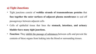
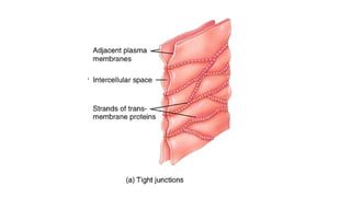
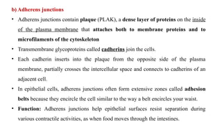
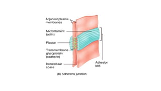
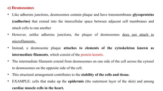
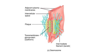
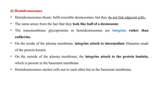
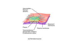
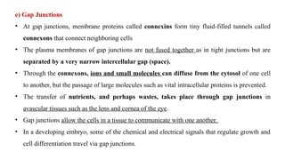
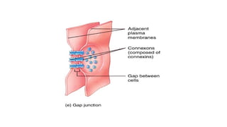
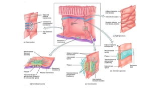
CELL
JUNCTION
Presented By
Ms. P. A. Chandurkar
Assistant Professor
DRGIOP, Amravati
• Cell junctions are contact points between the plasma membranes of
tissue cells.
• Types of Cell Junction
a) Tight junction
b) Adherens junctions
c) Desmosomes
d) Hemidesmosomes
e) Gap junctions
a) Tight Junctions
• Tight junctions consist of weblike strands of transmembrane proteins that
fuse together the outer surfaces of adjacent plasma membranes to seal off
passageways between adjacent cells
• Cells of epithelial tissue that lines the stomach, intestines, and urinary
bladder have many tight junctions.
• Function: They inhibit the passage of substances between cells and prevent the
contents of these organs from leaking into the blood or surrounding tissues.
b) Adherens junctions
• Adherens junctions contain plaque (PLAK), a dense layer of proteins on the inside
of the plasma membrane that attaches both to membrane proteins and to
microfilaments of the cytoskeleton
• Transmembrane glycoproteins called cadherins join the cells.
• Each cadherin inserts into the plaque from the opposite side of the plasma
membrane, partially crosses the intercellular space and connects to cadherins of an
adjacent cell.
• In epithelial cells, adherens junctions often form extensive zones called adhesion
belts because they encircle the cell similar to the way a belt encircles your waist.
• Function: Adherens junctions help epithelial surfaces resist separation during
various contractile activities, as when food moves through the intestines.
c) Desmosomes
• Like adherens junctions, desmosomes contain plaque and have transmembrane glycoproteins
(cadherins) that extend into the intercellular space between adjacent cell membranes and
attach cells to one another
• However, unlike adherens junctions, the plaque of desmosomes does not attach to
microfilaments.
• Instead, a desmosome plaque attaches to elements of the cytoskeleton known as
intermediate filaments, which consist of the protein keratin.
• The intermediate filaments extend from desmosomes on one side of the cell across the cytosol
to desmosomes on the opposite side of the cell.
• This structural arrangement contributes to the stability of the cells and tissue.
• EXAMPLE: cells that make up the epidermis (the outermost layer of the skin) and among
cardiac muscle cells in the heart.
d) Hemidesmosomes
• Hemidesmosomes (hemi- half) resemble desmosomes, but they do not link adjacent cells.
• The name arises from the fact that they look like half of a desmosome
• The transmembrane glycoproteins in hemidesmosomes are integrins rather than
cadherins.
• On the inside of the plasma membrane, integrins attach to intermediate filaments made
of the protein keratin.
• On the outside of the plasma membrane, the integrins attach to the protein laminin,
which is present in the basement membrane
• Hemidesmosomes anchor cells not to each other but to the basement membrane.
e) Gap Junctions
• At gap junctions, membrane proteins called connexins form tiny fluid-filled tunnels called
connexons that connect neighboring cells
• The plasma membranes of gap junctions are not fused together as in tight junctions but are
separated by a very narrow intercellular gap (space).
• Through the connexons, ions and small molecules can diffuse from the cytosol of one cell
to another, but the passage of large molecules such as vital intracellular proteins is prevented.
• The transfer of nutrients, and perhaps wastes, takes place through gap junctions in
avascular tissues such as the lens and cornea of the eye.
• Gap junctions allow the cells in a tissue to communicate with one another.
• In a developing embryo, some of the chemical and electrical signals that regulate growth and
cell differentiation travel via gap junctions.
THANK YOU

Cell Junction (Human Anatomy & Physiology)

  • 1.
    CELL JUNCTION Presented By Ms. P.A. Chandurkar Assistant Professor DRGIOP, Amravati
  • 2.
    • Cell junctionsare contact points between the plasma membranes of tissue cells. • Types of Cell Junction a) Tight junction b) Adherens junctions c) Desmosomes d) Hemidesmosomes e) Gap junctions
  • 3.
    a) Tight Junctions •Tight junctions consist of weblike strands of transmembrane proteins that fuse together the outer surfaces of adjacent plasma membranes to seal off passageways between adjacent cells • Cells of epithelial tissue that lines the stomach, intestines, and urinary bladder have many tight junctions. • Function: They inhibit the passage of substances between cells and prevent the contents of these organs from leaking into the blood or surrounding tissues.
  • 5.
    b) Adherens junctions •Adherens junctions contain plaque (PLAK), a dense layer of proteins on the inside of the plasma membrane that attaches both to membrane proteins and to microfilaments of the cytoskeleton • Transmembrane glycoproteins called cadherins join the cells. • Each cadherin inserts into the plaque from the opposite side of the plasma membrane, partially crosses the intercellular space and connects to cadherins of an adjacent cell. • In epithelial cells, adherens junctions often form extensive zones called adhesion belts because they encircle the cell similar to the way a belt encircles your waist. • Function: Adherens junctions help epithelial surfaces resist separation during various contractile activities, as when food moves through the intestines.
  • 7.
    c) Desmosomes • Likeadherens junctions, desmosomes contain plaque and have transmembrane glycoproteins (cadherins) that extend into the intercellular space between adjacent cell membranes and attach cells to one another • However, unlike adherens junctions, the plaque of desmosomes does not attach to microfilaments. • Instead, a desmosome plaque attaches to elements of the cytoskeleton known as intermediate filaments, which consist of the protein keratin. • The intermediate filaments extend from desmosomes on one side of the cell across the cytosol to desmosomes on the opposite side of the cell. • This structural arrangement contributes to the stability of the cells and tissue. • EXAMPLE: cells that make up the epidermis (the outermost layer of the skin) and among cardiac muscle cells in the heart.
  • 9.
    d) Hemidesmosomes • Hemidesmosomes(hemi- half) resemble desmosomes, but they do not link adjacent cells. • The name arises from the fact that they look like half of a desmosome • The transmembrane glycoproteins in hemidesmosomes are integrins rather than cadherins. • On the inside of the plasma membrane, integrins attach to intermediate filaments made of the protein keratin. • On the outside of the plasma membrane, the integrins attach to the protein laminin, which is present in the basement membrane • Hemidesmosomes anchor cells not to each other but to the basement membrane.
  • 11.
    e) Gap Junctions •At gap junctions, membrane proteins called connexins form tiny fluid-filled tunnels called connexons that connect neighboring cells • The plasma membranes of gap junctions are not fused together as in tight junctions but are separated by a very narrow intercellular gap (space). • Through the connexons, ions and small molecules can diffuse from the cytosol of one cell to another, but the passage of large molecules such as vital intracellular proteins is prevented. • The transfer of nutrients, and perhaps wastes, takes place through gap junctions in avascular tissues such as the lens and cornea of the eye. • Gap junctions allow the cells in a tissue to communicate with one another. • In a developing embryo, some of the chemical and electrical signals that regulate growth and cell differentiation travel via gap junctions.
  • 14.