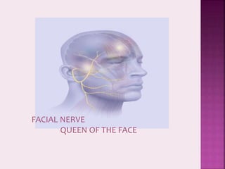
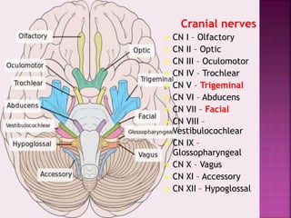
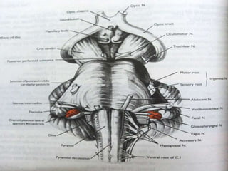
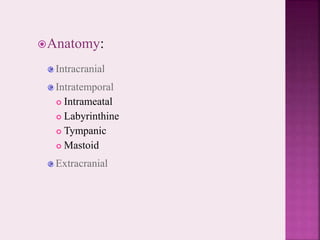
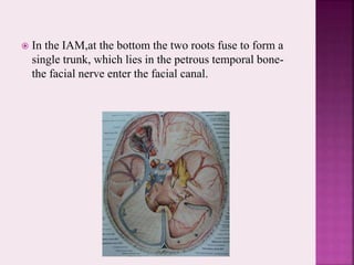
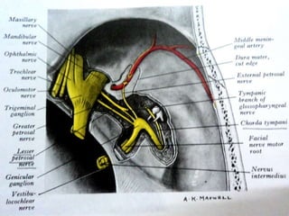
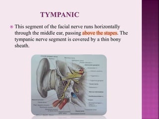
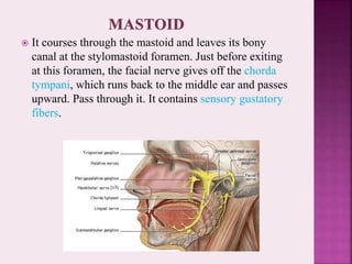
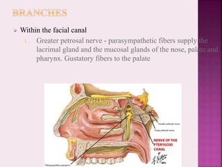
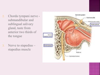
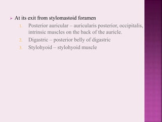
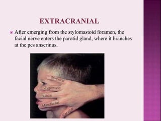
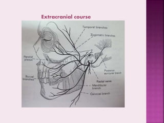
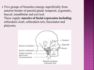
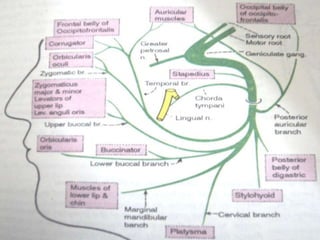
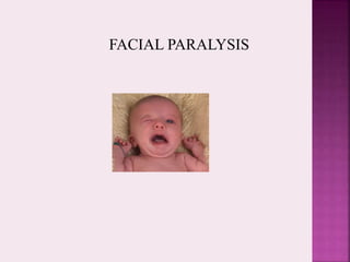
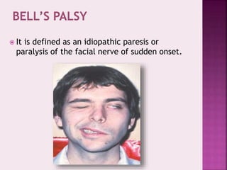
FACIAL NERVE
QUEEN OF THE FACE
 Introduction
 Embryology
 Origin
 Anatomy
 Blood Supply
 Ganglia
 Functional Components
 Nuclei
 Function
 Facial Paralysis
 Bell’s Palsy
 Degrees of nerve injury
 Evaluation of nerve function
 Syndromes
 Clinical Notes
 Conclusion
 Previous Year Questions
 References
 In humans- twelve pairs of cranial nerves.
 First and the second pair emerge from the
cerebrum; the remaining ten pairs emerge from
the brainstem.
 The cranial nerves are part of the peripheral
nervous system (PNS) with the exception of
cranial nerve II or optic nerve.
Cranial nerves
 CN I – Olfactory
 CN II – Optic
 CN III – Oculomotor
 CN IV – Trochlear
 CN V – Trigeminal
 CN VI – Abducens
 CN VII – Facial
 CN VIII –
Vestibulocochlear
 CN IX –
Glossopharyngeal
 CN X – Vagus
 CN XI – Accessory
 CN XII – Hypoglossal
 The entire nervous system is of ectodermal
origin
 The nuclei for all 12 cranial nerves are present
by the fourth week of I.U development.
 The sensory nerves are derived from neural
crest cells; central processes grow into the
brain and form the roots of the nerves, while
the peripheral processes extend outward and
constitute their fibers of distribution
 it is the 7th cranial nerve.
 It is the nerve of the second branchial
arch.
 Facial nerve course, branching pattern, and
anatomical relationships are established
during the first 3 months of prenatal life
 The nerve is not fully developed until about
4 years of age
 The first identifiable FN tissue is seen at the
third week of gestation-facioacoustic
primordium or crest
The facial portion of the crest cells extend to a
placode on the 2nd pharyngeal arch.
Two branches form from this area, one courses
caudally into the mesenchyme of the 2nd
pharyngeal arch to eventually become the main
trunk of the facial nerve, and the second branch
turns rostrally into the 1st branchial arch to
become the chorda tympani nerve.
The facial motor nucleus forms near the nucleus
of the 6th cranial nerve, but as the
metencephalon elongates, the nucleus of the 6th
cranial nerve ascends relative to that of the 7th,
displacing fibres of the 7th nucleus.
Thus the internal genu of the facial nerve is
formed.
This relationship also explains the many
disorders that affect both the 6th and 7th cranial
nerves.
Origin
• It originates from cerebellopontine angle –
lateral part of pontomedullary junction.
• Two adjacent roots:
motor root (larger, more medial);
nervus intermedius (smaller, more lateral) – so
called because it is found between two larger
nerves (main root of VII and VIII). Nervus
intermedius conveys parasympathetic and
sensory fibres and may be part of VIII initially.
Anatomy:
Intracranial
Intratemporal
 Intrameatal
 Labyrinthine
 Tympanic
 Mastoid
Extracranial
 Before the facial nerve leaves the brainstem, its motor
fibers wind around the abducens nucleus and form the
internal genu of the nerve.
Intracranial course and branches
 From cerebellopontine angle, crosses posterior cranial
fossa,enters internal acoustic meatus (IAM; with VIII).
• Nervus intermedius joins main root of facial nerve in IAM.
 In the IAM,at the bottom the two roots fuse to form a
single trunk, which lies in the petrous temporal bone-
the facial nerve enter the facial canal.
 Accompanied by cranial nerve VIII, the facial nerve
travels through the internal auditory canal to the
fundus; there it passes anterosuperiorly through the
meatal foramen, leaving the meatus.
This is the narrowest point
in the bony fallopian canal
(facial canal) and is the site
where the nerve is most likely
to become entrapped due to
inflammatory swelling
 After running a short distance anteriorly, the facial
nerve gives off the greater petrosal nerve with its
secretory fibers to the lacrimal glands and nasal
mucosal glands. The facial nerve turns sharply
downward and posteriorly at the geniculate ganglion,
forming the external genu.
 This segment of the facial nerve runs horizontally
through the middle ear, passing above the stapes. The
tympanic nerve segment is covered by a thin bony
sheath.
 It courses through the mastoid and leaves its bony
canal at the stylomastoid foramen. Just before exiting
at this foramen, the facial nerve gives off the chorda
tympani, which runs back to the middle ear and passes
upward. Pass through it. It contains sensory gustatory
fibers.
 BRANCHES
1.IN FACIAL CANAL
2.AT STYLOMASTOID FORAMEN
3.TERMINAL BRANCHES FOR FACIAL MUSCLES
BRANCHES IN FACIAL CANAL
1.GREATER PETROSAL
2. NERVE TO STAPEDIUS
3.CHORDA TYMPANI
AT STYLOMASTOID FORAMEN
1.POSTERIOR AURICULAR
2.BR.TO POSTERIOR BELLY OF DIGASTRIC
3.BRANCH TO STYLOHYOID
TERMINAL BRANCHES FOR FACIAL
MUSCLES:
1.TEMPORAL
2.BUCCAL
3.ZYGOMATIC
4.MANDIBULAR
5.CERVICAL
 Within the facial canal
1. Greater petrosal nerve - parasympathetic fibers supply the
lacrimal gland and the mucosal glands of the nose, palate and
pharynx. Gustatory fibers to the palate
2. Chorda tympani nerve -
submandibular and
sublingual salivary
gland, taste from
anterior two thirds of
the tongue
3. Nerve to stapedius –
stapedius muscle
 At its exit from stylomastoid foramen
1. Posterior auricular – auricularis posterior, occipitalis,
intrinsic muscles on the back of the auricle.
2. Digastric – posterior belly of digastric
3. Stylohyoid – stylohyoid muscle
 After emerging from the stylomastoid foramen, the
facial nerve enters the parotid gland, where it branches
at the pes anserinus.
Extracranial course and branches
• Outside stylomastoid foramen, small branches of VII supply
occipital belly of occipitofrontalis, stylohyoid and posterior
belly of digastric, and a variable amount of cutaneous sensation
from skin of external auditory meatus.
• Nerve enters parotid gland where it forms intricate plexus.
Branches of VII are superficial in the gland.
Extracranial course
• Five groups of branches emerge superficially from
anterior border of parotid gland: temporal, zygomatic,
buccal, mandibular and cervical.
These supply muscles of facial expression including
orbicularis oculi, orbicularis oris, buccinator and
platysma.
 Intracranial/Meatal: labyrinthine branches from ant inf
cerebellar artery
 Perigeniculate: superficial petrosal branch of middle
meningeal artery
 Tympanic/Mastoid: stylomastoid branch of posterior
auricular artery
The most important thing about the intracranial
course of VII is its relationship to the middle ear.
The most important thing about the extracranial
course is its relationship to the parotid gland.
There are three ganglia associated with the facial nerve.
 The genniculate ganglion
 The Submandibular ganglion
 The pterygopalatine ganglion.
 Geniculate ganglion-it is sensory ganglion and is located
in relation to the medial wall of the middle ear. The taste
fibres present in the nerve are peripheral processes of
pseudounipolar neurons present in geniculate ganglion.
SUBMANDIBULAR GANGLION-It is a parasympathetic
ganglion.It relays secretomotor fibres to the submandibular
and sublingual glands.functionally connected to facial
nerve,lies superficial to hyoglossus
muscle just above deep part of submandibular salivary
gland,suspended from lingual nerve.
 The pterygopalatine ganglion- it is the largest
parasympathetic peripheral ganglion. It serves as relay
station for secretomotor fibres to lacrimal gland and to
the mucous glands of the nose, the paranasal sinuses,
the palate and the pharynx.
 Brancial motor(special visceral efferent)-Supplies the
muscles of facial expression; posterior belly of
digastric muscle; stylohyoid, and stapedius.
 Visceral motor(general visceral efferent)
Parasympathetic innervation of the lacrimal,
submandibular, and sublingual glands, as well as
mucous membranes of nasopharynx, hard and soft
palate.
 Special sensory(special afferent)-Taste sensation from
the anterior 2/3 of tongue; hard and soft palates.
 General sensory(general somatic afferent)-General
sensation from the skin of the concha of the auricle and
from a small area behind the ear.
 Motor nucleus/branchiomotor
 Superior salivatory /parasympathetic
 Lacrimatory/Parasympathetic
 Nucleus of tractus solitaries-gustatory and also
receives afferent fibres from the glands
The facial nerve is responsible for:
I. Contraction of the muscles of the face
II. Production of tears from a gland (Lacrimal gland)
III. Conveying the sense of taste from the front part
of the tongue (via the Chorda tympani nerve)
IV. The sense of touch at auricular conchae
FACIAL PARALYSIS
SUPRANUCLEAR FACIAL PARALYSIS
 It is usually a part of hemiplegia-is the lower part of
the face that is chiefly affected, while the upper part
remains unaffected,i.e.,the frontalis and orbicularis oculi
muscles escape.
In upper motor neuron (UMN) lesions of the
facial nerve, the forehead and orbicularis oculi
muscles are largely spared. This is because
there is bilateral cortical control of the upper
facial muscles, and so if corticonuclear fibres
on one side of the brain are interrupted (e.g. in
the internal capsule) those of the other side are
unaffected. For the lower facial muscles this is
not so: the normal pattern prevails with only
contralateral control.
INFRANUCLEAR FACIAL
PARALYSIS
 The lower motor neuron lesion of facial
nerve cause paralysis of all facial muscles on
the same side.
An LMN lesion, whether of cell bodies in the facial
motor nucleus,or of any part of the peripheral course of
the facial nerve, intracranial or extracranial, would
result in a complete ipsilateral LMN lesion of the facial
nerve, irrespective of which part of the facial nucleus
was involved. The bilateral UMN innervation to the
upper part of the face would be of no significance since
the lesion affects the more distal LMN.
 Vascular abnormalities
 CNS degenerative diseases
 Tumours of the intracranial cavity
 Trauma to the brain
 Congenital abnormalities and agenesis
INTRACRANIAL (CENTRAL) CAUSES
 Bacterial and Viral infection
 Cholesteatoma
 Trauma- blunt temporal bone trauma,
longitudinal and horizontal fractures of the
temporal bone and gunshot wounds.
 Tumours invading the middle ear, mastoid
and facial nerve
 Iatrogenic causes
INTRATEMPORAL CAUSES
 Malignant tumours of the parotid gland
 Trauma
 Iatrogenic causes
 Primary tumours of the facial nerve
 Malignant tumours of the ascending ramus
of the mandible, pterygoid region and skin.
EXTRACRANIAL CAUSES
 It is defined as an idiopathic paresis or
paralysis of the facial nerve of sudden onset.
 INCIDENCE-15-40 cases per 1 lakh cases
 SEX PREDILECTION- women more affected
than men.3.3 more times common in
pregnancy and in the third trimester.
 AGE- can occur at any age, common in
middle aged people.
 SIDE INVOLVMENT- can be equally seen,
usually unilateral.
CLINICAL FEATURES
 There is sudden onset,usually pt gives h/o
occurrence after awakening early morning.
 Unilateral involvement of entire side of the
face.
 Abrupt loss of muscular on one side of face.
 Inability to smile, close the eye or wink or
raise the eyebrow on affected side.
 Whistling is not possible.
 In an attempt to close eyelid,the eyeball rolls
upward.
 Inability to wrinkle forehead or elevate upper or
lower lip.
 The eye waters due to inability to close.
 Obliteration of nasolabial fold.
 Face appears distorted and mask like appearance
to the facial features.
 Speech becomes slurred.
 Occasionally there is loss or alternative of taste.
Partial paralysis always resolves completely within a
few weeks.
Recovery from complete paralysis takes longer
(months) and is complete in only about 60-70% of
cases.
Approximately 15% of patients are left with
troublesome residual palsy and or synkinesis.
Treatment
 Oral antivirals - Acyclovir
 Corticosteroid taper
 Eye protection
 Follow progression with serial exams
 Physiotherapy
The most serious complication is corneal damage.
 Nerve decompression
Internally or externally
 Nerve anastomosis
 Nerve grafting
 Three degrees of facial nerve fiber injury:
Neurapraxia
Without degeneration
Axonotmesis
Wallerian degeneration of the myelin sheath
Intact perineurium
Compelete paralysis
Regeneration of the axon is also complete
Neurotmesis
Regeneration is unpredictable
1) residual dysfunction with synkinesis and persistent palsy
Axon
Epineurium
Perineurium
Endoneurium
NEUROPRAXIA
Axon
Epineurium
Perineurium
Endoneurium
Axonotmesis
Neuronotmesis
 HISTORY is of vital importance to establish
the onset characteristics,duration and
degree of recovery.
 Previous trauma, surgery or infection may
help in arriving at a diagnosis
 Examination of the face at rest and
movement.
 Radiolologic evaluations
 Nerve excitability tests.
 Open injuries of facial nerve should be
repaired surgically as soon as possible.
 Delayed primary repair or early secondary
repair is acceptable when delay is for certain
reasons like contaminated wound,
compromised medical status, extensive
tissue destruction, etc.
 Direct Micronerve Reconstruction/Nerve Grafts
 REANIMATION TECHNIQUES
Transfer of the 12th nerve to the stump of the 7th
nerve
Cross face transfer of 7th nerve
 MASKING PROCEDURES
Transfer of facial muscles
Free vascularised muscle grafts.
Reanimation/cross nerve grafting
Temporalis transportation
 Melkersson-Rosenthal syndrome
 Crocodile tear syndrome
 Guillain Barre syndrome
 James Ramsey Hunt syndrome
 Parotid disease:Parotid tumours, trauma or surgery
may damage branches of the facial nerve. This would
result in an ipsilateral facial palsy with wasting and
functional loss. It would be unlikely to recover.
 Stapedius: hyperacusis Dysfunction of the smallest
muscle supplied by the facial nerve can cause a
distressing symptom. Stapedius dampens the
movements of the ossicular chain and if it is inactive,
sounds will be distorted and echoing. This is
hyperacusis.
 Bell’s palsy:This is a facial palsy, usually of unknown
aetiology. It has been suggested that vascular spasm of
the arteries in the facial canal supplying the nerve
might be responsible, or inflammation and swelling of
the nerve within the bony canal.
The marginal mandibular branch of the facial
nerve:This branch passes on or just below the lower
margin of the mandible. It is superficial even to the
palpable facial arterial pulse and is thus liable to injury.
Section of this nerve would
result in paralysis of the muscles of the corner of the
mouth:drooling would occur.
 Facial nerve injury in babies
As the mastoid process is rudimentary at birth, the
facial nerve is more easily damaged in babies. Birth
injuries, or other trauma, can therefore cause an
ipsilateral facial palsy. This is serious since buccinator,
supplied by VII, is necessary for sucking(feeding)
 Cerebellopontine angle tumours :
Tumours in this region would cause signs and
symptoms of damage to the facial and
vestibulocochlear nerves and cerebellar signs.These
include facial palsy,deafness, vertigo and poor
coordination.
 Acoustic neuroma:This is a tumour of Schwann cells
on the vestibular nerve in the IAM. Since the tumour
grows within a bony canal it may compress the facial
and vestibulocochlear nerves causing a particular type
of deafness (nerve deafness) and an ipsilateral facial
palsy.
 Brain stem lesions:The relationship between the
nucleus of the abducens nerve and the axons of the
facial nerve means that a brain stem lesion may cause a
paralysis of the facial nerve in association with a
paralysis of the ipsilateral lateral rectus muscle of the
eye.
facial nerve is preserved by
removing gland in two parts,superficial and deep
separately.The plane of cleavage is defined by tracing
the nerve from behind backwards
 INJECTION OF L.A into capsule of parotid during
IANB causes transient facial palsy
The facial nerve is responsible for:
I. Contraction of the muscles of the face
II. Production of tears from the lacrimal gland
III. Conveying the sense of taste from the anterior
2/3rd of the tongue (via the Chorda tympani
nerve)
IV. The sense of touch at part of the skin of the
auricule
 FACIAL NERVE (10 MARKS –RGUHS MAY 2011)
 B.D CHAURASIA’S HUMAN ANATOMY VOLUME 3-
5TH EDITION
 LANGMANS MEDICAL EMBRYOLOGY
 CRANIAL NERVES-FUNCTIONAL ANATOMY –
STANLEY MONKHOUSE
 ORAL PATHOLOGY- REGEZ
 TEXTBOOK OF ORAL SURGERY – NEELIMA
MALIK
 GRAYS ANATOMY
 ANATOMY BY ROYLCE
1.facial nerve

1.facial nerve

  • 3.
  • 4.
     Introduction  Embryology Origin  Anatomy  Blood Supply  Ganglia  Functional Components  Nuclei  Function  Facial Paralysis  Bell’s Palsy
  • 5.
     Degrees ofnerve injury  Evaluation of nerve function  Syndromes  Clinical Notes  Conclusion  Previous Year Questions  References
  • 6.
     In humans-twelve pairs of cranial nerves.  First and the second pair emerge from the cerebrum; the remaining ten pairs emerge from the brainstem.  The cranial nerves are part of the peripheral nervous system (PNS) with the exception of cranial nerve II or optic nerve.
  • 7.
    Cranial nerves  CNI – Olfactory  CN II – Optic  CN III – Oculomotor  CN IV – Trochlear  CN V – Trigeminal  CN VI – Abducens  CN VII – Facial  CN VIII – Vestibulocochlear  CN IX – Glossopharyngeal  CN X – Vagus  CN XI – Accessory  CN XII – Hypoglossal
  • 8.
     The entirenervous system is of ectodermal origin  The nuclei for all 12 cranial nerves are present by the fourth week of I.U development.  The sensory nerves are derived from neural crest cells; central processes grow into the brain and form the roots of the nerves, while the peripheral processes extend outward and constitute their fibers of distribution
  • 9.
     it isthe 7th cranial nerve.  It is the nerve of the second branchial arch.
  • 10.
     Facial nervecourse, branching pattern, and anatomical relationships are established during the first 3 months of prenatal life  The nerve is not fully developed until about 4 years of age  The first identifiable FN tissue is seen at the third week of gestation-facioacoustic primordium or crest
  • 12.
    The facial portionof the crest cells extend to a placode on the 2nd pharyngeal arch. Two branches form from this area, one courses caudally into the mesenchyme of the 2nd pharyngeal arch to eventually become the main trunk of the facial nerve, and the second branch turns rostrally into the 1st branchial arch to become the chorda tympani nerve.
  • 13.
    The facial motornucleus forms near the nucleus of the 6th cranial nerve, but as the metencephalon elongates, the nucleus of the 6th cranial nerve ascends relative to that of the 7th, displacing fibres of the 7th nucleus. Thus the internal genu of the facial nerve is formed. This relationship also explains the many disorders that affect both the 6th and 7th cranial nerves.
  • 14.
    Origin • It originatesfrom cerebellopontine angle – lateral part of pontomedullary junction. • Two adjacent roots: motor root (larger, more medial); nervus intermedius (smaller, more lateral) – so called because it is found between two larger nerves (main root of VII and VIII). Nervus intermedius conveys parasympathetic and sensory fibres and may be part of VIII initially.
  • 16.
  • 17.
     Before thefacial nerve leaves the brainstem, its motor fibers wind around the abducens nucleus and form the internal genu of the nerve.
  • 18.
    Intracranial course andbranches  From cerebellopontine angle, crosses posterior cranial fossa,enters internal acoustic meatus (IAM; with VIII). • Nervus intermedius joins main root of facial nerve in IAM.
  • 19.
     In theIAM,at the bottom the two roots fuse to form a single trunk, which lies in the petrous temporal bone- the facial nerve enter the facial canal.
  • 21.
     Accompanied bycranial nerve VIII, the facial nerve travels through the internal auditory canal to the fundus; there it passes anterosuperiorly through the meatal foramen, leaving the meatus. This is the narrowest point in the bony fallopian canal (facial canal) and is the site where the nerve is most likely to become entrapped due to inflammatory swelling
  • 22.
     After runninga short distance anteriorly, the facial nerve gives off the greater petrosal nerve with its secretory fibers to the lacrimal glands and nasal mucosal glands. The facial nerve turns sharply downward and posteriorly at the geniculate ganglion, forming the external genu.
  • 23.
     This segmentof the facial nerve runs horizontally through the middle ear, passing above the stapes. The tympanic nerve segment is covered by a thin bony sheath.
  • 24.
     It coursesthrough the mastoid and leaves its bony canal at the stylomastoid foramen. Just before exiting at this foramen, the facial nerve gives off the chorda tympani, which runs back to the middle ear and passes upward. Pass through it. It contains sensory gustatory fibers.
  • 25.
     BRANCHES 1.IN FACIALCANAL 2.AT STYLOMASTOID FORAMEN 3.TERMINAL BRANCHES FOR FACIAL MUSCLES
  • 26.
    BRANCHES IN FACIALCANAL 1.GREATER PETROSAL 2. NERVE TO STAPEDIUS 3.CHORDA TYMPANI
  • 27.
    AT STYLOMASTOID FORAMEN 1.POSTERIORAURICULAR 2.BR.TO POSTERIOR BELLY OF DIGASTRIC 3.BRANCH TO STYLOHYOID
  • 28.
    TERMINAL BRANCHES FORFACIAL MUSCLES: 1.TEMPORAL 2.BUCCAL 3.ZYGOMATIC 4.MANDIBULAR 5.CERVICAL
  • 29.
     Within thefacial canal 1. Greater petrosal nerve - parasympathetic fibers supply the lacrimal gland and the mucosal glands of the nose, palate and pharynx. Gustatory fibers to the palate
  • 30.
    2. Chorda tympaninerve - submandibular and sublingual salivary gland, taste from anterior two thirds of the tongue 3. Nerve to stapedius – stapedius muscle
  • 31.
     At itsexit from stylomastoid foramen 1. Posterior auricular – auricularis posterior, occipitalis, intrinsic muscles on the back of the auricle. 2. Digastric – posterior belly of digastric 3. Stylohyoid – stylohyoid muscle
  • 32.
     After emergingfrom the stylomastoid foramen, the facial nerve enters the parotid gland, where it branches at the pes anserinus.
  • 33.
    Extracranial course andbranches • Outside stylomastoid foramen, small branches of VII supply occipital belly of occipitofrontalis, stylohyoid and posterior belly of digastric, and a variable amount of cutaneous sensation from skin of external auditory meatus. • Nerve enters parotid gland where it forms intricate plexus. Branches of VII are superficial in the gland.
  • 34.
  • 35.
    • Five groupsof branches emerge superficially from anterior border of parotid gland: temporal, zygomatic, buccal, mandibular and cervical. These supply muscles of facial expression including orbicularis oculi, orbicularis oris, buccinator and platysma.
  • 37.
     Intracranial/Meatal: labyrinthinebranches from ant inf cerebellar artery  Perigeniculate: superficial petrosal branch of middle meningeal artery  Tympanic/Mastoid: stylomastoid branch of posterior auricular artery
  • 38.
    The most importantthing about the intracranial course of VII is its relationship to the middle ear. The most important thing about the extracranial course is its relationship to the parotid gland.
  • 39.
    There are threeganglia associated with the facial nerve.  The genniculate ganglion  The Submandibular ganglion  The pterygopalatine ganglion.
  • 40.
     Geniculate ganglion-itis sensory ganglion and is located in relation to the medial wall of the middle ear. The taste fibres present in the nerve are peripheral processes of pseudounipolar neurons present in geniculate ganglion.
  • 41.
    SUBMANDIBULAR GANGLION-It isa parasympathetic ganglion.It relays secretomotor fibres to the submandibular and sublingual glands.functionally connected to facial nerve,lies superficial to hyoglossus muscle just above deep part of submandibular salivary gland,suspended from lingual nerve.
  • 42.
     The pterygopalatineganglion- it is the largest parasympathetic peripheral ganglion. It serves as relay station for secretomotor fibres to lacrimal gland and to the mucous glands of the nose, the paranasal sinuses, the palate and the pharynx.
  • 43.
     Brancial motor(specialvisceral efferent)-Supplies the muscles of facial expression; posterior belly of digastric muscle; stylohyoid, and stapedius.  Visceral motor(general visceral efferent) Parasympathetic innervation of the lacrimal, submandibular, and sublingual glands, as well as mucous membranes of nasopharynx, hard and soft palate.  Special sensory(special afferent)-Taste sensation from the anterior 2/3 of tongue; hard and soft palates.  General sensory(general somatic afferent)-General sensation from the skin of the concha of the auricle and from a small area behind the ear.
  • 44.
     Motor nucleus/branchiomotor Superior salivatory /parasympathetic  Lacrimatory/Parasympathetic  Nucleus of tractus solitaries-gustatory and also receives afferent fibres from the glands
  • 45.
    The facial nerveis responsible for: I. Contraction of the muscles of the face II. Production of tears from a gland (Lacrimal gland) III. Conveying the sense of taste from the front part of the tongue (via the Chorda tympani nerve) IV. The sense of touch at auricular conchae
  • 47.
  • 48.
    SUPRANUCLEAR FACIAL PARALYSIS It is usually a part of hemiplegia-is the lower part of the face that is chiefly affected, while the upper part remains unaffected,i.e.,the frontalis and orbicularis oculi muscles escape.
  • 49.
    In upper motorneuron (UMN) lesions of the facial nerve, the forehead and orbicularis oculi muscles are largely spared. This is because there is bilateral cortical control of the upper facial muscles, and so if corticonuclear fibres on one side of the brain are interrupted (e.g. in the internal capsule) those of the other side are unaffected. For the lower facial muscles this is not so: the normal pattern prevails with only contralateral control.
  • 51.
    INFRANUCLEAR FACIAL PARALYSIS  Thelower motor neuron lesion of facial nerve cause paralysis of all facial muscles on the same side.
  • 52.
    An LMN lesion,whether of cell bodies in the facial motor nucleus,or of any part of the peripheral course of the facial nerve, intracranial or extracranial, would result in a complete ipsilateral LMN lesion of the facial nerve, irrespective of which part of the facial nucleus was involved. The bilateral UMN innervation to the upper part of the face would be of no significance since the lesion affects the more distal LMN.
  • 53.
     Vascular abnormalities CNS degenerative diseases  Tumours of the intracranial cavity  Trauma to the brain  Congenital abnormalities and agenesis INTRACRANIAL (CENTRAL) CAUSES
  • 54.
     Bacterial andViral infection  Cholesteatoma  Trauma- blunt temporal bone trauma, longitudinal and horizontal fractures of the temporal bone and gunshot wounds.  Tumours invading the middle ear, mastoid and facial nerve  Iatrogenic causes INTRATEMPORAL CAUSES
  • 55.
     Malignant tumoursof the parotid gland  Trauma  Iatrogenic causes  Primary tumours of the facial nerve  Malignant tumours of the ascending ramus of the mandible, pterygoid region and skin. EXTRACRANIAL CAUSES
  • 56.
     It isdefined as an idiopathic paresis or paralysis of the facial nerve of sudden onset.
  • 57.
     INCIDENCE-15-40 casesper 1 lakh cases  SEX PREDILECTION- women more affected than men.3.3 more times common in pregnancy and in the third trimester.  AGE- can occur at any age, common in middle aged people.  SIDE INVOLVMENT- can be equally seen, usually unilateral.
  • 58.
    CLINICAL FEATURES  Thereis sudden onset,usually pt gives h/o occurrence after awakening early morning.  Unilateral involvement of entire side of the face.  Abrupt loss of muscular on one side of face.  Inability to smile, close the eye or wink or raise the eyebrow on affected side.  Whistling is not possible.
  • 59.
     In anattempt to close eyelid,the eyeball rolls upward.  Inability to wrinkle forehead or elevate upper or lower lip.  The eye waters due to inability to close.  Obliteration of nasolabial fold.  Face appears distorted and mask like appearance to the facial features.  Speech becomes slurred.  Occasionally there is loss or alternative of taste.
  • 60.
    Partial paralysis alwaysresolves completely within a few weeks. Recovery from complete paralysis takes longer (months) and is complete in only about 60-70% of cases. Approximately 15% of patients are left with troublesome residual palsy and or synkinesis.
  • 61.
    Treatment  Oral antivirals- Acyclovir  Corticosteroid taper  Eye protection  Follow progression with serial exams  Physiotherapy The most serious complication is corneal damage.
  • 62.
     Nerve decompression Internallyor externally  Nerve anastomosis  Nerve grafting
  • 63.
     Three degreesof facial nerve fiber injury: Neurapraxia Without degeneration Axonotmesis Wallerian degeneration of the myelin sheath Intact perineurium Compelete paralysis Regeneration of the axon is also complete Neurotmesis Regeneration is unpredictable 1) residual dysfunction with synkinesis and persistent palsy
  • 64.
  • 65.
  • 67.
     HISTORY isof vital importance to establish the onset characteristics,duration and degree of recovery.  Previous trauma, surgery or infection may help in arriving at a diagnosis  Examination of the face at rest and movement.  Radiolologic evaluations  Nerve excitability tests.
  • 68.
     Open injuriesof facial nerve should be repaired surgically as soon as possible.  Delayed primary repair or early secondary repair is acceptable when delay is for certain reasons like contaminated wound, compromised medical status, extensive tissue destruction, etc.
  • 69.
     Direct MicronerveReconstruction/Nerve Grafts  REANIMATION TECHNIQUES Transfer of the 12th nerve to the stump of the 7th nerve Cross face transfer of 7th nerve  MASKING PROCEDURES Transfer of facial muscles Free vascularised muscle grafts. Reanimation/cross nerve grafting Temporalis transportation
  • 70.
     Melkersson-Rosenthal syndrome Crocodile tear syndrome  Guillain Barre syndrome  James Ramsey Hunt syndrome
  • 71.
     Parotid disease:Parotidtumours, trauma or surgery may damage branches of the facial nerve. This would result in an ipsilateral facial palsy with wasting and functional loss. It would be unlikely to recover.  Stapedius: hyperacusis Dysfunction of the smallest muscle supplied by the facial nerve can cause a distressing symptom. Stapedius dampens the movements of the ossicular chain and if it is inactive, sounds will be distorted and echoing. This is hyperacusis.
  • 72.
     Bell’s palsy:Thisis a facial palsy, usually of unknown aetiology. It has been suggested that vascular spasm of the arteries in the facial canal supplying the nerve might be responsible, or inflammation and swelling of the nerve within the bony canal. The marginal mandibular branch of the facial nerve:This branch passes on or just below the lower margin of the mandible. It is superficial even to the palpable facial arterial pulse and is thus liable to injury. Section of this nerve would result in paralysis of the muscles of the corner of the mouth:drooling would occur.
  • 73.
     Facial nerveinjury in babies As the mastoid process is rudimentary at birth, the facial nerve is more easily damaged in babies. Birth injuries, or other trauma, can therefore cause an ipsilateral facial palsy. This is serious since buccinator, supplied by VII, is necessary for sucking(feeding)  Cerebellopontine angle tumours : Tumours in this region would cause signs and symptoms of damage to the facial and vestibulocochlear nerves and cerebellar signs.These include facial palsy,deafness, vertigo and poor coordination.
  • 74.
     Acoustic neuroma:Thisis a tumour of Schwann cells on the vestibular nerve in the IAM. Since the tumour grows within a bony canal it may compress the facial and vestibulocochlear nerves causing a particular type of deafness (nerve deafness) and an ipsilateral facial palsy.  Brain stem lesions:The relationship between the nucleus of the abducens nerve and the axons of the facial nerve means that a brain stem lesion may cause a paralysis of the facial nerve in association with a paralysis of the ipsilateral lateral rectus muscle of the eye.
  • 75.
    facial nerve ispreserved by removing gland in two parts,superficial and deep separately.The plane of cleavage is defined by tracing the nerve from behind backwards  INJECTION OF L.A into capsule of parotid during IANB causes transient facial palsy
  • 76.
    The facial nerveis responsible for: I. Contraction of the muscles of the face II. Production of tears from the lacrimal gland III. Conveying the sense of taste from the anterior 2/3rd of the tongue (via the Chorda tympani nerve) IV. The sense of touch at part of the skin of the auricule
  • 77.
     FACIAL NERVE(10 MARKS –RGUHS MAY 2011)
  • 78.
     B.D CHAURASIA’SHUMAN ANATOMY VOLUME 3- 5TH EDITION  LANGMANS MEDICAL EMBRYOLOGY  CRANIAL NERVES-FUNCTIONAL ANATOMY – STANLEY MONKHOUSE  ORAL PATHOLOGY- REGEZ  TEXTBOOK OF ORAL SURGERY – NEELIMA MALIK  GRAYS ANATOMY  ANATOMY BY ROYLCE

Editor's Notes

  • #7 Cranial nerves- emerge directly from the brain. Spinal nerves- emerge from segments of the spinal cord.
  • #30 GPN arise from geniculate ganglion, carries gustatory and parasympathetic fibers. joins with deep petrosal nerve at foramen lacerum to form nerve of pterygoid canal, which reach the pterygopalatine ganglion. Post ganglionic parasympathetic fibers supply the lacrimal gland and the mucosal glands of the nose, palate and pharynx. The gustatory fibers are distributed to the palate
  • #31 It joins with the lingual nerve in the infra temporal fossa through which it is distributed.
  • #32 MOTOR, MYLOHYOID AND ANT BELLY OF DIGASTRIC SUPPLIED MANDIBULAR NERVE