Objectives
Identify the maincauses of outbreak in the
work zone
Know the different types of samples and
diagnostic tests that can be conducted, based
on the infection dynamics
Review the role of the laboratory coordinated
with surveillance to characterize outbreaks
Most used testsfor diagnosing
etiologic agents
Serological:
– ELISA (IgM
and IgG)
– Direct and
indirect
immunofluor
escence
Molecular:
– Polymerase chain
reaction (PCR)
– Real time PCR
– Sequencing
Microscopic:
– Optical
microscopy
-Fluorescent
M
- Electron M
Specimen type, collection, and conservation
– Examples of type of sample and appropriate conservation
Culture:
– isolation
• Identificatio
n
• Antibiotic
sensitivity
testing
5.
1- Microscopy
Microorganisms canbe examined microscopically for:
a- Bacterial motility:
Hanging drop method:
A drop of bacterial suspension is placed between a cover slip and glass slid
b- Morphology and staining reactions of bacteria:
Simple stain: methylene blue stain
Gram stain: differentiation between Gm+ve and Gm–ve bacteria
. Primary stain (Crystal violet)
. Mordant (Grams Iodine mixture)
. Decolorization (ethyl alcohol)
. Secondary stain ( Saffranin)
Ziehl-Neelsen stain: staining acid fast bacilli
. Apply strong carbol fuchsin with heat
. Decolorization (H2SO4 20% and ethyl alcohol
. Counter stain (methylen blue)
6.
2- Culture Techniques
*Culture media are used for:
- Isolation and identification of pathogenic organisms
- Antimicrobial sensitivity tests
* Types of culture media:
a- Liquid media:
- Nutrient broth: meat extract and peptone
- Peptone water for preparation sugar media
- Growth of bacteria detected by turbidity
b- Solid media:
- Colonial appearance
- Hemolytic activity
- Pigment production
7.
1- simple media:
Nutrientagar
2- Enriched media: media of high nutritive value
. Blood agar
. Chocolate agar
. Loffler’s serum
3- Selective media: allow needed bacteria to grow
. Lowenstein–Jensen medium
. MacConkeys agar
. Mannitol Salt Agar
4- Indicator media: to different. between lact. and non lact. ferment
. MacConkeys medium
. Eosine Methlyne blue Agar
5- Anaerobic media: for anaerobic cultivation
. Deep agar, Robertson’s Cooked Meat Medium
Types of solid media
8.
Colonial appearance onculture media
* Colony morphology:
. Shape . Size . Edge of colony . Color
* Growth pattern in broth:
. Uniform turbidity
. Sediment or surface pellicle
* Pigment production:
. Endopigment production (Staph. aureus)
. Exopigment production (Ps. aeruginosa)
* Haemolysis on blood agar:
. Complete haemolysis (Strept. Pyogenes)
. Partial haemolysis (Strept. Viridans)
* Growth on MacConkey’s medium:
. Rose pink colonies (Lactose fermenters)
. Pale yellow colonies (Non lactose fermenters)
9.
3- Biochemical Reaction
Useof substrates and sugars to identify pathogens:
a- Sugar fermentation:
Organisms ferment sugar with production of acid only
Organisms ferment sugar with production of acid and gas
Organisms do not ferment sugar
b- Production of indole:
Depends on production of indole from amino acid tryptophan
Indole is detected by addition of Kovac’s reagent
Appearance of red ring on the surface
e- H2S production:
Depends on production H2S from protein or polypeptides
Detection by using a strip of filter paper containing lead acetate
10.
3- Biochemical Reaction(cont.)
c- Methyl red reaction (MR):
Fermentation of glucose with production of huge amount of acid
Lowering pH is detected by methyl red indicator
d- Voges proskaur’s reaction (VP):
Production of acetyl methyl carbinol from glucose fermentation
Acetyl methyl carbinol is detected by addition KOH
Color of medium turns pink (positive)
e- Action on milk:
Fermentation of lactose with acid production
Red color if litmus indicator is added
11.
f- Oxidase test:
Somebacteria produce Oxidase enzyme
Detection by adding few drops of colorless oxidase reagent
Colonies turn deep purple in color (positive)
g- Catalase test:
Some bacteria produce catalase enzyme
Addition of H2O2 lead to production of gas bubbles (O2 production)
h- Coagulase test:
Some bacteria produce coagulase enzyme
Coagulase enzyme converts fibrinogen to fibrin (plasma clot)
Detected by slide or test tube method
i- Urease test:
Some bacteria produce urease enzyme
Urease enzyme hydrolyze urea with production of NH3
Alklinity of mediaand change color of indicator from yellow to pink
13.
Serological tests (Serology)
Twotypes: direct and
indirect tests
– Direct tests: based on the antigen
antibody reaction to investigate the
presence of antigen. That is, these
tests investigate the presence of
the etiologic agent or one of its
components. Example: direct
immunofluorescence.
– Indirect tests: based on detecting
specific antibodies against the
agent or one of its components,
which indirectly allow us to assume
that the pathogen being
investigated was present in the host
at some point
Detect the presence of
antibodies
Cheaper than molecular tests
Based on the antigen antibody
reaction and constitute a
valuable tool in diagnosing
infectious disease
14.
Serological tests
(Serology)
Important conceptsfor interpreting serological
results:
• Positive IgM is related to a recent infection
• Increased antibody titer in paired samples
indicates a recent infection
– First sample: negative
– Second sample: antibodies present
– Antibody titer in the second sample > 4x the antibody titer in
the first sample
• Positive IgG indicates (generally) a past
infection
15.
Types of samplesand appropriate
collection
1 3 5 7 9
Days of symptoms
Viral
Charge
5 15 5 15
Antibodies
Primary
infection
Secondary
infection
16.
Serological tests
(Serology)
• Immunofluorescence
–Immunofluorescence techniques may be applied as techniques to
investigate the presence of viral, parasitic, bacterial, or mycotic
antigens in clinical samples or antibodies produced against them in
the patient’s serum.
• ELISA (enzyme-linked immunosorbent assay)
– Immunoenzymatic techniques are tests based on antibody antigen
reaction, making it possible to detect the presence of antibodies
produced against viral, parasitic, bacterial, or mycotic antigens in
clinical specimens in the patient’s serum.
– The ELISA technique may be used to detect IgM or IgG.
17.
Molecular tests
• Arebased on detecting the etiologic agent’s genome.
• Make it possible to detect the etiologic agent’s RNA
or DNA with high sensitivity and specificity.
• Are more costly than serology.
• Make it possible to confirm the presence of the
etiologic agent in clinical samples.
• Make it possible to detect RNA or DNA even in very
small sample volumes.
18.
Molecular tests
• Polymerasechain reaction (end point PCR)
– The end point PCR is based on amplifying and visualizing the
complementary DNA or ADNA (cDNA). This technique consists of
amplifying and visualizing a specific DNA fragment from the
etiologic agent.
• Real time PCR
– Also known as quantitative PCR, this test is a variant of the
polymerase chain reaction (PCR).
– Real time PCR makes it possible to amplify and at the same time
absolutely quantify specific DNA or cDNA molecules
19.
Microscopic
• Consists ofusing special instruments such as
magnifying glasses and microscopes to magnify the
size of structures that are invisible to the naked
human eye, such as viruses, bacteria, and parasites.
• Microscopic observation is necessary in the field of
parasitology.
• Modes of observation vary based on strategies
employed and range from micrometric to nanometric
observation.
• Therefore, various types of microscopes are required,
such as photonic and electronic (scanning,
transmission, and atomic energy).
20.
Microscopic
• Optical microscopytechnique allows us to visualize
bacteria and parasites simply by examining the
sample with a microscope.
• For bacteria, a Gram stain (a purple stain) is often
performed first. Bacteria is classified as follows:
– Gram positive (appear blue because they retain the Gram stain)
– Gram negative (appear red because they do not capture the stain)
• A blood swab is performed to detect parasites found
in the blood, such as filariasis, malaria, or babesiosis.
For this test, a drop of blood is placed on a
microscope slide, stained, and examined under the
microscope to visualize the parasite.
Optical microscopy
21.
Sample type, collection,and
conservation
Successful laboratory diagnosis depends on two factors prior to the
laboratory phase:
Selecting the type of sample:
• It is important to select the correct type
of sample, based on clinical material
and days since the onset of symptoms,
whenever it is possible to obtain a
positive result (serologically or
molecularly)
Sample quality:
• There are three factors that have a
direct influence on the quality of the
sample to be received by the
laboratory:
– Correct collection
– Conservation of the cold chain
during transport
– Conservation at the appropriate
temperature based on the
processing time
22.
Examples of sampletype and
appropriate conservation
Dengue, Chikungunya,
yellow fever, hantavirus,
leptospirosis
Zika, measles, and rubella
Hepatitis
Influenza and other respiratory
viruses
Bacterial and viral encephalitis
Rotavirus, norovirus,
enterobacteria
Malaria Meningeal disease
23.
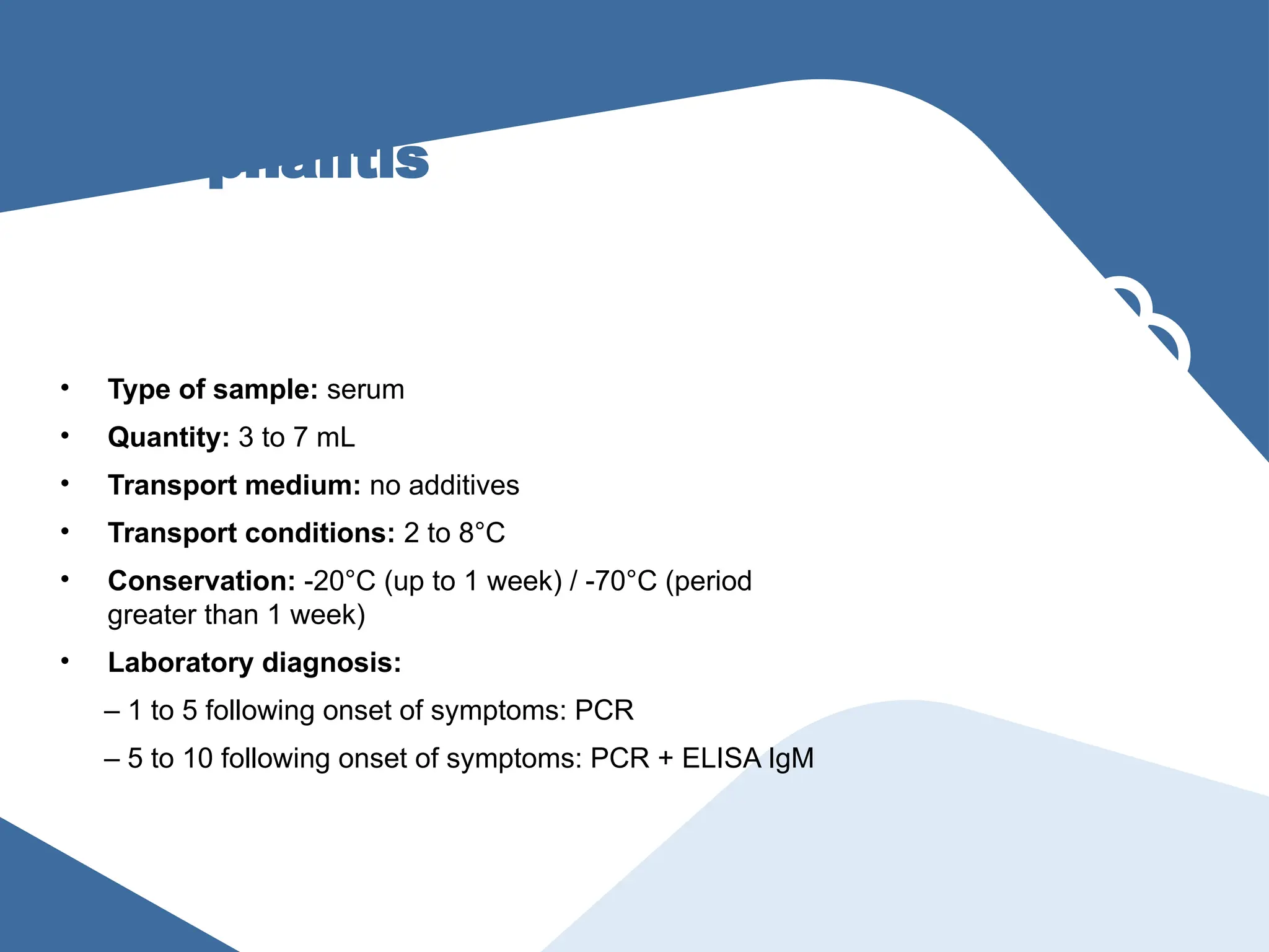
Dengue, Chikungunya,
yellow fever,hantavirus,
leptospirosis
• Type of sample: serum
• Quantity: 3 to 7 mL
• Transport medium: no additives
• Transport conditions: 2 to 8°C
• Conservation: -20°C (up to 1 week) / -70°C (period
greater than 1 week)
• Laboratory diagnosis:
– 1 to 5 days following onset of symptoms: PCR
– 5 to 10 days following the onset of symptoms: PCR +
ELISA IgM
24.
Zika, measles, and
rubella
•Type of sample: serum
• Quantity: 3 to 7 mL
• Transport medium: no additives
• Transport conditions: 2 to 8°C
• Conservation: -20°C (up to 1 week) / -70°C (period
greater than one week)
• Laboratory diagnosis:
– 1 to 5 following the onset of symptoms: PCR
– 5 to 10 days following the onset of symptoms: PCR +
ELISA IgM
25.
Zika, measles, and
rubella
•Type of sample: urine
• Quantity: 3 to 7 mL
• Transport medium: no additives
• Transport conditions: 2 to 8°C
• Conservation: -20°C (up to 1 week) / -70°C (period
greater than 1 week)
• Laboratory diagnosis:
– 1 to 15 days following the onset of symptoms: PCR
26.
Zika – congenitalsyndrome or fatal
cases
Sample Quantity
Transport
medium
Transport
conditions
Conservation
>1 week
Laboratory test
Mother’s serum 5–7 mL No additives 4 / 8 °C -20 / -70 °C PCR, ELISA IgM, PRNT, others
Umbilical cord blood 5–7 mL No additives 4 / 8 °C -20 / -70 °C PCR, ELISA IgM, PRNT, others
Placenta 0,5–1 mL Buffered formalin 4 °C – TA* 4 °C – TA* Immunohistochemical
Placenta 5–7 mL Saline solution 4 / 8 °C -20 / -70 °C PCR
Umbilical cord (tissue) Buffered formalin 4 °C – TA* 4 °C – TA* Immunohistochemical
Umbilical cord (tissue) Saline solution 4 / 8 °C -20 / -70 °C PCR
Newborn’s blood 0,5–1 mL No additives 4 / 8 °C -20 / -70 °C PCR, ELISA IgM, PRNT, others
Newborn’s CSF** 0,5 mL No additives 4 / 8 °C -20 / -70 °C PCR, ELISA IgM, PRNT, others
Mother’s total blood 5–7 mL EDTA, others 4 / 8 °C 4 °C Biochemical, others
Newborn’s total blood 2–5 mL EDTA, others 4 / 8 °C 4 °C Biochemical, others
Tissue** 3x3 cm (approx) Buffered formalin 4 °C – TA* 4 °C Immunohistochemical
Tissue** 3x3 cm (approx) Saline solution 4 / 8 °C -20 / -70 °C PCR
* Ambient temperature
** Under medical indication for suspected neurological syndrome
*** Fatal cases: brain, liver, kidney, others
27.
Hepatitis
• Type ofsample: serum
• Quantity: 3 to 7 mL
• Transport medium: no additives
• Transport conditions: 2 to 8°C
• Conservation: -20°C (up to 1 week) / -70°C (period
greater than 1 week)
• Laboratory diagnosis:
– 1 to 5 days after onset of symptoms: PCR + Elisa
HBsAg
– 5 to 10 after onset of symptoms: PCR + ELISA IgM
28.
Influenza and other
respiratoryviruses
• Type of sample: nasopharyngeal swab
• Quantity: 2 nylon swabs
• Transport medium: viral transport medium or saline
solution (3 mL)
• Transport conditions: 2 to 8°C
• Conservation: 4°C until aliquots are prepared; aliquots to
a -20°C (up to 48 hours) and -70°C (period greater than
48 hours)
• Laboratory diagnosis:
– PCR or IF (only typification) followed by PCR
29.
Influenza and other
respiratoryviruses
• Type of sample: nasopharyngeal aspiration;
nasopharyngeal wash
• Material: suction device
• Transport medium: saline solution
• Transport conditions: 2 to 8°C
• Conservation: 4°C until aliquots are prepared; aliquots to
a -20°C (up to 48 hours) and -70°C (period greater than
48 hours)
• Laboratory diagnosis:
– PCR or IF (only typification) followed by PCR
30.
Bacterial and viral
encephalitis
•Type of sample: serum
• Quantity: 3 to 7 mL
• Transport medium: no additives
• Transport conditions: 2 to 8°C
• Conservation: -20°C (up to 1 week) / -70°C (period
greater than 1 week)
• Laboratory diagnosis:
– 1 to 5 following onset of symptoms: PCR
– 5 to 10 following onset of symptoms: PCR + ELISA IgM
31.
Bacterial and viral
encephalitis
•Type of sample: blood
• Quantity: 3 to 7 mL
• Transport medium: no additives
• Transport conditions: 2 to 8°C
• Conservation: -20°C (up to 1 week) / -70°C (period
greater than 1 week)
• Laboratory diagnosis:
– 1 to 5 following onset of symptoms: PCR
– 5 to 10 following onset of symptoms: PCR + ELISA IgM
32.
Rotavirus, norovirus,
enterobacteria
• Typeof sample: fecal material
• Conservation: 2 to 8°C
• Laboratory diagnosis:
– Generally, molecular methods are used based
on PCR and ELISA kits for antigen detection
33.
Malaria
• Type ofsample: total blood
• Conservation: 2 to 4°C
• Laboratory diagnosis:
– Large drop (extended) for microscopy
Specimen Selection, Collection,and Processing
• The quantity material must be adequate
• Specimens are selected on the basis of signs and
symptoms, should be representative of the disease
process
• Contamination of the specimen must be avoided by
using only sterile equipment and aseptic
precautions
• The specimen must be taken to the laboratory and
examined promptly. Special transport media may
be helpful.
• Meaningful specimens to diagnose bacterial
infections must be secured before antimicrobial
drugs are administered.
36.
4- Animal pathogenicity
*Animal pathogenicity test:
Animals commonly used are guinea pigs, rabbits, mice
* Importance of pathogenicity test:
- Differentiate pathogenic and non pathogenic
- Isolation organism in pure form
- To test ability of toxin production
- Evaluation of vaccines and antibiotics
37.
Serological identification
A- Directserological tests:
- Identification of unknown organism
- Detection of microbial antigens by using specific
known antibodies
- Serogrouping and serotyping of isolated organism
B- Indirect serological tests:
- Detection of specific and non specific antibodies
(IgM & IgG) by using antigens or organisms
RAPID DIAGNOSTIC TESTS
Highsensitivity and specificity
High negative and positive predictive values
High accuracy compared to gold standard
Simple to perform
Rapid turn around time
Cost effective
40.
LIMITATIONS OF CONVENTIONALCLINICAL
MICROBIOLOGY
Culture
Labor intensive
Need for special media
Prolonged period of time to culture
Some organisms are uncultivable on artificial media
Potential health hazards
Antigen Detection
Negative tests require confirmation
Effected by poor specimen collection
Low microbe burden
Serology
Unhelpful during early stage of infection
Not quite useful in immunocompromised patients
41.
Molecular Biology Techniques
A-Genetic probes (DNA or RNA probes):
Detection of a segment of DNA sequence (gene) in unknown
organism using a labeled probe
Probe: consists of specific short sequence of labeled single-
stranded DNA or RNA that form strong covalently
bonded hybrid with specific complementary strand of
nucleic acid of organism in question
B- Polymerase chain reaction (PCR):
Amplification of a short sequence of target DNA or RNA Then
It is detected by a labeled probe
C- Plasmid profile analysis:
Isolation of plasmids from bacteria and determination of their
size and number compared with standard strains by agarose
gel electrophoresis
Polymerase Chain Reaction
*SpecificPCR: Uses primers to known DNA targets.
Use when conventional diagnostics are inadequate, time consuming, difficult and hazardous
*Broad range PCR: uses complementary primers to conserved regions shared by a given taxonomic group
Used in cases of B. henselae and Mycobacterium spp
• *Multiplex PCR
– Uses single clinical specimen to investigate several potential
pathogens simultaneously
• Encephalitis/meningitis panel: HSV,VZV, CMV HHV-6, EBV, Enteroviruses
• *Real-time PCR
– Utilizes a fluorescent labeled probe
– Requires small volumes thus takes 30-60 minutes to complete
44.
Real time PCRfor Diagnosis of Infectious
Disease
] Detect PCR product during synthesis
] Requires fluorescence-based detection and specialized
detection instrumentation
] Advantages
Less time for results
Improved analytical sensitivity
Broad applicability (target characterization, load determination etc)
LIMITATION OF PCRTECHNOLOGIES
Specimen should be frozen until amplification
No antimicrobial sensitivity is available
Needs the clinician to name the suspect
Cost
False positives caused by amplification of contaminants
Only sample from normally sterile sites should be considered for
broad-range PCR
Specimen is required to be refrigerated or stored in alcohol before
processing
47.
Antimicrobial Susceptibility testing
•Introduction:
• Identification of a bacterial isolate from a patient provides
guidance in the choice of an appropriate antibiotic for
treatment
• Many bacterial species are not uniformly susceptible to a
particular anti-bacterial compound
• This is particularly evident among the Enterobacteriaceae,
Staphylococcus spp., and Pseudomonas spp.
• The wide variation in susceptibility and high frequencies of
drug resistance among strains in many bacterial species
necessitates the determination of levels of resistance or
susceptibility as a basis for the selection of the proper
antibiotic for chemotherapy
48.
• Antimicrobial Susceptibilitytesting can be down by three ways:
1. Minimum Inhibitory Concentration (MIC)
2. Disk Diffusion Method
3. Minimum Bactericidal Concentration (MBC)
49.
Most importantaspect of laboratory medicine
• Insufficient quantity
• Contamination
• Improper transport media
• Delay in transportation
• Inappropriate storage
Editor's Notes
#15 Stage of infection when the sample is collected. This refers to the stage of infection when the sample is taken.
It is very important that we know what we can find and when we can find what we will detect in the laboratory.
The curve above shows a typical viral load of a person infected with influenza virus. Viral load is present approximately 10 days after the onset of symptoms. However, its concentration drops considerably after 5 days. Due to the high concentration of viruses, the preferable time for sample collection is between the first 2 to 3 days.
The lower curve shows the antibody stimulation curves in a primary and secondary infection. Collection of serum samples for antibody identification should be performed in the first 7 days for the acute sample (first sample in the acute phase of infection) and the second or convalescence phase taken approximately 2 to 3 weeks after the acute phase.


![Real time PCR for Diagnosis of Infectious
Disease
] Detect PCR product during synthesis
] Requires fluorescence-based detection and specialized
detection instrumentation
] Advantages
Less time for results
Improved analytical sensitivity
Broad applicability (target characterization, load determination etc)](https://image.slidesharecdn.com/p10-laboratory-tests0-251101063803-57ca78b9/75/p10-laboratory-diagnosis-bacterial-_0-pptx-44-2048.jpg)