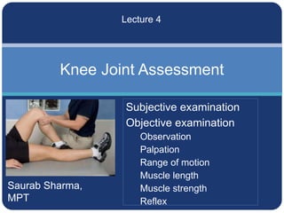
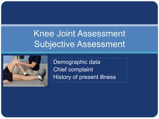
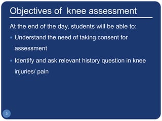
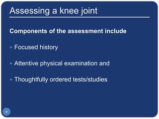
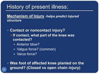
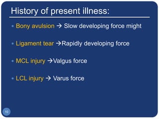
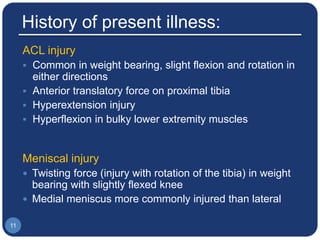
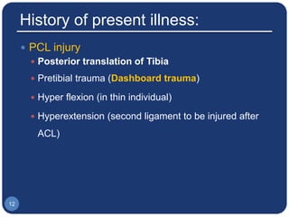
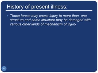
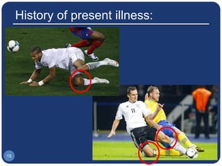
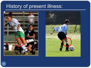
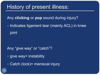
Saurab Sharma,
MPT
Knee Joint Assessment
Lecture 4
Subjective examination
Objective examination
Observation
Palpation
Range of motion
Muscle length
Muscle strength
Reflex
Knee Joint Assessment
Subjective Assessment
Demographic data
Chief complaint
History of present illness
Objectives of knee assessment
At the end of the day, students will be able to:
 Understand the need of taking consent for
assessment
 Identify and ask relevant history question in knee
injuries/ pain
3
Assessing a knee joint
Components of the assessment include
 Focused history
 Attentive physical examination and
 Thoughtfully ordered tests/studies
4
Subjective Assessment
Demographic data
 Name
 Age
 Gender
 Dominance
 Address
 Occupation
6
Chief complaint
In patient’s own word
 Pain
 Swelling: immediate, delayed
 Giving way
 Locking
 Deformity
 Functional difficulty
7
History of present illness:
 Onset of Pain
 Date of injury or when symptoms started
 Gradual/ sudden
 Progression: getting better, getting worse, no
change
 Location of pain
 Anterior
 Medial
 Lateral
 Posterior
8
History of present illness:
Mechanism of Injury -helps predict injured
structure
 Contact or noncontact injury?
 If contact, what part of the knee was
contacted?
 Anterior blow?
 Valgus force? (common)
 Varus force?
 Was foot of affected knee planted on the
ground? (Closed vs open chain injury)9
History of present illness:
 Bony avulsion  Slow developing force might
 Ligament tear Rapidly developing force
 MCL injury Valgus force
 LCL injury  Varus force
10
History of present illness:
ACL injury
 Common in weight bearing, slight flexion and rotation in
either directions
 Anterior translatory force on proximal tibia
 Hyperextension injury
 Hyperflexion in bulky lower extremity muscles
Meniscal injury
 Twisting force (injury with rotation of the tibia) in weight
bearing with slightly flexed knee
 Medial meniscus more commonly injured than lateral
11
History of present illness:
 PCL injury
 Posterior translation of Tibia
 Pretibial trauma (Dashboard trauma)
 Hyper flexion (in thin individual)
 Hyperextension (second ligament to be injured after
ACL)
12
History of present illness:
 These forces may cause injury to more than one
structure and same structure may be damaged with
various other kinds of mechanism of injury
13
History of present illness:
14
History of present illness:
15
History of present illness:
16
History of present illness:
17
History of present illness:
Any clicking or pop sound during injury?
 Indicates ligament tear (mainly ACL) in knee
joint
Any “give way” or “catch”?
 give way= instability
 Catch (lock)= meniscal injury
18
History of present illness:
 History of Swelling?
 Was the swelling immediate or delayed?
 Synovial swelling takes 8-24 hours to
develop
 Haemarthrosis  Immediate swelling
19
History of present illness:
 Site of swelling?
 Area of swelling?
 Localized swelling Extracapsular injury
 Generalized swelling Intracapsular injury
 Inflammed bursae?
20
History of present illness (HOPI):
 History of type of shoes patient normally
uses?
 Flat shoes?
 High heels?
 Duration of usage of shoes?
 Functional ability of the patient?
 Any disability in running, climbing stairs etc21
History of present illness (HOPI):
Sports specific history:
 Total duration of play
 Total practice/training session
 Exercise protocol- warm up/ cool down
 Competition
22
Summary:
23
Plan for next class
 Pain assessment
 Observation
 Prerequisites:
 Pain assessment
 Observation
24

4 knee assessment - History

  • 1.
    Saurab Sharma, MPT Knee JointAssessment Lecture 4 Subjective examination Objective examination Observation Palpation Range of motion Muscle length Muscle strength Reflex
  • 2.
    Knee Joint Assessment SubjectiveAssessment Demographic data Chief complaint History of present illness
  • 3.
    Objectives of kneeassessment At the end of the day, students will be able to:  Understand the need of taking consent for assessment  Identify and ask relevant history question in knee injuries/ pain 3
  • 4.
    Assessing a kneejoint Components of the assessment include  Focused history  Attentive physical examination and  Thoughtfully ordered tests/studies 4
  • 5.
  • 6.
    Demographic data  Name Age  Gender  Dominance  Address  Occupation 6
  • 7.
    Chief complaint In patient’sown word  Pain  Swelling: immediate, delayed  Giving way  Locking  Deformity  Functional difficulty 7
  • 8.
    History of presentillness:  Onset of Pain  Date of injury or when symptoms started  Gradual/ sudden  Progression: getting better, getting worse, no change  Location of pain  Anterior  Medial  Lateral  Posterior 8
  • 9.
    History of presentillness: Mechanism of Injury -helps predict injured structure  Contact or noncontact injury?  If contact, what part of the knee was contacted?  Anterior blow?  Valgus force? (common)  Varus force?  Was foot of affected knee planted on the ground? (Closed vs open chain injury)9
  • 10.
    History of presentillness:  Bony avulsion  Slow developing force might  Ligament tear Rapidly developing force  MCL injury Valgus force  LCL injury  Varus force 10
  • 11.
    History of presentillness: ACL injury  Common in weight bearing, slight flexion and rotation in either directions  Anterior translatory force on proximal tibia  Hyperextension injury  Hyperflexion in bulky lower extremity muscles Meniscal injury  Twisting force (injury with rotation of the tibia) in weight bearing with slightly flexed knee  Medial meniscus more commonly injured than lateral 11
  • 12.
    History of presentillness:  PCL injury  Posterior translation of Tibia  Pretibial trauma (Dashboard trauma)  Hyper flexion (in thin individual)  Hyperextension (second ligament to be injured after ACL) 12
  • 13.
    History of presentillness:  These forces may cause injury to more than one structure and same structure may be damaged with various other kinds of mechanism of injury 13
  • 14.
    History of presentillness: 14
  • 15.
    History of presentillness: 15
  • 16.
    History of presentillness: 16
  • 17.
    History of presentillness: 17
  • 18.
    History of presentillness: Any clicking or pop sound during injury?  Indicates ligament tear (mainly ACL) in knee joint Any “give way” or “catch”?  give way= instability  Catch (lock)= meniscal injury 18
  • 19.
    History of presentillness:  History of Swelling?  Was the swelling immediate or delayed?  Synovial swelling takes 8-24 hours to develop  Haemarthrosis  Immediate swelling 19
  • 20.
    History of presentillness:  Site of swelling?  Area of swelling?  Localized swelling Extracapsular injury  Generalized swelling Intracapsular injury  Inflammed bursae? 20
  • 21.
    History of presentillness (HOPI):  History of type of shoes patient normally uses?  Flat shoes?  High heels?  Duration of usage of shoes?  Functional ability of the patient?  Any disability in running, climbing stairs etc21
  • 22.
    History of presentillness (HOPI): Sports specific history:  Total duration of play  Total practice/training session  Exercise protocol- warm up/ cool down  Competition 22
  • 23.
  • 24.
    Plan for nextclass  Pain assessment  Observation  Prerequisites:  Pain assessment  Observation 24

Editor's Notes

  • #9 *Differential diagnosis by LOCATION: Anterior – Patellofemoral pain syndrome, bursitis, Osgood-Schlatter’s disease, patellar tendinitis, patellar fracture Medial – meniscus, MCL, DJD, pes anserine bursitis Lateral – Meniscus, LCL, DJD, iliotibial band friction syndrome, fibular head dysfunction Posterior – hamstring injury, tear of posterior horn of medial or lateral meniscus, Baker’s cyst, neurovascular injury (popliteal artery or nerve)
  • #10 *CONTACT INJURIES/DIRECT BLOWS: Commonly cause injury to: collateral ligaments, patellar dislocation, epiphyseal fractures in children with open growth plates Valgus forces are more common than varus-directed forces Blow to lateral aspect of knee resulting in stretch injury to soft tissues of medial knee (MCL more prone to injury than LCL) Pearl to help remember the difference between varus and valgus stress, Valgus has “L” as in lateral and patella. NONCONTACT INJURIES: Vulnerable structures: Cruciate ligaments (most common) Menisci Joint capsule **Think ACL INJURY any time you have a patient with a significant NON-CONTACT injury with foot planed on the ground (foot planted then knee twisted or body changed direction, felt a pop, immediate swelling, could not continue playing)
  • #11 Mechanism of injury: Was the injury in Closed or open kinematic chain? More prone for injury in closed kinematic chain
  • #22 high heels can cause excess stress to knee joint and lead to hyperextension of the knee