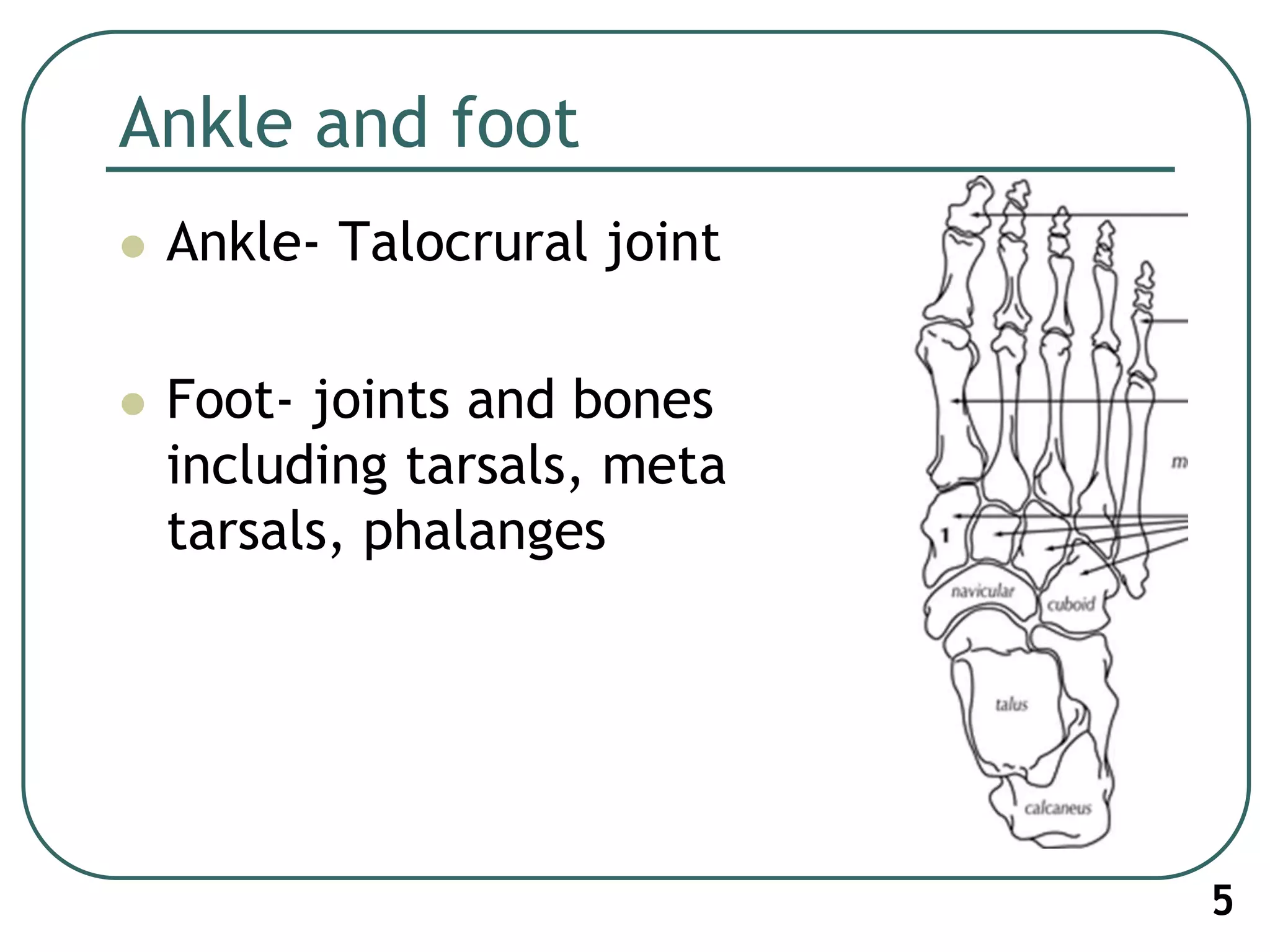
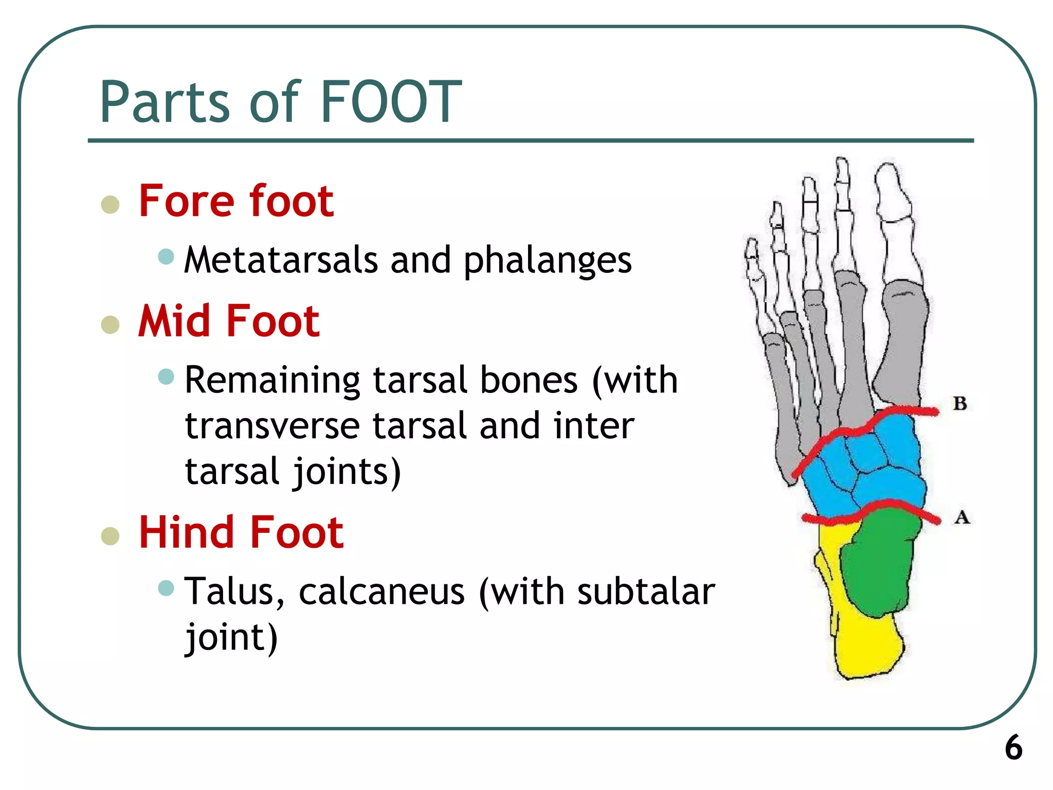
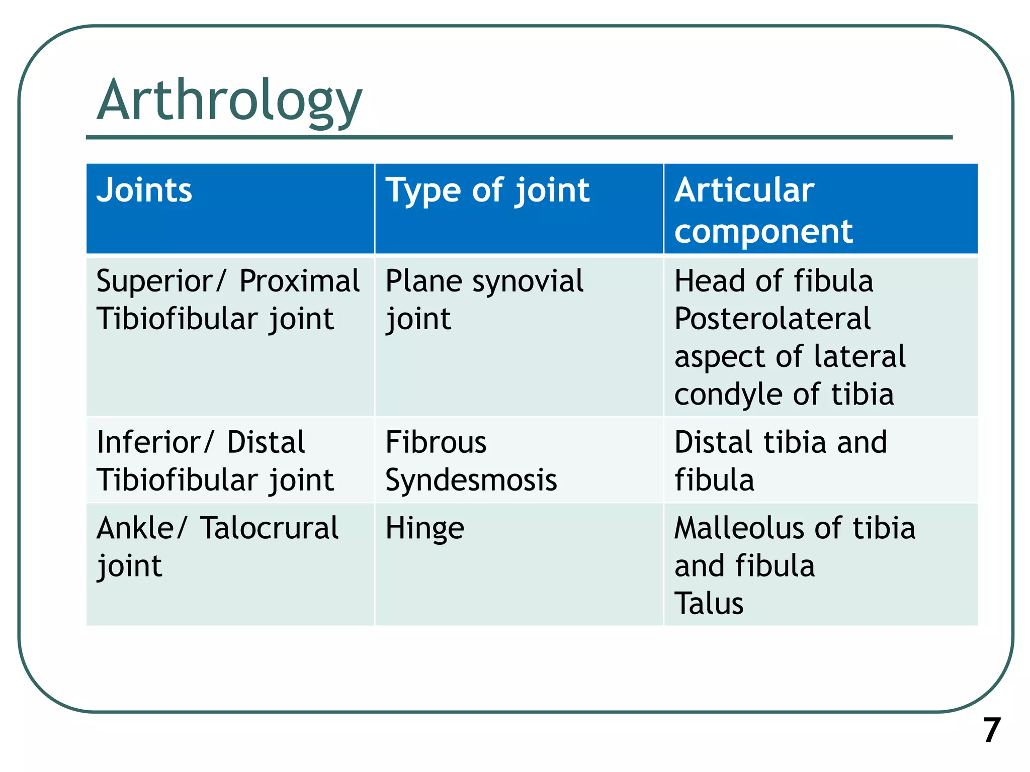
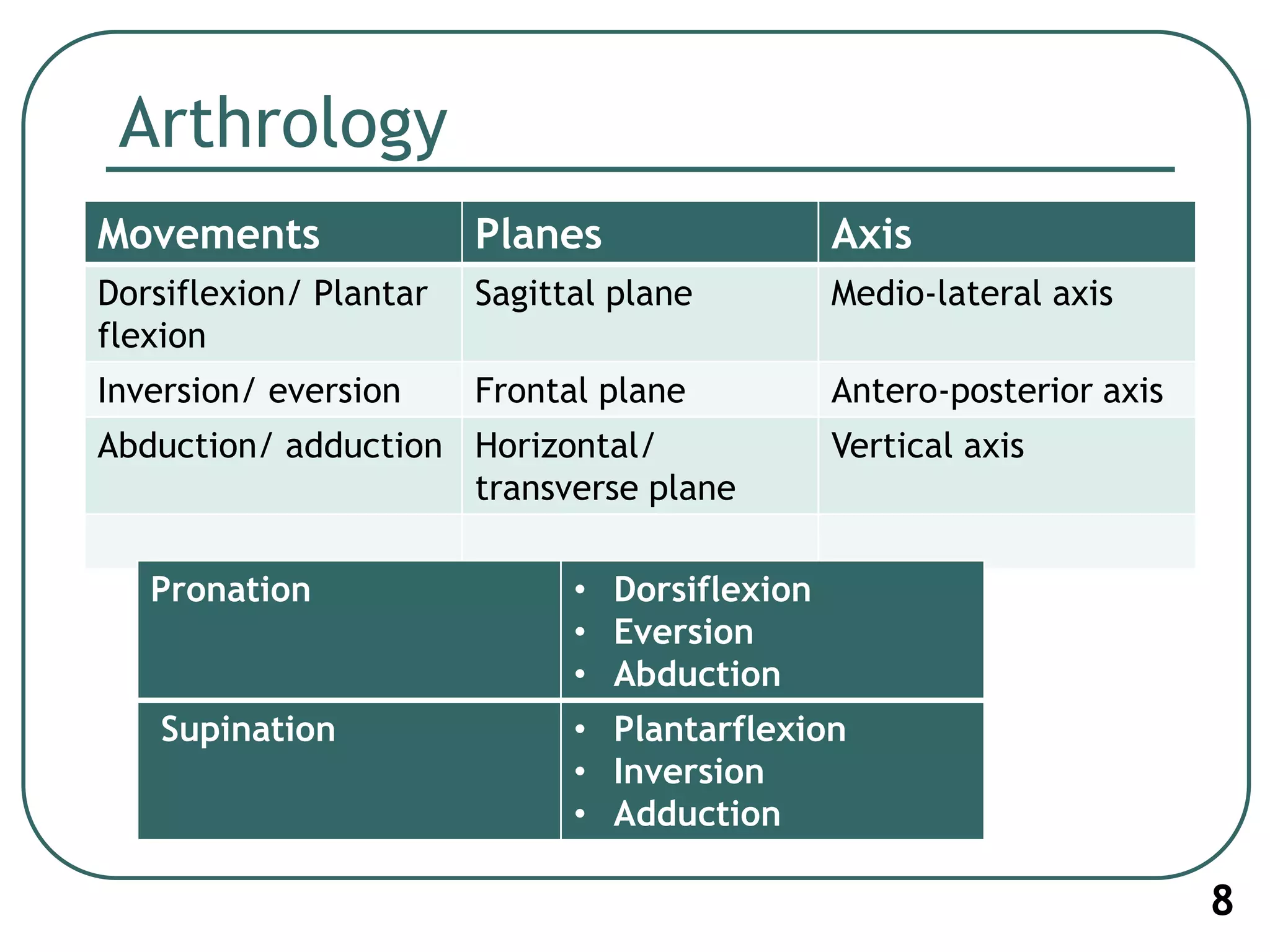
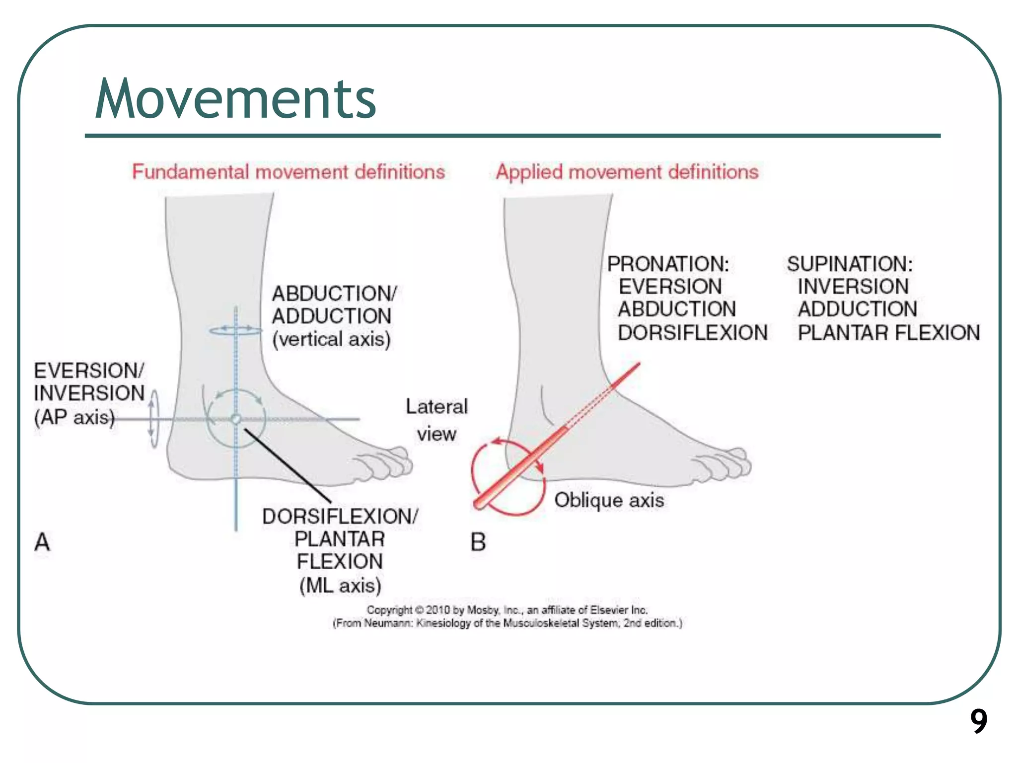
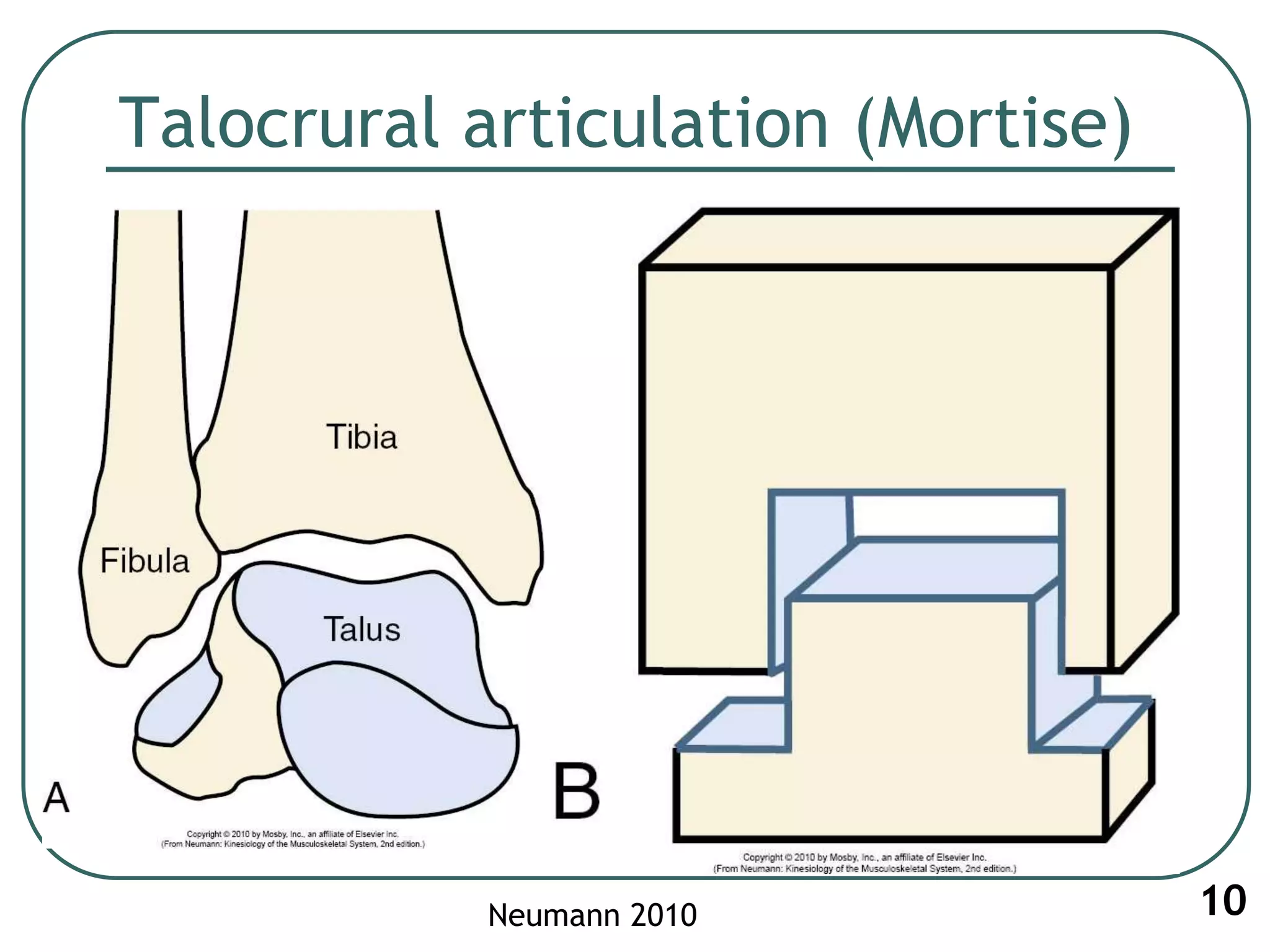
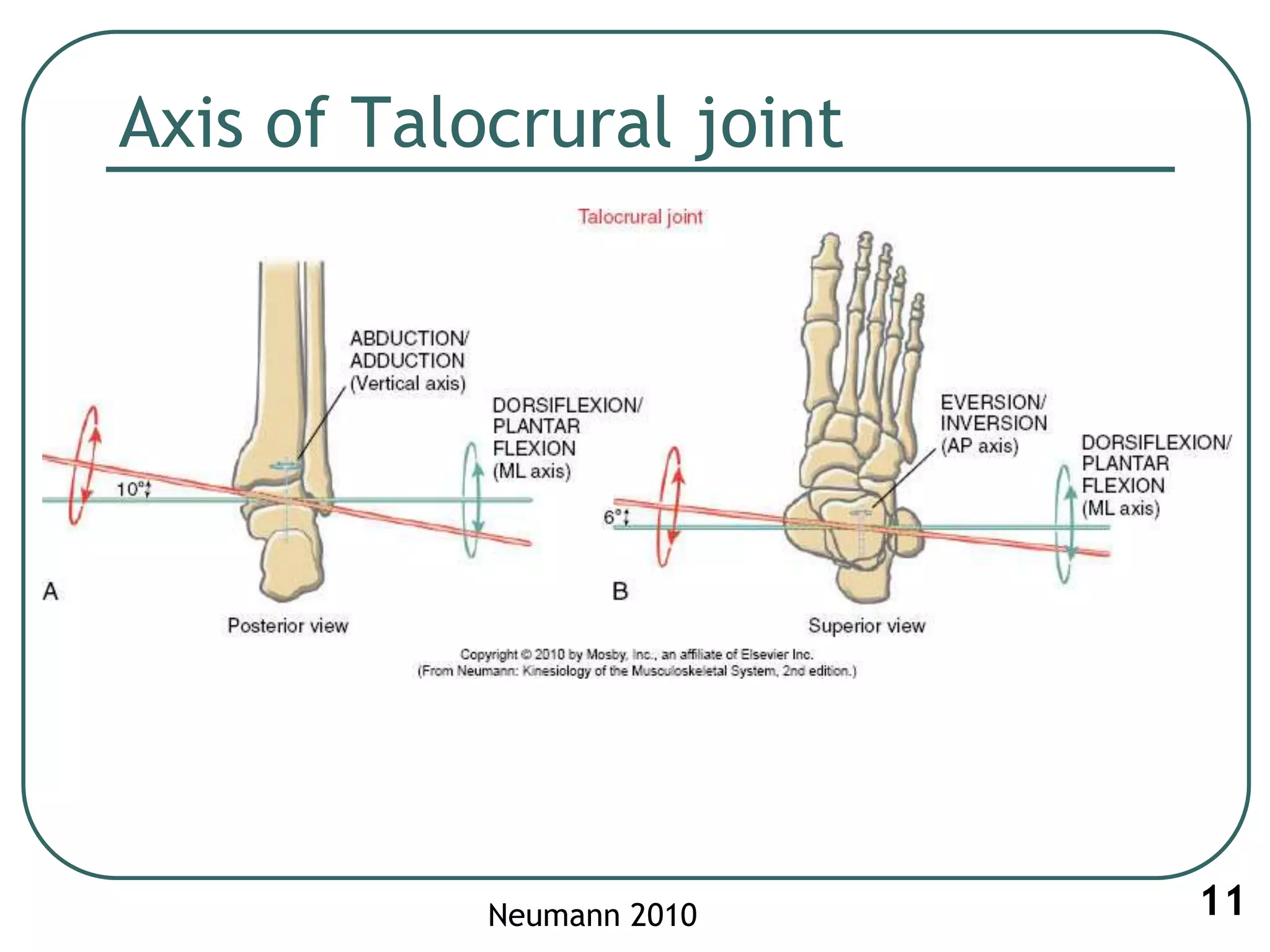
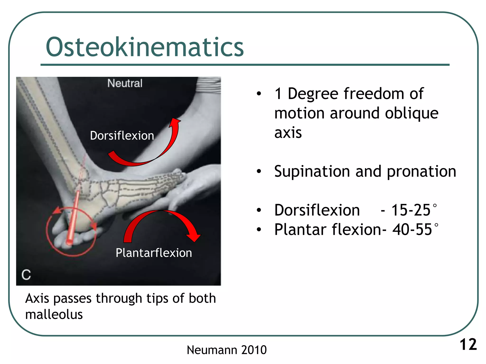
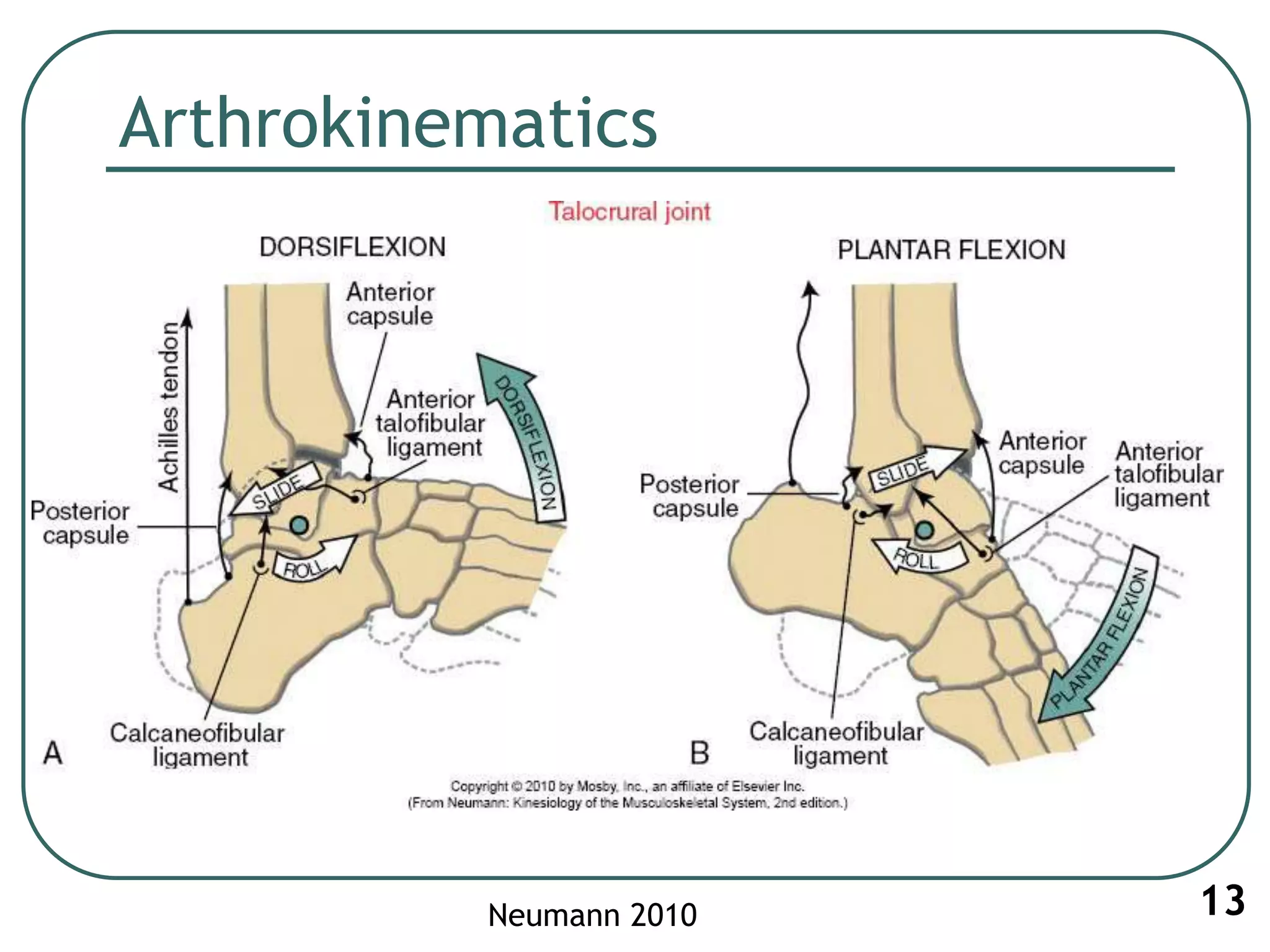
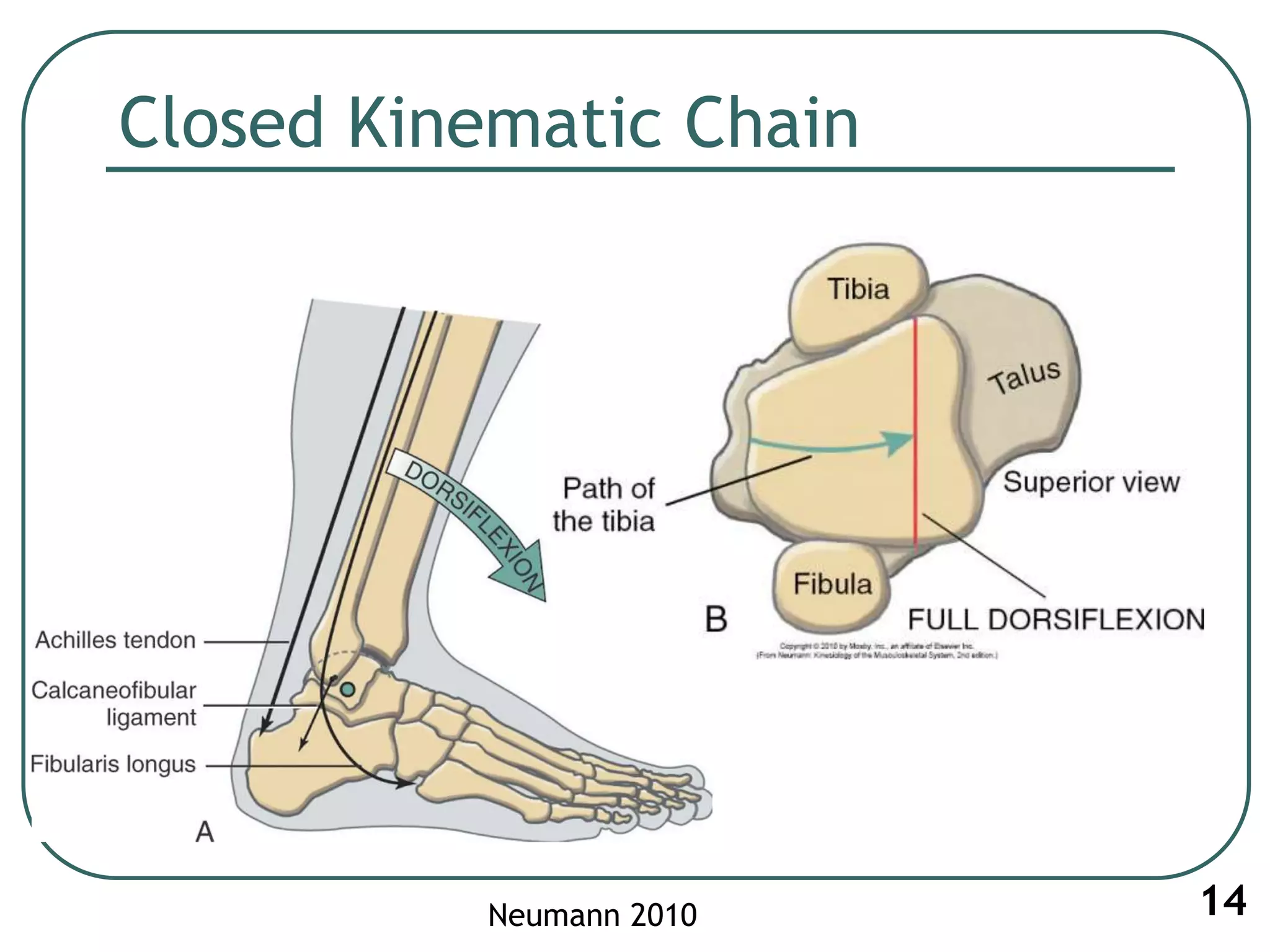
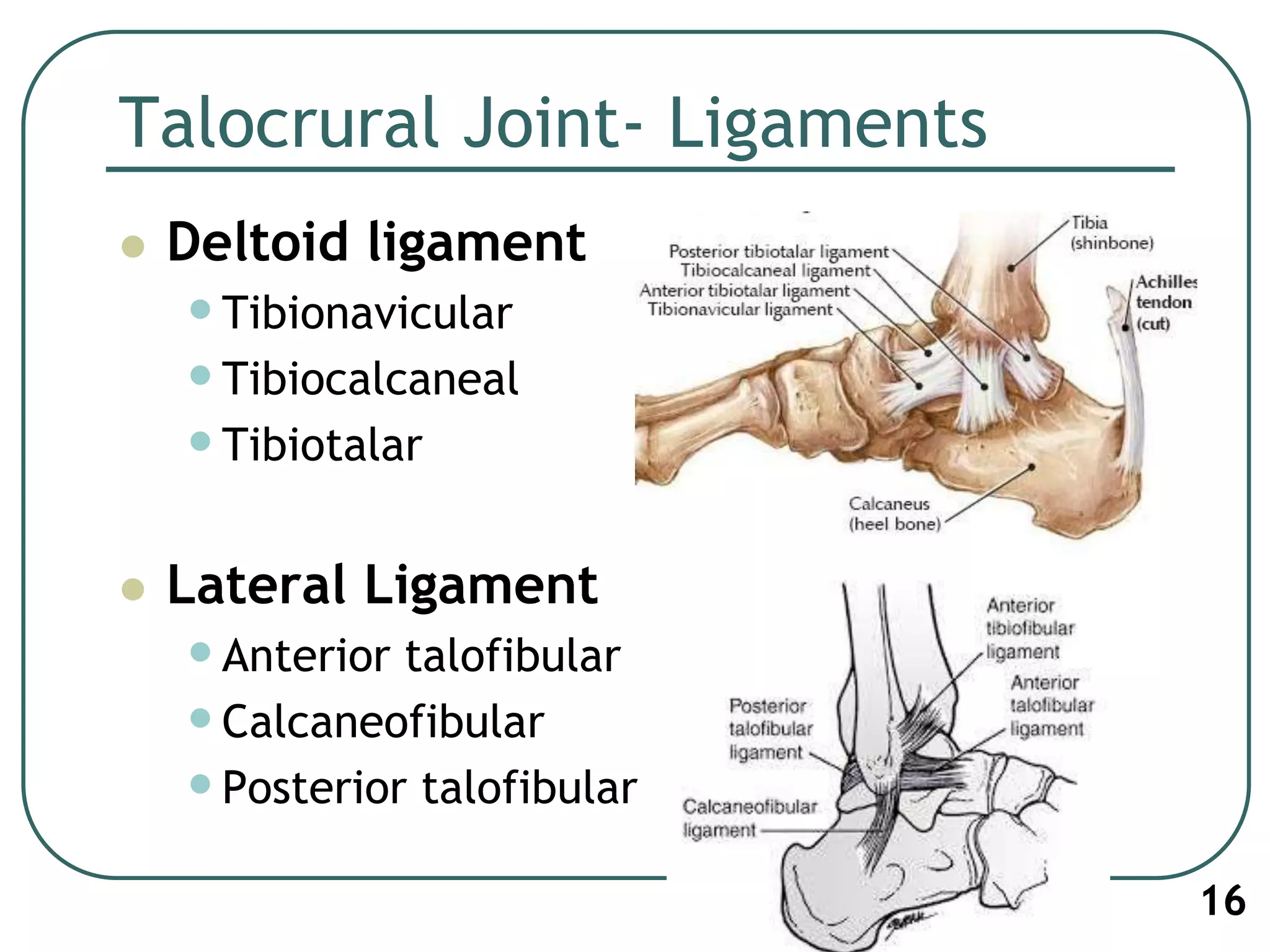
Ankle and Foot
Saurab Sharma, MPT
Lecturer
Contents
 Anatomy review
•Joints of ankle and foot
•Ligaments
•Muscles
 Biomechanics: artho & osteokinematics
2
Functions of ankle and foot
 Absorb stress or shock – pliability of foot
 Propulsion of body in walking and
running- rigid foot
 Protection – sensation of sole of foot
3
Bones of ankle and foot
 26 bones and 2 sesamoids
 Forefoot
•Metatarsals- 5
•Phalanges- 14
 Midfoot
•5 tarsals
 Rearfoot
•Talus and Calcaneus
4
Ankle and foot
 Ankle- Talocrural joint
 Foot- joints and bones
including tarsals, meta
tarsals, phalanges
5
Parts of FOOT
 Fore foot
•Metatarsals and phalanges
 Mid Foot
•Remaining tarsal bones (with
transverse tarsal and inter
tarsal joints)
 Hind Foot
•Talus, calcaneus (with subtalar
joint)
6
Arthrology
7
Joints Type of joint Articular
component
Superior/ Proximal
Tibiofibular joint
Plane synovial
joint
Head of fibula
Posterolateral
aspect of lateral
condyle of tibia
Inferior/ Distal
Tibiofibular joint
Fibrous
Syndesmosis
Distal tibia and
fibula
Ankle/ Talocrural
joint
Hinge Malleolus of tibia
and fibula
Talus
Arthrology
Movements Planes Axis
Dorsiflexion/ Plantar
flexion
Sagittal plane Medio-lateral axis
Inversion/ eversion Frontal plane Antero-posterior axis
Abduction/ adduction Horizontal/
transverse plane
Vertical axis
Pronation • Dorsiflexion
• Eversion
• Abduction
Supination • Plantarflexion
• Inversion
• Adduction
8
Movements
9
Talocrural articulation (Mortise)
10Neumann 2010
Axis of Talocrural joint
11Neumann 2010
Osteokinematics
12Neumann 2010
• 1 Degree freedom of
motion around oblique
axis
• Supination and pronation
• Dorsiflexion - 15-25°
• Plantar flexion- 40-55°
Axis passes through tips of both
malleolus
Dorsiflexion
Plantarflexion
Arthrokinematics
13Neumann 2010
Closed Kinematic Chain
14Neumann 2010
Stability of Joints associated with Ankle
 Superior Tibio-fibular joint: Synovial
•Joint Capsule
•Popliteus
 Inferior Tibio-fibular joint: Syndesmosis
•Ant and post tibiofibular joint
•Interosseous ligament
15
Talocrural Joint- Ligaments
 Deltoid ligament
•Tibionavicular
•Tibiocalcaneal
•Tibiotalar
 Lateral Ligament
•Anterior talofibular
•Calcaneofibular
•Posterior talofibular
16
Subtalar Joint
 Pronation and supination
 Closed chain and open chain motion
 Important for walking uphill, standing with
feel kept wide apart, agility activities,
balancing on unstable surface
17
Subtalar Joint
 Articulation: posterior, middle
and anterior facets of
calcaneus and talus.
 Ligaments- medial, posterior
and lateral talocalcaneal
ligament, cervical ligament
 Calcaneofibular lig limits
excessive inversion, deltoid lig
limits eversion
18
Subtalar Joint Axis
19
Motion at Subtalar
20
anteriorly,
medially and
superiorly
Inversion = 2 X eversion?
(22.6°, 12.5°)
• Lateral malleolus
• Deltoid ligament
Transverse Tarsal (Mid tarsal) Joint
 Talonavicular + Calcaneocuboid joint
 Connects rearfoot and mid foot
 Equal through all the planes
 Role to adapt to variety of surface contours
21
Talonavicular joint
 Resembles a Ball and socket joint
 Provides substantial mobility to medial
longitudinal arch
 Much movement as inversion and eversion
(twisting)
 Head of talus (convex); proximal navicular
(concave)
22
Talonavicular joint (TNJ)
Spring ligament:
 Plantar calcaneo-navicular
 From sustentaculum talus
(of calcaneus) to medial
plantar surface of navicular
bone
 Forms floor and medial wall
of TNJ
23
Calcaneocuboid Joint
 Anterior (distal) surface of
calcaneus and proximal
surface of cuboid
 Dorsal calcaneocuboid
ligament
 Bifurcated ligament (Y-
shaped)
 Long and short plantar
ligament
24
Transverse Tarsal joint
 Axes of Rotation-
•Longitudinal- inversion/ eversion
•Oblique – combination of abduction+ dorsiflexion
and adduction+ plantarflexion
 Midfoot allows twice as much as supination
(20-25° of inversion) compared to pronation
(10- 15° of eversion)
25
Transverse Tarsal Joint:
Longitudinal axis (Inversion/Eversion)
26
Transverse tarsal joint
Oblique axis (DF/PF + Abd/Add)
27
Pronation of the foot
28
Supination of the foot
29
Medial Longitudinal Arch of foot
 Load bearing and
shock absorbing
 Keystone-
talonavicular joint
30
Passive support of Medial Longitudinal Arch
 Bones- 3 medial metatarsals, 3 cuneiforms,
navicular bone, talus, calcaneus
 TNJ, plantar fascia, spring ligament, 1st TMT
joint
31
Arch of Foot
Normal arch of Foot Dropped arch of Foot
32
Pes Cavus (Increased Arch)
33
Summary
 Ankle joint
 Subtalar joint
 Transverse tarsal joint
 Arch of foot
34
Foot posture and tibial rotation
 Hindfoot
Pronation
35
Foot posture and tibial rotation
36
Rarefoot
Supination
Distal Inter tarsal Joints
 Cuneonavicular Joint
 Cuboideonavicular Joint
 Intercuneiform and cuneocuboid joint
complex
Functions:
 Assist TTJ in pronation and supination
 Stability across midfoot
 Formation of transverse arch of foot
37
Distal Inter tarsal Joints
38
TMT joint (Lisfranc’s Joint)
 Separates forefoot and midfoot
 Base of metatarsals + distal surfaces of
cuneiform and cuboid
 Ligaments: dorsal, plantar and iterosseous
ligaments
 Capsule: well developed for 1st TMT joint
39
TMT joint
41
Function of Mid and forefoot
42
1st MTP joint
44
(Flexor Hallucis Brevis)
Hallux Valgus
45
Intrinsic muscles
46
Windlass Effect
Summary!!!!
47

1. Biomechanics of ankle joint subtalar joint and foot

  • 1.
    Ankle and Foot SaurabSharma, MPT Lecturer
  • 2.
    Contents  Anatomy review •Jointsof ankle and foot •Ligaments •Muscles  Biomechanics: artho & osteokinematics 2
  • 3.
    Functions of ankleand foot  Absorb stress or shock – pliability of foot  Propulsion of body in walking and running- rigid foot  Protection – sensation of sole of foot 3
  • 4.
    Bones of ankleand foot  26 bones and 2 sesamoids  Forefoot •Metatarsals- 5 •Phalanges- 14  Midfoot •5 tarsals  Rearfoot •Talus and Calcaneus 4
  • 5.
    Ankle and foot Ankle- Talocrural joint  Foot- joints and bones including tarsals, meta tarsals, phalanges 5
  • 6.
    Parts of FOOT Fore foot •Metatarsals and phalanges  Mid Foot •Remaining tarsal bones (with transverse tarsal and inter tarsal joints)  Hind Foot •Talus, calcaneus (with subtalar joint) 6
  • 7.
    Arthrology 7 Joints Type ofjoint Articular component Superior/ Proximal Tibiofibular joint Plane synovial joint Head of fibula Posterolateral aspect of lateral condyle of tibia Inferior/ Distal Tibiofibular joint Fibrous Syndesmosis Distal tibia and fibula Ankle/ Talocrural joint Hinge Malleolus of tibia and fibula Talus
  • 8.
    Arthrology Movements Planes Axis Dorsiflexion/Plantar flexion Sagittal plane Medio-lateral axis Inversion/ eversion Frontal plane Antero-posterior axis Abduction/ adduction Horizontal/ transverse plane Vertical axis Pronation • Dorsiflexion • Eversion • Abduction Supination • Plantarflexion • Inversion • Adduction 8
  • 9.
  • 10.
  • 11.
    Axis of Talocruraljoint 11Neumann 2010
  • 12.
    Osteokinematics 12Neumann 2010 • 1Degree freedom of motion around oblique axis • Supination and pronation • Dorsiflexion - 15-25° • Plantar flexion- 40-55° Axis passes through tips of both malleolus Dorsiflexion Plantarflexion
  • 13.
  • 14.
  • 15.
    Stability of Jointsassociated with Ankle  Superior Tibio-fibular joint: Synovial •Joint Capsule •Popliteus  Inferior Tibio-fibular joint: Syndesmosis •Ant and post tibiofibular joint •Interosseous ligament 15
  • 16.
    Talocrural Joint- Ligaments Deltoid ligament •Tibionavicular •Tibiocalcaneal •Tibiotalar  Lateral Ligament •Anterior talofibular •Calcaneofibular •Posterior talofibular 16
  • 17.
    Subtalar Joint  Pronationand supination  Closed chain and open chain motion  Important for walking uphill, standing with feel kept wide apart, agility activities, balancing on unstable surface 17
  • 18.
    Subtalar Joint  Articulation:posterior, middle and anterior facets of calcaneus and talus.  Ligaments- medial, posterior and lateral talocalcaneal ligament, cervical ligament  Calcaneofibular lig limits excessive inversion, deltoid lig limits eversion 18
  • 19.
  • 20.
    Motion at Subtalar 20 anteriorly, mediallyand superiorly Inversion = 2 X eversion? (22.6°, 12.5°) • Lateral malleolus • Deltoid ligament
  • 21.
    Transverse Tarsal (Midtarsal) Joint  Talonavicular + Calcaneocuboid joint  Connects rearfoot and mid foot  Equal through all the planes  Role to adapt to variety of surface contours 21
  • 22.
    Talonavicular joint  Resemblesa Ball and socket joint  Provides substantial mobility to medial longitudinal arch  Much movement as inversion and eversion (twisting)  Head of talus (convex); proximal navicular (concave) 22
  • 23.
    Talonavicular joint (TNJ) Springligament:  Plantar calcaneo-navicular  From sustentaculum talus (of calcaneus) to medial plantar surface of navicular bone  Forms floor and medial wall of TNJ 23
  • 24.
    Calcaneocuboid Joint  Anterior(distal) surface of calcaneus and proximal surface of cuboid  Dorsal calcaneocuboid ligament  Bifurcated ligament (Y- shaped)  Long and short plantar ligament 24
  • 25.
    Transverse Tarsal joint Axes of Rotation- •Longitudinal- inversion/ eversion •Oblique – combination of abduction+ dorsiflexion and adduction+ plantarflexion  Midfoot allows twice as much as supination (20-25° of inversion) compared to pronation (10- 15° of eversion) 25
  • 26.
    Transverse Tarsal Joint: Longitudinalaxis (Inversion/Eversion) 26
  • 27.
    Transverse tarsal joint Obliqueaxis (DF/PF + Abd/Add) 27
  • 28.
  • 29.
  • 30.
    Medial Longitudinal Archof foot  Load bearing and shock absorbing  Keystone- talonavicular joint 30
  • 31.
    Passive support ofMedial Longitudinal Arch  Bones- 3 medial metatarsals, 3 cuneiforms, navicular bone, talus, calcaneus  TNJ, plantar fascia, spring ligament, 1st TMT joint 31
  • 32.
    Arch of Foot Normalarch of Foot Dropped arch of Foot 32
  • 33.
  • 34.
    Summary  Ankle joint Subtalar joint  Transverse tarsal joint  Arch of foot 34
  • 35.
    Foot posture andtibial rotation  Hindfoot Pronation 35
  • 36.
    Foot posture andtibial rotation 36 Rarefoot Supination
  • 37.
    Distal Inter tarsalJoints  Cuneonavicular Joint  Cuboideonavicular Joint  Intercuneiform and cuneocuboid joint complex Functions:  Assist TTJ in pronation and supination  Stability across midfoot  Formation of transverse arch of foot 37
  • 38.
  • 39.
    TMT joint (Lisfranc’sJoint)  Separates forefoot and midfoot  Base of metatarsals + distal surfaces of cuneiform and cuboid  Ligaments: dorsal, plantar and iterosseous ligaments  Capsule: well developed for 1st TMT joint 39
  • 40.
  • 41.
    Function of Midand forefoot 42
  • 42.
    1st MTP joint 44 (FlexorHallucis Brevis)
  • 43.
  • 44.
  • 45.

Editor's Notes

  • #11 Like a carpenters mortise
  • #13 Single axis of rotation for Dorsi and plantar flexion/ one degree freedom of motion
  • #15 Tibia rolls anteriorly in CKC Dorsiflexion, and broader anterior part of talus distracts the distal TF joint causing high ankle sprain
  • #16 Firm stabilization is needed at Proximal TF joint so that the force is transmitted from biceps femoris and LCL of knee to fibula then to tibia …
  • #17 Posterior talofibular lig resists abduction of talus with dorsiflexion
  • #19 Circles denote separate capsules for each circle Ligaments reinforce the joint capsules
  • #20 Pronation supination at subtalar has two evident movements- inversion/eversion and adduction abduction. DF and PF is small and is ignored clinically
  • #21 Measurement of pronation and supination by rarefoot inversion and eversion by placing the subtalar in neural
  • #25 (concavoconvex surface in each)
  • #26 (concavoconvex surface in each)
  • #29 Peroneus longus lowers the medial longitudinal arch of the foot while pronation
  • #31 Stability and resilency of foot during wt bearing
  • #32 3 medial metatarsals, 3 cuneiforms, navicular bone, talus, calcaneus
  • #33 Normally rear foot receives twice as much load as forefoot!!!
  • #38 Small motion
  • #40 Base joint for forefoot. Least mobility at 2nd and 3rd joint. Provides longitudinal stability
  • #43 A- transverse arch of foot B- stable 2nd metatarsal joint C- Medial Cuneiform