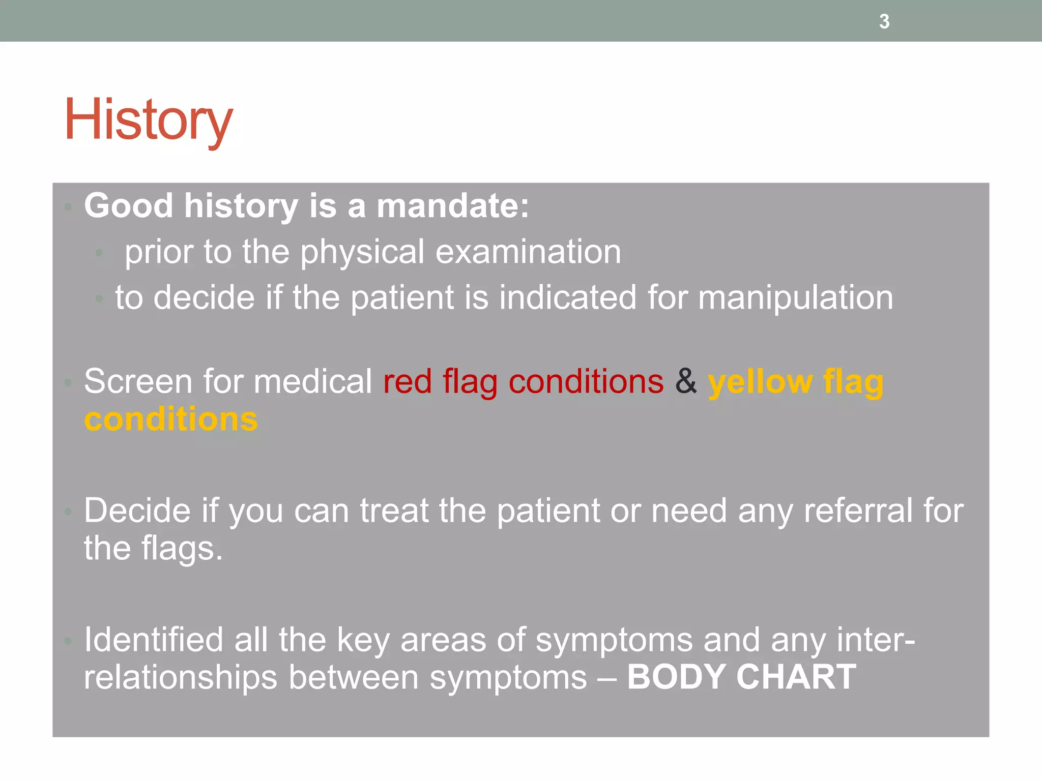
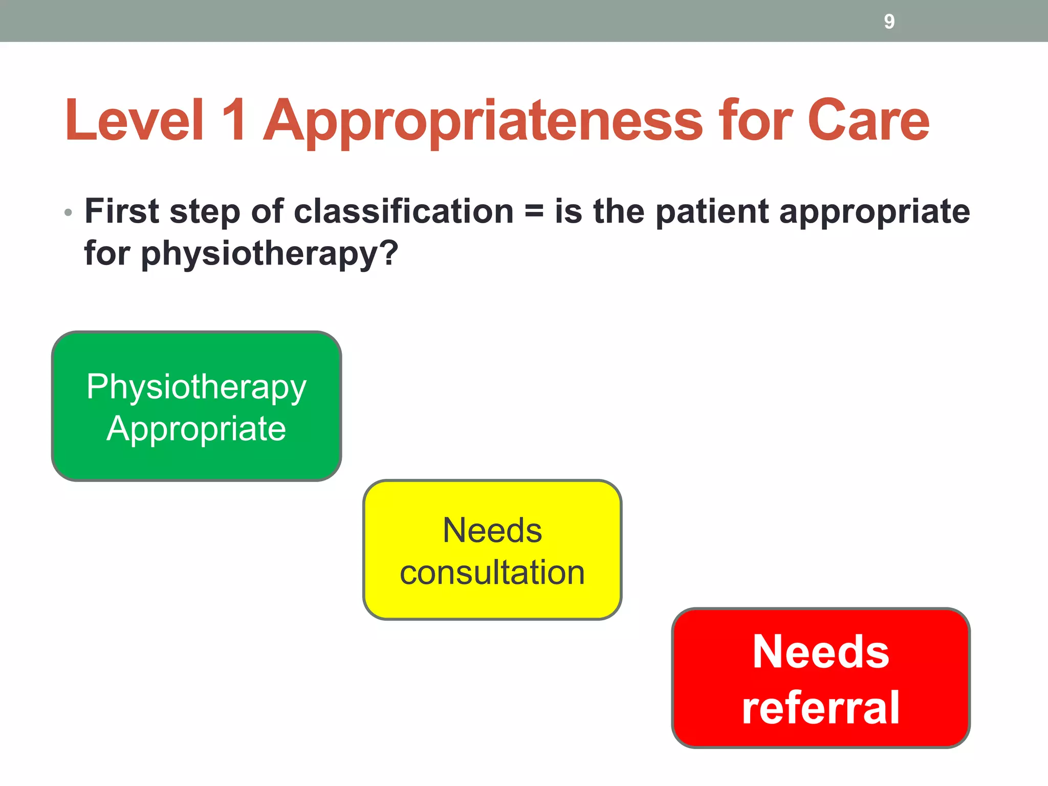
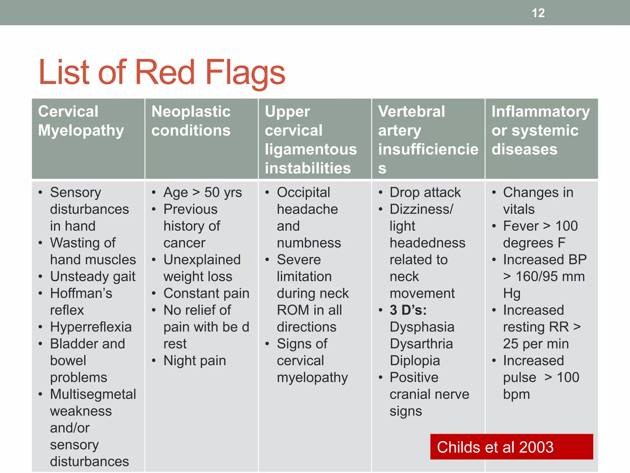
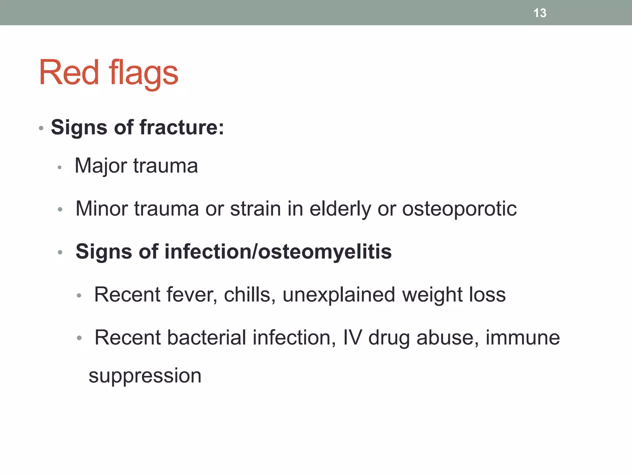
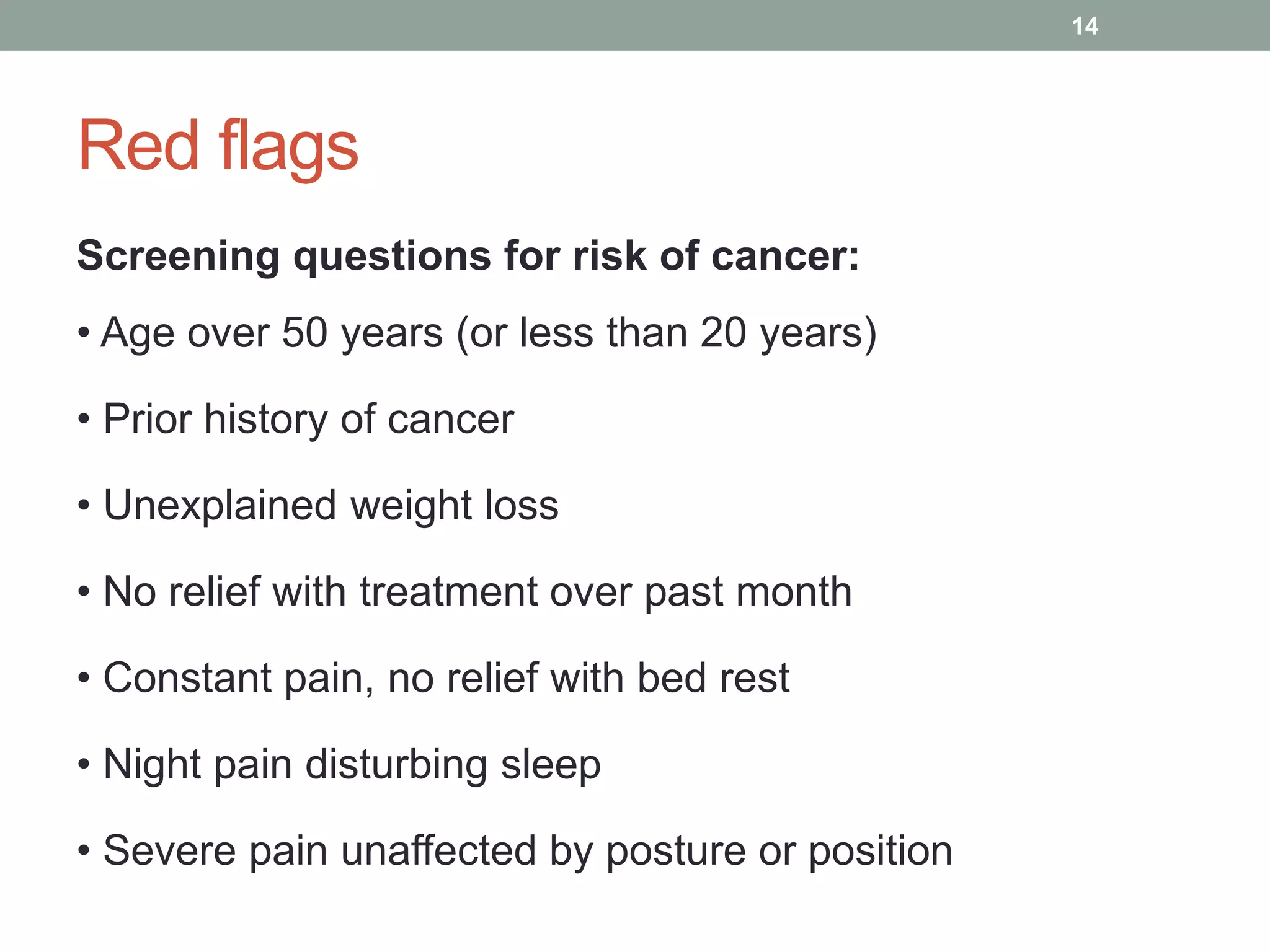
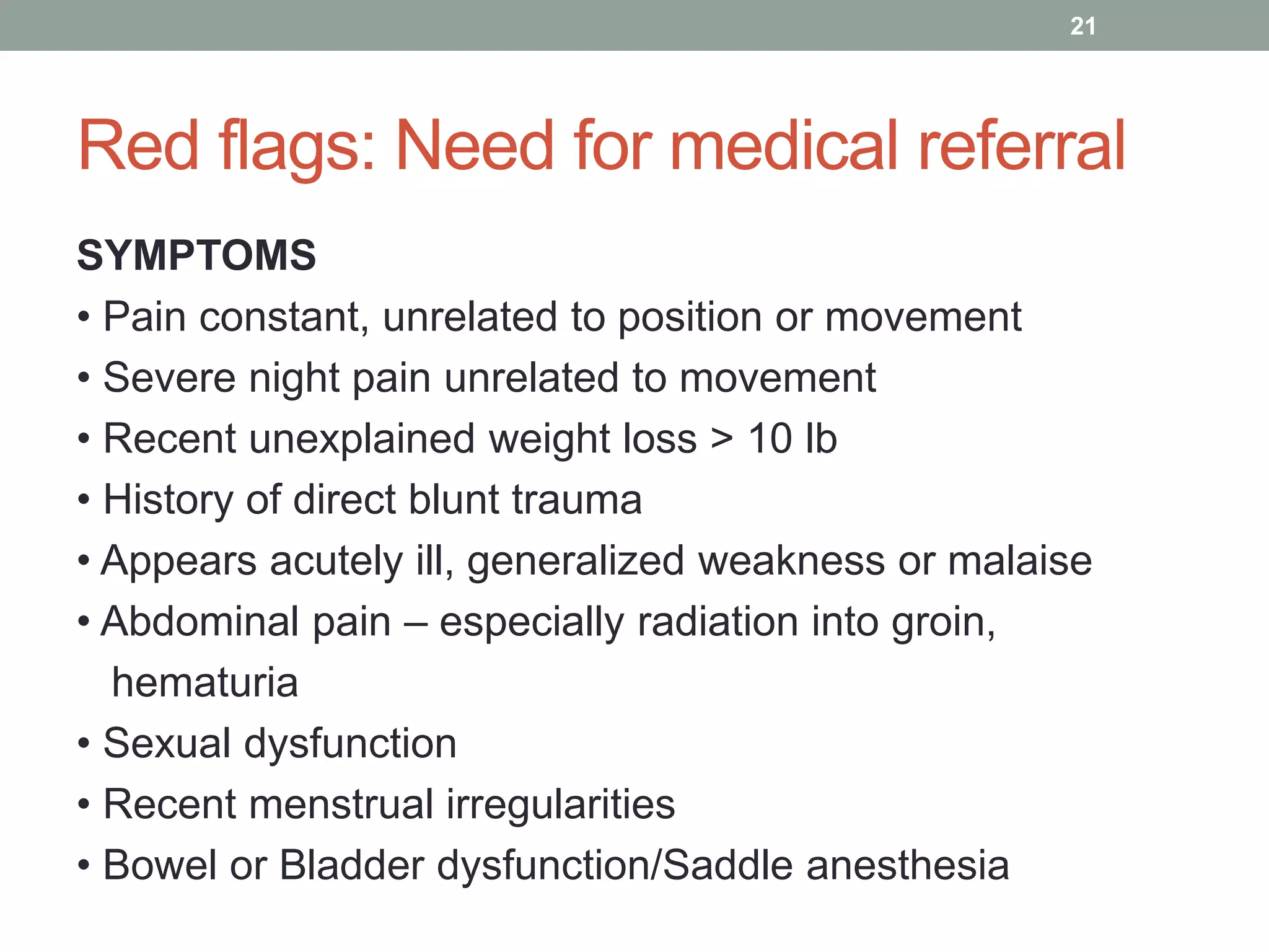
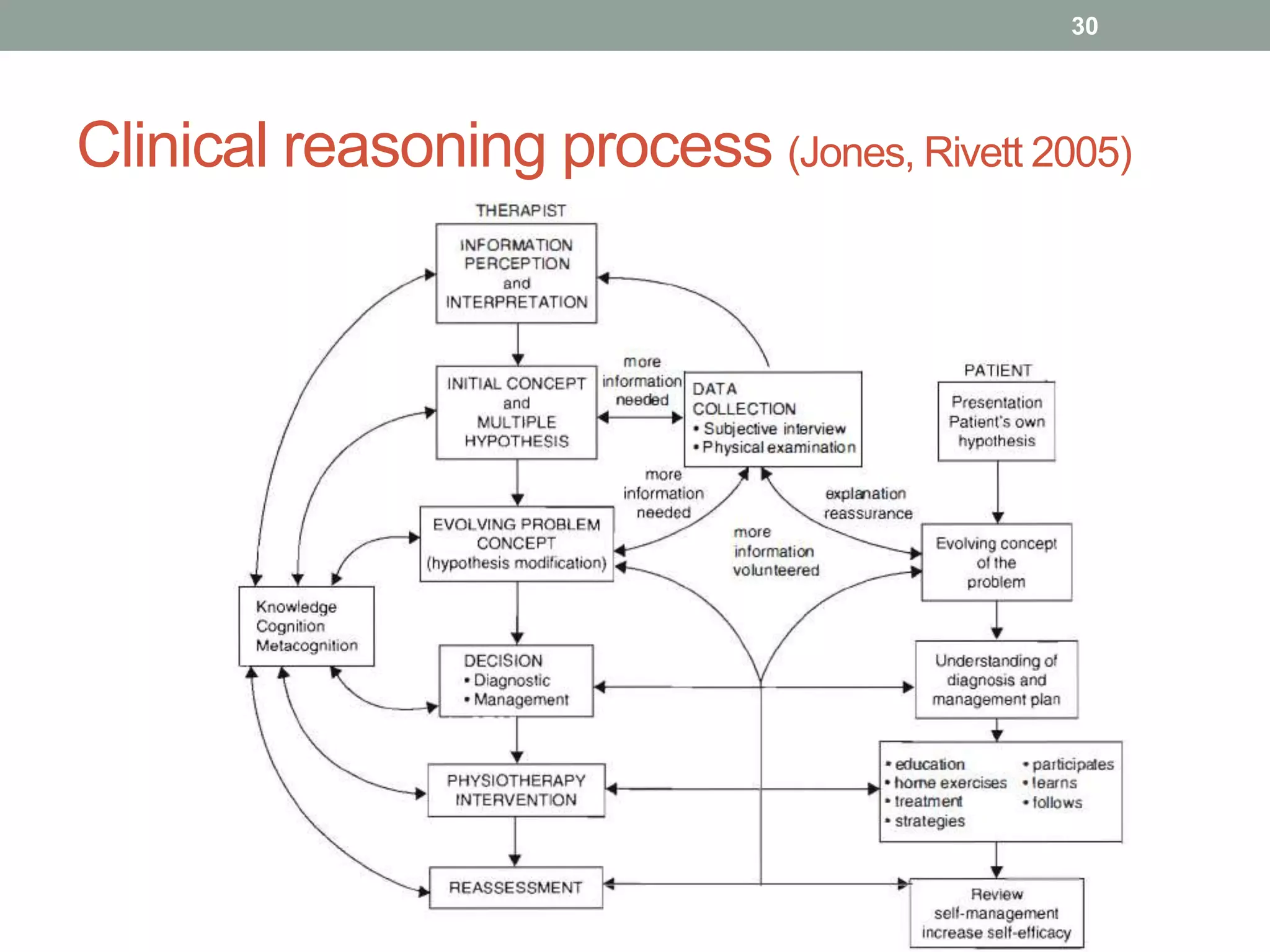
MANIPULATION
PRINCIPLES
Saurab Sharma, MPT
Lecturer, KUSMS
Contents
• Background assessment for manipulation
• Subjective history including flags
• Objective assessment
• Screening tools/ measures
• Maitland’s grades of mobilization and manipulation
• Appropriateness for care
• Overview of treatment based classification
• Manipulation- Indications / Clinical prediction rule
• Precautions and contraindications
• Risk/ side effects/ dangers of manipulation
2
History
• Good history is a mandate:
• prior to the physical examination
• to decide if the patient is indicated for manipulation
• Screen for medical red flag conditions & yellow flag
conditions
• Decide if you can treat the patient or need any referral for
the flags.
• Identified all the key areas of symptoms and any inter-
relationships between symptoms – BODY CHART
3
History
• Ask questions to help differentiate between symptoms
coming from the thoracic spine, lumbar spine/pelvis, hip,
neural tissues etc.
• Determine a primary hypothesis (& 1- 2 additional)
• Decide:
1) what symptoms you want to bring on in the exam and
why
2) what symptoms you don’t want to bring on and why, and
3) What tests/measures are needed to rule in/out your
hypotheses
4
Terminologies
• Mobilization ‐ A passive therapeutic movement within a
range of motion at variable amplitudes and speed. (Note‐
not always at the end of the available range)
• Manipulation‐ A passive therapeutic movement, of small
amplitude and high velocity at the end of the available
range of motion.
• Active Physiologic movements ‐ Voluntary motion by
the patient such as standard flexion, abduction and
rotation.
5
Terms specific to spine
• PPIVM
– Passive Physiological Inter-Vertebral Movements
• PAIVM
– Passive Accessory Inter-Vertebral Movements
6
GRADES OF MOBILIZATION (Maitland)
I Small amplitude out of resistance
II Large amplitude out of resistance
III Large amplitude into resistance
IV Small amplitude into resistance
V High velocity thrust
Resistance
7
What determines force?
• Stage of healing
• SINSS
• Patient response to intervention
• Grades I and II used to treat pain prior to reaching
resistance
• Grades III and IV used to treat resistance (joint
restrictions) When pain is not a limitation
• Grade V used to treat resistance historically but may have
other neurophysiological benefits
8
Level 1 Appropriateness for Care
• First step of classification = is the patient appropriate
for physiotherapy?
Physiotherapy
Appropriate
Needs
consultation
Needs
referral
9
Level 1 Appropriateness for Care
Physiotherapy
PT +
Consultation Referral
Spinal
Symptoms of
mechanical
original Psychological
Medical
Psychological
Medical/
surgical
10
11
List of Red Flags
Cervical
Myelopathy
Neoplastic
conditions
Upper
cervical
ligamentous
instabilities
Vertebral
artery
insufficiencie
s
Inflammatory
or systemic
diseases
• Sensory
disturbances
in hand
• Wasting of
hand muscles
• Unsteady gait
• Hoffman’s
reflex
• Hyperreflexia
• Bladder and
bowel
problems
• Multisegmetal
weakness
and/or
sensory
disturbances
• Age > 50 yrs
• Previous
history of
cancer
• Unexplained
weight loss
• Constant pain
• No relief of
pain with be d
rest
• Night pain
• Occipital
headache
and
numbness
• Severe
limitation
during neck
ROM in all
directions
• Signs of
cervical
myelopathy
• Drop attack
• Dizziness/
light
headedness
related to
neck
movement
• 3 D’s:
Dysphasia
Dysarthria
Diplopia
• Positive
cranial nerve
signs
• Changes in
vitals
• Fever > 100
degrees F
• Increased BP
> 160/95 mm
Hg
• Increased
resting RR >
25 per min
• Increased
pulse > 100
bpm
Childs et al 2003
12
Red flags
• Signs of fracture:
• Major trauma
• Minor trauma or strain in elderly or osteoporotic
• Signs of infection/osteomyelitis
• Recent fever, chills, unexplained weight loss
• Recent bacterial infection, IV drug abuse, immune
suppression
13
Red flags
Screening questions for risk of cancer:
• Age over 50 years (or less than 20 years)
• Prior history of cancer
• Unexplained weight loss
• No relief with treatment over past month
• Constant pain, no relief with bed rest
• Night pain disturbing sleep
• Severe pain unaffected by posture or position
14
Red flags
Signs of cauda equina syndrome:
• Paresthesia of 4th sacral dermatome (saddle region)
• Alteration in bowel or bladder function (increased
frequency, overflow incontinence, etc.)
• Severe or progressive neurological deficits
Cauda Equina Syndrome Necessitates
Immediate Referral!
16
Referred pain to shoulder
• Liver R
• Stomach R
• Pancreas R
• Pancoast’s tumor L/R
• Myocardium L
• Spleen L
17
Other causes of low back pain
• ?
18
Red flags
Screening questions for risk of ankylosing spondylitis:
• Morning stiffness
• Improvement with activity
• Age < 40 years
• Local SIJ tenderness
• Pain not relieved when supine
• Paraspinal muscle spasm
19
Red flags: Need for medical referral
SIGNS
• Temp > 100° F
• BP > 160/95 mmHg
• Resting Pulse > 100/min
• Resting Respiration > 25/min
20
Red flags: Need for medical referral
SYMPTOMS
• Pain constant, unrelated to position or movement
• Severe night pain unrelated to movement
• Recent unexplained weight loss > 10 lb
• History of direct blunt trauma
• Appears acutely ill, generalized weakness or malaise
• Abdominal pain – especially radiation into groin,
hematuria
• Sexual dysfunction
• Recent menstrual irregularities
• Bowel or Bladder dysfunction/Saddle anesthesia
21
Yellow flags
• “Yellow flags are factors that increase the risk of
developing, or perpetuating long-term disability
and work loss associated with low back pain.”
(Kendall et al, 1997)
22
Level 2
• Staging not based on time since onset
• Based upon symptoms and functional limitations
Tools
• Oswestry Disability Questionnaire
• Numeric Pain Rating Index
23
24
Level 3
• Treatment Based Classification System
• Diagnosis: “The process of determining the cause of a
patient’s illness or discomfort”
• Classification: “The process of classifying clinical data
into named categories of clinical entities for the purpose
of making clinical decisions regarding therapeutic
• management”
(Rose, 1989)
28
29
Clinical reasoning process (Jones, Rivett 2005)
30
LUMBOPELVIC EXAMINATION
In Order of Exam Sequence
A thorough competently performed examination is
therapeutic.
The examination is an important ritual.
31
SCREENING/ OUTCOME MEASURES
• Medical History Form
• Modified Oswestry Questionnaire (OSW)
• Fear-avoidance Beliefs Questionnaire (FABQ)
• Pain Diagram
32
Oswestry disability index (ODI)
• 10 questions related to function
• Modified changes sex question
• Max of 5 points per question
• Score is reported as a percentage: (Score X 2)%
• 0-20% mild
• 20-40% moderately impaired
• 40-75% severely impaired
• >75% likely non-movement component if not hospitalized
• Clinically Meaningful Change = 6 - 10
• <12% can safely return to work and normal activities
33
Subjective Examination
• The patient’s story
• Provides most (80%) of the information needed to
• clarify the cause or establish a hypothesis
Components of the SE:
• Patient profile
• Chief complaint
• Body chart
• AGG/Ease factors
• 24-hour behavior
• Special questions
• Present episode
• Past history
34
Mobilization principles (Maitland and Greenmann)
• Patient must be completely relaxed
• Operator must be relaxed
• Patient must be comfortable and have complete
confidence in the operator’s grasp
• Embrace the joint to be moved, hold around the joint
to feel movement
35
Mobilization principles (Maitland and Greenmann)
• Move one joint, one motion at one time
• Patient must be confident that the joint will not be hurt
• Operator’s position must be comfortable and easy to
maintain
• Operator’s position must afford him/her complete
control
36
Contraindications to thrust manipulations
• Vertebral Malignancy
• Local Infection
• Severe Inflammation
• Osteoporosis
• Aneurysm
• Myelopathy
• Multiple Adjacent
• Radiculopathies
• Cauda Equina Syndrome
• Vertebral Bone Disease
37
• Bony Joint Instability
• Cervical Rheumatoid Disease
• Vertebral Fractures
• Systemic Anticoagulation
• Severe Diabetes
• Atherosclerotic Disease
• Vertebral Basilar Disease
• Active Spondyloarthropathies
• Ligamentous Joint Instability
• Congenital Joint Laxity
• Aseptic Necrosis
• Local Osteoporosis,
• Osteomalacia
• Acute Disc Herniation
Precautions to thrust
• Unhealed fracture
• Excessive pain or irritability
• Hypermobility: Do they need it?
• Total joint replacements
• Pregnancy? 1st trimester
• No evidence that it is dangerous, but don’t want to be
associated with miscarriage
• Spondylolisthesis
• Muscle Guarding
• Anticoagulants
38
Risks of Manipulation
• Haldeman and Rubenstein (Spine, 1992) Reviewed the
literature over 77 year period
• Ten episodes of cauda equina syndrome following lumbar
manipulation reported
• Estimated risk: < 1 per 10 million manipulations
39
What are the side effects?
• Senstad et al (Spine, 1997)
• Surveyed 1058 patients treated with spinal manipulation
in Norway
• 75% of all treatments included manipulation to the Lx
Spine
• No severe complications noted
55% reported at least one side effect
- Local discomfort-53%
- Headache-12%
- Fatigue-11%
- Radiating discomfort- 10%
40
What are the side effects?
• LeBoeuf-Yde et al (J Manip Physiol Ther, 1997)
surveyed 625 patients treated with 1856 spinal
manipulations in Sweden
• No severe complications / injuries noted
• 44% reported at least one side effect
• Local discomfort, fatigue, headache
• Symptoms resolved < 48 hours in 81%
41
How does this risk compare to risk associated
with other medical interventions for patients
with low back pain?
42
43
Sensory Examination
44
Motor examination
45
Reflexes
46
Vascular screening
47
Functional Quick Tests
• Patient demonstrates activity that causes symptoms or
therapist identifies functional activity that is problematic
Frequent ‘functional quick tests”
• Step-Up,
• Step-Down,
• Squat,
• gait,
48
bending/lifting,
Sit-to-stand,
on/off socks,
crossing legs, etc
Functional Quick Tests
• Assess quality, ROM, pain (0-10), symptom location
Use for:
• Re-assessment after interventions (‘test/retest’)
• Differential diagnosis of primary pain generator
49
Postural examination with plumb line
50
Lumbar AROM
(w or w/o overpressure)
• Flexion
• Extension
• Side bending
• Quadrant – sustained
• Identify a Comparable Sign **
• Remember to re-test after treatment!
51
Test re-test
• Recheck the impairment, comparable
• Sign immediately following intervention
• Assess change
• Reinforcement with home program
52
ROM - inclinometer
Flexion
• Patient assumes standardized foot position, goniometer
placed
• Patient fully flexes trunk without bending knees.
• Therapist records measurement at end-range to nearest
degree
Extension
• From starting position, patient fully extends trunk without
bending knees (therapist may support)
• Therapist records measurement at end-range to nearest
degree
53
Goniometry: Side bending
Side bending
• Patient assumes standardized foot position, goniometer
placed
• Patient instructed to slide hand down thigh and fully side-
bends trunk without bending knees
• Therapist records measurement at end-range to nearest
degree
• Repeat on opposite side
54
AROM
Flexion With Overpressure
• Standardize patient positioning
• Ask the patient to fully flex the lumbar spine while keeping
the knees straight
• Apply overpressure by adducting your arms
• Add neck flexion to differentiate adverse neurodynamics
from other sources of pain or decreased ROM
• Note end-feel, range, pain and resistance
55
AROM
Extension With Overpressure
• Standardize patient positioning
• Ask the patient to fully extent his lumbar spine
• Apply overpressure as indicated
• Note end-feel, range, pain and resistance
56
Lumbar Quadrant
• Standardize patient positioning
• Stabilize the pelvis
• Guide the patient into Left Rotation, left side flexion,
extension
• Sustain for 5 seconds if needed
• Note end-feel, range, pain and resistance
57
Thoracic Screening
• The therapist stabilizes the pelvis and hips by supporting
the patients knees as shown
• Passively rotate the patient’s trunk in both directions
• Apply overpressure at end range.
• Positive Finding:
• Reproduction of pain or familiar symptoms. If positive, a
detailed exam of the thoracic spine and rib cage should
be considered.
58
Spine vs. Hip Differentiation
• The therapist can localize movement to hip by ensuring
trunk and pelvis move as a unit.
• Repeat rotation again, but this time the therapist localizes
movement to the lumbo-pelvic region by stabilizing the
pelvis.
Positive findings:
1) Reproduction of symptoms when the lumbo-pelvic region
rotates as a unit implicates a hip dysfunction
2) Reproduction of symptoms when the pelvis was
stabilized implicates a dysfunction originating primarily from
the spine
59
Hip Screening
• Therapist stabilizes the iliac crest opposite the tested
lower extremity (LE)
FABER (flexion, abduction, & external rotation)
• Rest ankle of tested LE on opposite knee.
• Apply downward pressure over knee of tested LE, apply
overpressure when endpoint reached
F/Add (flexion, adduction)
• Rest knee/posterior thigh of tested LE on opposite knee.
• Apply adduction force over lateral knee of tested LE,
apply overpressure when endpoint reached
60
Hip Internal & External Rotation
• The patient sits with his hands under his thighs so that his
arms stabilize the thighs laterally
• The therapist sights between knees and passively
internally rotates (IR) the hips bilaterally
• Passively external rotation (ER) of each hip is performed
individually
• Apply overpressure at end-range for both IR & ER
Positive Findings: Judgments
• regarding pain and/or limited motion are made.
• Examine further if positive
61
Prone Lumbar Central/Unilateral PA
• Segmentally palpate lumbar spine
• Note end-feel, range, pain and resistance
• Rate as hypomobile, hypermobile, or normal
• Comparable sign **
62
Five-Factor Prediction Rule
• Duration of symptoms < 16 days
• FABQ work subscale 18 or less
• Symptoms not distal to the knee
• At least one hip internal rotation PROM > 35 degrees
• Hypomobility at one or more lumbar levels with spring
testing
Flynn, et al. Spine 2002; Childs et al. Annals Int Med 2004
• 4/5 met: +LR = 13.2
63
Summary
• Flags
• Levels of assessment
• Treatment based classification
• Contraindications and precautions
• Examination key points
• Prediction rule
64

Principles of Manipulation or manipulative therapy

  • 1.
  • 2.
    Contents • Background assessmentfor manipulation • Subjective history including flags • Objective assessment • Screening tools/ measures • Maitland’s grades of mobilization and manipulation • Appropriateness for care • Overview of treatment based classification • Manipulation- Indications / Clinical prediction rule • Precautions and contraindications • Risk/ side effects/ dangers of manipulation 2
  • 3.
    History • Good historyis a mandate: • prior to the physical examination • to decide if the patient is indicated for manipulation • Screen for medical red flag conditions & yellow flag conditions • Decide if you can treat the patient or need any referral for the flags. • Identified all the key areas of symptoms and any inter- relationships between symptoms – BODY CHART 3
  • 4.
    History • Ask questionsto help differentiate between symptoms coming from the thoracic spine, lumbar spine/pelvis, hip, neural tissues etc. • Determine a primary hypothesis (& 1- 2 additional) • Decide: 1) what symptoms you want to bring on in the exam and why 2) what symptoms you don’t want to bring on and why, and 3) What tests/measures are needed to rule in/out your hypotheses 4
  • 5.
    Terminologies • Mobilization ‐A passive therapeutic movement within a range of motion at variable amplitudes and speed. (Note‐ not always at the end of the available range) • Manipulation‐ A passive therapeutic movement, of small amplitude and high velocity at the end of the available range of motion. • Active Physiologic movements ‐ Voluntary motion by the patient such as standard flexion, abduction and rotation. 5
  • 6.
    Terms specific tospine • PPIVM – Passive Physiological Inter-Vertebral Movements • PAIVM – Passive Accessory Inter-Vertebral Movements 6
  • 7.
    GRADES OF MOBILIZATION(Maitland) I Small amplitude out of resistance II Large amplitude out of resistance III Large amplitude into resistance IV Small amplitude into resistance V High velocity thrust Resistance 7
  • 8.
    What determines force? •Stage of healing • SINSS • Patient response to intervention • Grades I and II used to treat pain prior to reaching resistance • Grades III and IV used to treat resistance (joint restrictions) When pain is not a limitation • Grade V used to treat resistance historically but may have other neurophysiological benefits 8
  • 9.
    Level 1 Appropriatenessfor Care • First step of classification = is the patient appropriate for physiotherapy? Physiotherapy Appropriate Needs consultation Needs referral 9
  • 10.
    Level 1 Appropriatenessfor Care Physiotherapy PT + Consultation Referral Spinal Symptoms of mechanical original Psychological Medical Psychological Medical/ surgical 10
  • 11.
  • 12.
    List of RedFlags Cervical Myelopathy Neoplastic conditions Upper cervical ligamentous instabilities Vertebral artery insufficiencie s Inflammatory or systemic diseases • Sensory disturbances in hand • Wasting of hand muscles • Unsteady gait • Hoffman’s reflex • Hyperreflexia • Bladder and bowel problems • Multisegmetal weakness and/or sensory disturbances • Age > 50 yrs • Previous history of cancer • Unexplained weight loss • Constant pain • No relief of pain with be d rest • Night pain • Occipital headache and numbness • Severe limitation during neck ROM in all directions • Signs of cervical myelopathy • Drop attack • Dizziness/ light headedness related to neck movement • 3 D’s: Dysphasia Dysarthria Diplopia • Positive cranial nerve signs • Changes in vitals • Fever > 100 degrees F • Increased BP > 160/95 mm Hg • Increased resting RR > 25 per min • Increased pulse > 100 bpm Childs et al 2003 12
  • 13.
    Red flags • Signsof fracture: • Major trauma • Minor trauma or strain in elderly or osteoporotic • Signs of infection/osteomyelitis • Recent fever, chills, unexplained weight loss • Recent bacterial infection, IV drug abuse, immune suppression 13
  • 14.
    Red flags Screening questionsfor risk of cancer: • Age over 50 years (or less than 20 years) • Prior history of cancer • Unexplained weight loss • No relief with treatment over past month • Constant pain, no relief with bed rest • Night pain disturbing sleep • Severe pain unaffected by posture or position 14
  • 15.
    Red flags Signs ofcauda equina syndrome: • Paresthesia of 4th sacral dermatome (saddle region) • Alteration in bowel or bladder function (increased frequency, overflow incontinence, etc.) • Severe or progressive neurological deficits Cauda Equina Syndrome Necessitates Immediate Referral! 16
  • 16.
    Referred pain toshoulder • Liver R • Stomach R • Pancreas R • Pancoast’s tumor L/R • Myocardium L • Spleen L 17
  • 17.
    Other causes oflow back pain • ? 18
  • 18.
    Red flags Screening questionsfor risk of ankylosing spondylitis: • Morning stiffness • Improvement with activity • Age < 40 years • Local SIJ tenderness • Pain not relieved when supine • Paraspinal muscle spasm 19
  • 19.
    Red flags: Needfor medical referral SIGNS • Temp > 100° F • BP > 160/95 mmHg • Resting Pulse > 100/min • Resting Respiration > 25/min 20
  • 20.
    Red flags: Needfor medical referral SYMPTOMS • Pain constant, unrelated to position or movement • Severe night pain unrelated to movement • Recent unexplained weight loss > 10 lb • History of direct blunt trauma • Appears acutely ill, generalized weakness or malaise • Abdominal pain – especially radiation into groin, hematuria • Sexual dysfunction • Recent menstrual irregularities • Bowel or Bladder dysfunction/Saddle anesthesia 21
  • 21.
    Yellow flags • “Yellowflags are factors that increase the risk of developing, or perpetuating long-term disability and work loss associated with low back pain.” (Kendall et al, 1997) 22
  • 22.
    Level 2 • Stagingnot based on time since onset • Based upon symptoms and functional limitations Tools • Oswestry Disability Questionnaire • Numeric Pain Rating Index 23
  • 23.
  • 24.
    Level 3 • TreatmentBased Classification System • Diagnosis: “The process of determining the cause of a patient’s illness or discomfort” • Classification: “The process of classifying clinical data into named categories of clinical entities for the purpose of making clinical decisions regarding therapeutic • management” (Rose, 1989) 28
  • 25.
  • 26.
    Clinical reasoning process(Jones, Rivett 2005) 30
  • 27.
    LUMBOPELVIC EXAMINATION In Orderof Exam Sequence A thorough competently performed examination is therapeutic. The examination is an important ritual. 31
  • 28.
    SCREENING/ OUTCOME MEASURES •Medical History Form • Modified Oswestry Questionnaire (OSW) • Fear-avoidance Beliefs Questionnaire (FABQ) • Pain Diagram 32
  • 29.
    Oswestry disability index(ODI) • 10 questions related to function • Modified changes sex question • Max of 5 points per question • Score is reported as a percentage: (Score X 2)% • 0-20% mild • 20-40% moderately impaired • 40-75% severely impaired • >75% likely non-movement component if not hospitalized • Clinically Meaningful Change = 6 - 10 • <12% can safely return to work and normal activities 33
  • 30.
    Subjective Examination • Thepatient’s story • Provides most (80%) of the information needed to • clarify the cause or establish a hypothesis Components of the SE: • Patient profile • Chief complaint • Body chart • AGG/Ease factors • 24-hour behavior • Special questions • Present episode • Past history 34
  • 31.
    Mobilization principles (Maitlandand Greenmann) • Patient must be completely relaxed • Operator must be relaxed • Patient must be comfortable and have complete confidence in the operator’s grasp • Embrace the joint to be moved, hold around the joint to feel movement 35
  • 32.
    Mobilization principles (Maitlandand Greenmann) • Move one joint, one motion at one time • Patient must be confident that the joint will not be hurt • Operator’s position must be comfortable and easy to maintain • Operator’s position must afford him/her complete control 36
  • 33.
    Contraindications to thrustmanipulations • Vertebral Malignancy • Local Infection • Severe Inflammation • Osteoporosis • Aneurysm • Myelopathy • Multiple Adjacent • Radiculopathies • Cauda Equina Syndrome • Vertebral Bone Disease 37 • Bony Joint Instability • Cervical Rheumatoid Disease • Vertebral Fractures • Systemic Anticoagulation • Severe Diabetes • Atherosclerotic Disease • Vertebral Basilar Disease • Active Spondyloarthropathies • Ligamentous Joint Instability • Congenital Joint Laxity • Aseptic Necrosis • Local Osteoporosis, • Osteomalacia • Acute Disc Herniation
  • 34.
    Precautions to thrust •Unhealed fracture • Excessive pain or irritability • Hypermobility: Do they need it? • Total joint replacements • Pregnancy? 1st trimester • No evidence that it is dangerous, but don’t want to be associated with miscarriage • Spondylolisthesis • Muscle Guarding • Anticoagulants 38
  • 35.
    Risks of Manipulation •Haldeman and Rubenstein (Spine, 1992) Reviewed the literature over 77 year period • Ten episodes of cauda equina syndrome following lumbar manipulation reported • Estimated risk: < 1 per 10 million manipulations 39
  • 36.
    What are theside effects? • Senstad et al (Spine, 1997) • Surveyed 1058 patients treated with spinal manipulation in Norway • 75% of all treatments included manipulation to the Lx Spine • No severe complications noted 55% reported at least one side effect - Local discomfort-53% - Headache-12% - Fatigue-11% - Radiating discomfort- 10% 40
  • 37.
    What are theside effects? • LeBoeuf-Yde et al (J Manip Physiol Ther, 1997) surveyed 625 patients treated with 1856 spinal manipulations in Sweden • No severe complications / injuries noted • 44% reported at least one side effect • Local discomfort, fatigue, headache • Symptoms resolved < 48 hours in 81% 41
  • 38.
    How does thisrisk compare to risk associated with other medical interventions for patients with low back pain? 42
  • 39.
  • 40.
  • 41.
  • 42.
  • 43.
  • 44.
    Functional Quick Tests •Patient demonstrates activity that causes symptoms or therapist identifies functional activity that is problematic Frequent ‘functional quick tests” • Step-Up, • Step-Down, • Squat, • gait, 48 bending/lifting, Sit-to-stand, on/off socks, crossing legs, etc
  • 45.
    Functional Quick Tests •Assess quality, ROM, pain (0-10), symptom location Use for: • Re-assessment after interventions (‘test/retest’) • Differential diagnosis of primary pain generator 49
  • 46.
  • 47.
    Lumbar AROM (w orw/o overpressure) • Flexion • Extension • Side bending • Quadrant – sustained • Identify a Comparable Sign ** • Remember to re-test after treatment! 51
  • 48.
    Test re-test • Recheckthe impairment, comparable • Sign immediately following intervention • Assess change • Reinforcement with home program 52
  • 49.
    ROM - inclinometer Flexion •Patient assumes standardized foot position, goniometer placed • Patient fully flexes trunk without bending knees. • Therapist records measurement at end-range to nearest degree Extension • From starting position, patient fully extends trunk without bending knees (therapist may support) • Therapist records measurement at end-range to nearest degree 53
  • 50.
    Goniometry: Side bending Sidebending • Patient assumes standardized foot position, goniometer placed • Patient instructed to slide hand down thigh and fully side- bends trunk without bending knees • Therapist records measurement at end-range to nearest degree • Repeat on opposite side 54
  • 51.
    AROM Flexion With Overpressure •Standardize patient positioning • Ask the patient to fully flex the lumbar spine while keeping the knees straight • Apply overpressure by adducting your arms • Add neck flexion to differentiate adverse neurodynamics from other sources of pain or decreased ROM • Note end-feel, range, pain and resistance 55
  • 52.
    AROM Extension With Overpressure •Standardize patient positioning • Ask the patient to fully extent his lumbar spine • Apply overpressure as indicated • Note end-feel, range, pain and resistance 56
  • 53.
    Lumbar Quadrant • Standardizepatient positioning • Stabilize the pelvis • Guide the patient into Left Rotation, left side flexion, extension • Sustain for 5 seconds if needed • Note end-feel, range, pain and resistance 57
  • 54.
    Thoracic Screening • Thetherapist stabilizes the pelvis and hips by supporting the patients knees as shown • Passively rotate the patient’s trunk in both directions • Apply overpressure at end range. • Positive Finding: • Reproduction of pain or familiar symptoms. If positive, a detailed exam of the thoracic spine and rib cage should be considered. 58
  • 55.
    Spine vs. HipDifferentiation • The therapist can localize movement to hip by ensuring trunk and pelvis move as a unit. • Repeat rotation again, but this time the therapist localizes movement to the lumbo-pelvic region by stabilizing the pelvis. Positive findings: 1) Reproduction of symptoms when the lumbo-pelvic region rotates as a unit implicates a hip dysfunction 2) Reproduction of symptoms when the pelvis was stabilized implicates a dysfunction originating primarily from the spine 59
  • 56.
    Hip Screening • Therapiststabilizes the iliac crest opposite the tested lower extremity (LE) FABER (flexion, abduction, & external rotation) • Rest ankle of tested LE on opposite knee. • Apply downward pressure over knee of tested LE, apply overpressure when endpoint reached F/Add (flexion, adduction) • Rest knee/posterior thigh of tested LE on opposite knee. • Apply adduction force over lateral knee of tested LE, apply overpressure when endpoint reached 60
  • 57.
    Hip Internal &External Rotation • The patient sits with his hands under his thighs so that his arms stabilize the thighs laterally • The therapist sights between knees and passively internally rotates (IR) the hips bilaterally • Passively external rotation (ER) of each hip is performed individually • Apply overpressure at end-range for both IR & ER Positive Findings: Judgments • regarding pain and/or limited motion are made. • Examine further if positive 61
  • 58.
    Prone Lumbar Central/UnilateralPA • Segmentally palpate lumbar spine • Note end-feel, range, pain and resistance • Rate as hypomobile, hypermobile, or normal • Comparable sign ** 62
  • 59.
    Five-Factor Prediction Rule •Duration of symptoms < 16 days • FABQ work subscale 18 or less • Symptoms not distal to the knee • At least one hip internal rotation PROM > 35 degrees • Hypomobility at one or more lumbar levels with spring testing Flynn, et al. Spine 2002; Childs et al. Annals Int Med 2004 • 4/5 met: +LR = 13.2 63
  • 60.
    Summary • Flags • Levelsof assessment • Treatment based classification • Contraindications and precautions • Examination key points • Prediction rule 64