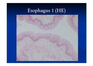
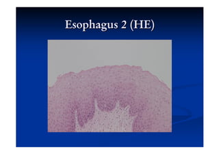
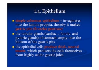
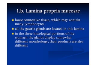
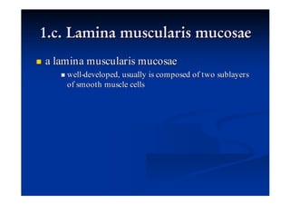
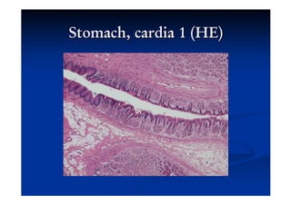
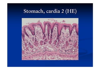
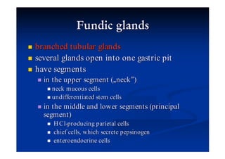
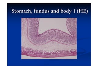
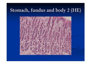
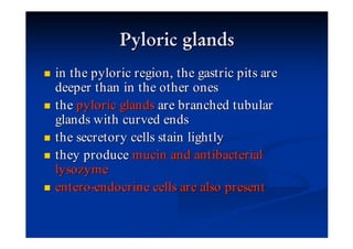
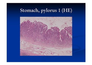
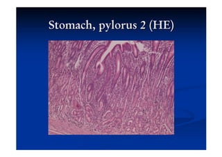
about 25-cm -long muscular tube
function : forwarding swallowed food from
the pharynx down into the stomach
its lumen is star -shaped, due to the
longitudinal reserve folds
the typical structure of the alimentary canal
appears in this segment
epithelium (lamina epithelialis mucosae):
stratified, nonkeratinized squamous
epithelium at its entire length
lamina propria mucosae comprises cardiac
esophageal glands (glandulae oesophageae
cardiacae) in its begining and terminal
portions; these glands resemble the cardiac
glands, hence the name
cardiac esophageal glands produce neutral mucin,
which protects the cells against the possible reflux
of gastric juice
in the lamina propria, there are scattered lymphatic
elements, frequently in the vicinity of the excretory
ducts
lamina muscularis mucosae is composed of mainly
longitudinal smooth muscle cells
is thrown into longitodinal folds, just like
mucous membrane
there are mixed or purely mucous small
salivary glands, which are called esophageal
glands proper (glandulae oesophageae
propriae)
in the upper one-third, striated muscle can be
found, only
in the middle approximately one-third
segments, the striated and smooth muscles
are intermingled
in the lower one-third, there are smooth
muscle cells, only
tunica adventitia is the outermost main layer
of the esophagus; this layer connects the
majority (cervical and thoracic portions) of
the organ to the surrounding tissue
the abdominal portion , however , is replaced
by tunica serosa (serous membrane)
bean -shaped hollow muscular organ of the
gastro -intestinal tract involved in the second
phase of digestion
it has four anatomical parts: cardia, fundus,
body and pylorus
it has three histological distinct parts: (1)
cardia, (2) the fundus and the body have the
same histological properties, (3) pylorus
simple columnar epithelium invaginates
into the lamina propria, thereby it makes
gastric pits (foveolae gastricae)
the tubular glands (cardiac -, fundic- and
pyloric glands) of stomach empty into the
bottom of the gastric pits
the epithelial cells produce thick , neutral
mucin , which protects the cells themselves
from highly acidic gastric juice
loose connective tissue, which may contain
many lymphocytes
all the gastric glands are located in this lamina
in the three histological portions of the
stomach the glands display somewhat
different morphology ; their products are also
different
a lamina muscularis mucosae
    well-developed, usually is composed of two sublayers
    of smooth muscle cells
loose coonecting tissue, containing
  blood vessels
  lymphatics
  lymphocytes
  solitary fat cells
  macrophages
  mast cells
contains smooth muscle cells
composed of sublayers:
  outermost sublayer : discontinuous; comprise mainly
  longitudinally oriented smooth muscles cells, which may
  be absent from areas within the pyloric portion
  middle sublayer : circularly arranged smooth muscle cells,
  well-developed throughout the stomach ; the middle
  sublayer is greatly enhanced in the pyloric portion , where
  it is called pyloric sphincter muscle
  innermost sublayer : oblique bundles of smooth muscle
  cells may occur
The stomach is an intraperitoneal organ . Its
 outermost main layer is a serous membrane
 of loose coonective tissue and mesothelium
 towards the peritoneal cavity .
located in the lamina propria, which are
different in the histological portions:
  cardiac glands
  fundic glands
  pyloric glands
simple or branched glands, located in the cardiac
portion
the secretory cells resemble the surface mucous cells
of the gastric pits
produce mucin
branched tubular glands
several glands open into one gastric pit
have segments
  in the upper segment ( neck )
    neck mucous cells
    undifferentiated stem cells
  in the middle and lower segments (principal
  segment )
     H Cl-producing parietal cells
     chief cells, which secrete pepsinogen
     enteroendocrine cells
in the pyloric region , the gastric pits are
deeper than in the other ones
the pyloric glands are branched tubular
glands with curved ends
the secretory cells stain lightly
they produce mucin and antibacterial
lysozyme
entero -endocrine cells are also present
Digestive system part 2

Digestive system part 2

  • 2.
    about 25-cm -longmuscular tube function : forwarding swallowed food from the pharynx down into the stomach its lumen is star -shaped, due to the longitudinal reserve folds the typical structure of the alimentary canal appears in this segment
  • 3.
    epithelium (lamina epithelialismucosae): stratified, nonkeratinized squamous epithelium at its entire length lamina propria mucosae comprises cardiac esophageal glands (glandulae oesophageae cardiacae) in its begining and terminal portions; these glands resemble the cardiac glands, hence the name
  • 4.
    cardiac esophageal glandsproduce neutral mucin, which protects the cells against the possible reflux of gastric juice in the lamina propria, there are scattered lymphatic elements, frequently in the vicinity of the excretory ducts lamina muscularis mucosae is composed of mainly longitudinal smooth muscle cells
  • 5.
    is thrown intolongitodinal folds, just like mucous membrane there are mixed or purely mucous small salivary glands, which are called esophageal glands proper (glandulae oesophageae propriae)
  • 6.
    in the upperone-third, striated muscle can be found, only in the middle approximately one-third segments, the striated and smooth muscles are intermingled in the lower one-third, there are smooth muscle cells, only
  • 7.
    tunica adventitia isthe outermost main layer of the esophagus; this layer connects the majority (cervical and thoracic portions) of the organ to the surrounding tissue the abdominal portion , however , is replaced by tunica serosa (serous membrane)
  • 10.
    bean -shaped hollowmuscular organ of the gastro -intestinal tract involved in the second phase of digestion it has four anatomical parts: cardia, fundus, body and pylorus it has three histological distinct parts: (1) cardia, (2) the fundus and the body have the same histological properties, (3) pylorus
  • 11.
    simple columnar epitheliuminvaginates into the lamina propria, thereby it makes gastric pits (foveolae gastricae) the tubular glands (cardiac -, fundic- and pyloric glands) of stomach empty into the bottom of the gastric pits the epithelial cells produce thick , neutral mucin , which protects the cells themselves from highly acidic gastric juice
  • 12.
    loose connective tissue,which may contain many lymphocytes all the gastric glands are located in this lamina in the three histological portions of the stomach the glands display somewhat different morphology ; their products are also different
  • 13.
    a lamina muscularismucosae well-developed, usually is composed of two sublayers of smooth muscle cells
  • 14.
    loose coonecting tissue,containing blood vessels lymphatics lymphocytes solitary fat cells macrophages mast cells
  • 15.
    contains smooth musclecells composed of sublayers: outermost sublayer : discontinuous; comprise mainly longitudinally oriented smooth muscles cells, which may be absent from areas within the pyloric portion middle sublayer : circularly arranged smooth muscle cells, well-developed throughout the stomach ; the middle sublayer is greatly enhanced in the pyloric portion , where it is called pyloric sphincter muscle innermost sublayer : oblique bundles of smooth muscle cells may occur
  • 16.
    The stomach isan intraperitoneal organ . Its outermost main layer is a serous membrane of loose coonective tissue and mesothelium towards the peritoneal cavity .
  • 17.
    located in thelamina propria, which are different in the histological portions: cardiac glands fundic glands pyloric glands
  • 18.
    simple or branchedglands, located in the cardiac portion the secretory cells resemble the surface mucous cells of the gastric pits produce mucin
  • 21.
    branched tubular glands severalglands open into one gastric pit have segments in the upper segment ( neck ) neck mucous cells undifferentiated stem cells in the middle and lower segments (principal segment ) H Cl-producing parietal cells chief cells, which secrete pepsinogen enteroendocrine cells
  • 24.
    in the pyloricregion , the gastric pits are deeper than in the other ones the pyloric glands are branched tubular glands with curved ends the secretory cells stain lightly they produce mucin and antibacterial lysozyme entero -endocrine cells are also present