Chapter 15 -The Digestive System
Irregular tube; open at both ends, called
“Alimentary canal” or “Gastrointestinal (GI) Tract”
29 feet long (adults) - 9 meters
Food & other substances that enter tube are not really
inside body
Passageway of food: broken down (digested) and
absorbed thru walls < entering body - cells
Both - Mechanical & Chemical Digestion
2.
Break Down ofFood
Teeth- first physical breakdown
Stomach-churning of food (physical)
Mouth- first chemical breakdown (salvia)
Digestive enzymes throughout GI tract
Digestion - Process where large food particles
reduced to absorbable molecules
Absorption - Process of small molecules passing thru
digestive system walls into body
Wall of DigestiveTract -
Mouth to anus
Four layers of tissue; surrounding the hollow
space within the tube “lumen”
May vary in structure in different organs
Mucosa or mucous membrane - tough in
esophagus, delicate, for absorption or
secretion in rest of tract
Submucosa - connective tissue, blood
vessels & nerves
5.
Muscularis - 2layers, responsible for
wavelike, rhythmic contractions
(peristalsis), moves contents, assists in
mixing & mechanical breakdown
Serosa - outermost covering, composed
of visceral peritoneum
Mesentery - double folded peritoneal
tissue, anchors loops of digestive tract to
posterior wall of abdominal cavity
6.
Mouth -
Oral cavity- hollow chamber (roof, a floor, & walls)
Entrance of food; digestion begins immediately
Mucous membranes > mucus, protects against
digestive juices & lubricates food passage
This mucous protects & lubricates
Hard palate - bony structure, front portion
Soft palate - posterior, chiefly muscles
Uvula - cone-shaped process hanging down from
soft palate. W/ help of soft pal., prevents food or
liquid from entering nasal cavity
7.
Floor of themouth -
Tongue - skeletal muscular structure, covered w/
mucous membrane
Anchored to bones in skull > hyoid bone
Frenulum- thin membrane; attaches tongue to floor
of mouth
Tongue-tied: too short
Papillae: small elevations on surface
Vallate type - largest, inverted V-shaped row of about
10-12 mushroomlike elevations - tastebuds
8.
Teeth -
Four majortypes -
Incisors - (sharp/cutting)
Canines - cuspids (pierce/tear)
Premolars - bicuspids & Molars - tricuspids
(grinding/crush)
Mastication > chewing of food
Forms a bolus > ready for swallowing
By age 2 - full set 20 teeth (cut 1st - 2 yrs.)
By age 17 to 24 - 32 permanent teeth (cut 1st - 6
yrs.)
9.
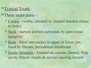
Typical Tooth -
Threemain parts -
Crown - visible, covered w/ enamel (hardest tissue
in body)
Neck - narrow portion surrounds by gum tissue
(gingiva)
Root - fitted into socket in upper or lower jaw,
lined by fibrous, periodontal membrane
Inside Structure - Enamel on outside, Dentin, Pulp
cavity (blood vessels & nerves) moving inward
10.
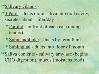
Salivary Glands -
3Pairs - ducts drain saliva into oral cavity,
secretes about 1 liter/day
Parotid - in front of each ear (mumps -
tender)
Submandibular - ducts by fernullum
Sublingual - ducts into floor of mouth
Saliva contents - salivary amylase (begins
CHO digestion), mucus (moistens food)
11.
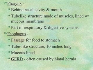
Pharynx -
Behind nasalcavity & mouth
Tubelike structure made of muscles, lined w/
mucous membrane
Part of respiratory & digestive systems
Esophagus -
Passage for food to stomach
Tube-like structure, 10 inches long
Mucous lined
GERD - often caused by hiatal hernia
12.
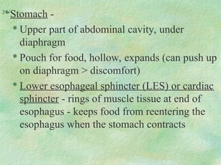
Stomach -
Upper partof abdominal cavity, under
diaphragm
Pouch for food, hollow, expands (can push up
on diaphragm > discomfort)
Lower esophageal sphincter (LES) or cardiac
sphincter - rings of muscle tissue at end of
esophagus - keeps food from reentering the
esophagus when the stomach contracts
13.
Chyme - semi-solidmixture, produced
by contraction of stomach muscles that
mixes food w/ gastric juices
Stomach contractions -
Created by 3 layers of muscles, run
lengthwise, around, obliquely
Makes stomach one of strongest organs >
peristalsis
Breaks food into tiny particles
14.
Mucous membranes linestomach -
contains gastric glands > secrete gastric
juice & hydrochloric acid
When empty, wrinkled folds - rugae
Three divisions of stomach -
Fundus, body, pylorus
Pyloric sphincter - holds food in stomach,
empties contents slowly into small
intestine
15.
Ulcer - carterlikewound or sore in membrane of
stomach
• 1 in 10 persons suffer in USA
• Helicobacter pylori bacterium (H. pylori)
Small Intestines -
Portion of digestion tract that extends from the pylorus to
the ileocecal valve
12- 20 feet in length, coiled, convoluted, and occupies
most of the abdominal cavity
Intestinal glands - secrete digestive juices
Smooth muscle wall - contracts > peristalsis
16.
Plicea - circularfolds covered w/ villi,
increases surface area > absorption
In or on the villi -
Blood capillaries - absorb CHO & protein end
products (glucose & amino acids)
Lacteal - lymphatic vessel - absorb lipids
Microvilli - brushlike border, > surface more
Chemical digestion - most occurs in 1st
subdivision of duodenum
Minor & major duodenal papillae - ducts where
pancreatic enzymes & bile enter small intestine
17.
Liver -
Large organ,fills R upper abdominal cavity
Exocrine gland - secretes bile into ducts
Hepatic - means liver
Bile - essential for breaking up or emulsification of
fats
CCK (cholecystokinin) - hormone secretion
triggered by lipids in chyme > makes gallbladder
contract & release bile
Drains from common bile duct into duodenum
Gallbladder - concentrates & stores bile
18.
Pancreas -
C-shaped, exocrinegland that lies behind the
stomach & duodenum
Pancreatic juice - most important digestive juice -
contains enzymes for all 3 food groups
Sodium bicarbonate (alkaline substance) -
neutralizes hydrochloric acid
Enters small intestine thru same duct as bile
Islets of Langerhans - hormones produced
Pancreatitis - inflammation (blockage, CF)
19.
Large Intestine -
Beginswith the ending of the ileum at the ileocecal
valve - called the cecum
Approximately 5 feet in length, much larger in
diameter than small intestine
Contents - not called chyme
Function - reabsorb water & salts
Material acted on by bacteria > more nutrients from
cellulose & other fibers
Synthesis Vit. K needed for blood clotting,
Production of some B-complex vit.
20.
Not as wellsuited for absorption as small intestine -
no villi
Normal passage of material thru large intestine - 3 to
5 days
Subdivisions - flow in GI Tract one-way
Cecum - pouchlike area
Ascending colon - right side of body
Bends at hepatic or right colic flexure
Transverse colon - extends across front
Bends at splenic or left colic flexure
Descending colon - left side abdomen
21.
Sigmond colon -S-shaped segment,
terminates in rectum
Anal canal - terminal end of rectum, ends at
external opening - anus
Inner anal sphincter - involuntary, smooth
muscle, keeps anus closed except during
defecation
Outer anal sphincter - striated, voluntary
muscle
22.
Appendix -
Vermiform appendix- “worm-shaped”, tubular structure,
blind tube
No important digestive fnc. - digest cellulose
Appendicitis - inflammation
Peritoneum -
Large, moist, slippery sheet of serous membrane
Peritoneal space - small space between parietal & visceral
layers - surfaces slide freely
Retroperitoneal - organs outside peritoneum
Extensions of peritoneum-mesentary, greater omentum -
both assist in anchoring abd. contents
23.
Digestion -Chemical &mechanical breakdown
CHO - amylase in mouth, slight effect
amylase from pancreas - into small intestine
Absorption of simple sugars (glucose)
Proteins - stomach (HCL/pepsinogen> pepsin)
Finished in small intestine by pancreatic (trypsin)
& peptidases in intestinal juice
Amino acids - basic protein units
24.
Fats - insmall intestine
Bile emulsification of fats > pancreatic
lipase > fatty acids & glycerol
Key digestive juices & enzymes
* page 410 Table 15-2
Absorption - taking food, breaking it
down in form for utilization of body
Just as important as digestion
Editor's Notes
#4 Mucosa of esophagus- tough and stratified abrasion-resistant epithelium
****Mucosa of reminder of GI tract- delicate layer of simple columnar epithelium (absorption & secretion)
*Mucus- coats the lining
Submucosa- connective tissue, below the mucosa; contains large amounts of bld. vessels & nerves
***Muscularis- two layers of muscles: wavelike, rhythmic contraction of muscular coat> peristalsis
Peristalsis: assists in the mixing of food> further mechanical breakdown of larger food particles
Serosa- outermost covering of digestive tract ; composed of the visceral peritoneum * anchored to posterior wall with double fold of peritoneal tissue> mesentary
#6 ***Uvula & soft palate- prevent any food & liquid from entering the nasal cavities above the mouth
#8 Baby : 2 years old> full set of (20) baby teeth *first @ 6 months Deciduous
Young adult: age 17-24 full set of permanent teeth (32) first @ 6 years
Parts of the typical tooth:
Crown: exposed/visable portion of tooth
Hard tissue (enamel & dentin/cementum)
Dentin- composed of tooth shell> center: pulp cavity consisting of connective tissue, blood & lymphatic vessels & sensory nerves
Cementum- over the neck & root; NECK: surrounded by pink gingiva (gum tissue) ROOT: fits down into socket of upper/lower jaw; lined by the periodontal membrane****
* Orthodonist/Peridontist
Clinical Application- Malocclusion page 482
#11 Provides active movement of food into the esophagus closing and sealing off the trachea
Nasopharynx
Oropharynx
laryngeal pharynx
The motor impulses from the swallowing act conroled by nervous stimulation of trigeminal, glossopharyneal, vagus, and hypoglossal cranial nerves
#12 *Contraction of the muscular walls mixes the food thoroughly with the gastric juice and breaks it down into a semisolid mixture called CHYME.
***Formation of CHYME: continuous of mechanical digestion> beginning in mouth
Gastric juices: ***hydrochloric acid and enzymes that function in the digestive process
Three layers of smooth muscles in the stomach wall: running lengthwise, around, and obliquely>>> one of the strongest internal organs of body>>able to break up the food into tiny particles > PERISTALSIS
Peristalsis- propels food down the digestive tract
Mucous membranes line stomach; contains thousands of microscopic gastric glands> HCL and gastric juice
Internal folds of stomach : RUGAE
*Intrinsic Factor- enables the absorption of Vit B-12; very important for blood cell formation > pernicious anemia
#15 Mucosa- small blood vessels, plasma, nerve cells, and blood cells
Submucosa- large blood vessels, connective tissue, nerves, ganglia, and lymphactics
VILLA- millions in number; “fingers” or folds projecting into the hallow interior of the intestine> rich with blood capillaries that absorb products of CHO/protein digestion ^ surface area for increased absorption
** Cryts of Lieberkunn- pitlike structures that lie in grooves between the villi & are composed of absorptive cells & mucous-producing goblet cells PH 7.0
** Peyer’s patches_ lymphoid follicles> important in immune response; antibody synthesis
Lacteal (lymph) > absorbs lipids or fatty materials from the chyme passing through the small intestines
Approximately 8 liters of water q day is absorbed by the small intestines into the portal blood
#18 Control of secretions: vagal control & hormonal control Hormones: beta cells_ secrete insulin- utilization of glucose
Alpha cells- secrete glucagon
Enymes: trypsin- breaks down proteins
Amylase- “”””’’ CHO tosimple sugars
Lipase- breaks down trigylercides
Pancreatitis- inflammation of pancreas
blockage of common bile duct
very serious/fata
CF- lack of enyzme (too much and too thick pancratic enzymes)
Cancer of pancreas- adenocarcinoma; life-span limited to around 5 years after diagnosis
#19 Flexures:
1) ***Hepatic: bend at the junction of the ascending and transverse colon; upper right quadrant of abd.
2) ***Splenic: bend at the juction of the transverse and descending colon; located at the left upper quadrant of abd.
Vitamin K synthesis: essential (blood clotting)
Normal passage of material through colon takes about 3-5 days
Anus- inner/outer anal sphincter- straited/voluntary muscle(outer) inner - smooth/involuntary muscle> opens during defecation process
Appenditis- inflammation of appendix; rupture > peritonitis: septecemia
#22 Ascites: abnormal accumulation of fluid in the peritoneal space
Osmotic pressure problem (low plasma proteins)
Accompanies> abdominal swelling, decreased urinary output
CHF, cirrhosis, kidney disease, cancer, malnutrition