SlideShare a Scribd company logo
1 of 60
T H E D I G E S T I V E S Y S T E M A N D
H O M EO S T A S I S
The gastrointestinal (GI) tract, or alimentary canal (alimentary nourishment).
Extends from the mouth to the anus through the thoracic and abdominopelvic cavities.
The length of the GI tract is about 5–7 meters (16.5–23 ft) in a living person.
It is longer in a cadaver (about 7–9meters or 23–29.5 ft) .
Because the muscles along the wall of the GI tract organs are in a state of tonus (sustained
contraction).
Functions
1. Ingestion: taking food into the mouth.
2. Secretion: release of water, acid, buffers, and enzymes into the
lumen of the GI tract.
3. Mixing and propulsion: churning and propulsion of food
through the GI tract.
4. Digestion: mechanical and chemical breakdown of food.
5. Absorption: passage of digested products from the GI tract
into the blood and lymph.
6. Defecation: the elimination of feces from the GI tract.
Organs of the digestive system
Layers of the gastrointestinal tract.
Mucosa Layers of the gastrointestinal tract.
A.The mucosa, or inner lining of the GI tract, is a mucous membrane.
It is composed of:
(1) Epithelium in direct contact with the contents of the GI tract.
(2) A layer of connective tissue called the lamina propria.88888
(3) A thin layer of smooth muscle (muscularis mucosae).
1. The epithelium:
In the mouth, pharynx, esophagus, and anal canal.
Nonkeratinized stratified squamous epithelium that serves a protective function.
Simple columnar epithelium, which functions in secretion and absorption.
Lines the stomach and intestines.
The rate of renewal of GI tract epithelial cells is rapid:
Every 5 to 7 days they slough off and are replaced by new cells.
Enteroendocrine cells, that secrete hormones.
2. The lamina propria :
Areolar connective tissue containing many blood and lymphatic vessels.
Which are the routes by which nutrients absorbed into the GI tract reach the
other tissues of the body.
The lamina propria also contains the majority of the cells of the mucosa-
associated lymphatic tissue (MALT).
These prominent lymphatic nodules contain immune system cells that protect
against disease .
3. A thin layer of smooth muscle fibers called the muscularis:
Mucosae throws the mucous membrane of the stomach and small intestine into many
small folds.
Which increase the surface area for digestion and absorption.
Movements of the muscularis mucosae ensure that all absorptive cells are fully exposed
to the contents of the GI tract.
B. Submucosa
Consists of areolar connective tissue that binds the mucosa to the muscularis.
It contains many blood and lymphatic vessels that receive absorbed food molecules.
Also located in the submucosa is an extensive network of neurons known as the
submucosal plexus .
The submucosa may also contain glands and lymphatic tissue.
Muscularis
The muscularis of the mouth, pharynx, and superior and middle parts of the
esophagus contains skeletal muscle that produces voluntary swallowing.
Skeletal muscle also forms the external anal sphincter, which permits voluntary control
of defecation.
The muscularis consists of smooth muscle that is generally found in two sheets:
Inner sheet of circular fibers.
Outer sheet of longitudinal fibers.
Between the layers of the muscularis is a second plexus of neurons—the myenteric
plexus.
C. Serosa
Those portions of the GI tract that are suspended in the abdominopelvic cavity
have a superficial layer called the serosa.
The serosa is a serous membrane composed of areolar connective tissue and
simple squamous epithelium (mesothelium).
The esophagus lacks a serosa.
Autonomic Nervous System
The vagus (X) nerves supply parasympathetic fibers to most parts of the GI tract.
The exception of the last half of the large intestine.
Which is supplied with parasympathetic fibers from the sacral spinal cord.
The parasympathetic nerves that supply the GI tract form neural connections with
the ENS.
Parasympathetic preganglionic neurons of the vagus or pelvic splanchnic nerves synapse
with parasympathetic postganglionic neurons located in the myenteric
and submucosal plexuses.
Some of the parasympathetic postganglionic neurons in turn synapse with neurons in the
ENS;
Others directly innervate smooth muscle and glands within the wall of the GI tract.
Stimulation of theparasympathetic nerves that innervate the GI tract causes an
increase in GI secretion and motility by increasing the activity of ENS neurons.
Sympathetic nerves that supply the GI tract arise from the thoracic and upper lumbar
regions of the spinal cord.
Sympathetic nerves form neural connections with the ENS.
Sympathetic postganglionic neurons synapse with neurons located in the myenteric
plexus and the submucosal plexus.
The sympathetic nerves that supply the GI tract cause a decrease in GI secretion and
motility by inhibiting the neurons of the ENS.
Emotions such as anger, fear, and anxiety may slow digestion because they
stimulate the sympathetic nerves that supply the GI tract.
Organization of the enteric nervous system.
The peritoneum
Largest serous membrane of the body;
It consists of a layer of ;
Simple squamous epithelium (mesothelium) with an underlying supporting layer
of areolar connective tissue.
The peritoneum is divided into the parietal peritoneum:
Which lines the wall of the abdomino pelvic cavity.
The visceral peritoneum:
Which covers some of the organs in the cavity and is their serosa
The slim space containing lubricating serous fluid that is between the parietal and
visceral portions of the peritoneum is called the peritoneal cavity.
In certain diseases the peritoneal cavity may become distended by the accu-
mulation of several liters of fluid, a condition called ascites.
Some organs are not lie in the peritoneal cavity.
Such organs, including the kidneys, ascending and descending colons of the
large intestine, duodenum of the small intestine, and pancreas, are said to be
retroperitoneal (retro- behind).
The peritoneum contains large folds that weave between the viscera.
The folds bind the organs to one another and to the walls of the abdominal cavity.
They also contain blood vessels, lymphatic vessels, and nerves that supply
the abdominal organs.
There are five major peritoneal folds:
The greater omentum,
Falciform ligament,
Lesser omentum,
Mesentery,
Mesocolon.
1. The greater omentum (o¯-MEN-tum fat skin), the largest peritoneal fold.
The greater omentum is a double sheet that folds back on itself, giving it a total of
four layers.
2. The falciform ligament (FAL-si-form; falc- sickleshaped) attaches the liver
to the anterior abdominal wall and diaphragm .
The liver is the only digestive organ that is attached to the anterior abdominal wall.
3. The lesser omentum arises as an anterior fold in the serosa of the stomach
and duodenum, and it suspends the stomach and duodenum from the liver .
It is the pathway for blood vessels entering the liver and contains the hepatic portal
vein, common hepatic artery, and common bile duct, along with some lymph
nodes.
4. A fan-shaped fold of the peritoneum, called the mesentery binds the jejunum
and ileum of the small intestine to the posterior abdominal wall .
5. Two separate folds of peritoneum, called the mesocolon bind the transverse
colon (transverse mesocolon) and sigmoid colon (sigmoid mesocolon) of the large
intestine to the posterior abdominal wall .
Structures of the mouth (oral cavity).
The mouth
Also referred to as the oral or buccal cavity (BUK-al; bucca cheeks):
is formed by the cheeks, hard and soft palates, and tongue .
The cheeks form the lateral walls of the oral cavity.
They are covered externally by skin and internally by a mucous membrane.
Which consists of nonkeratinized stratified squamous epithelium.
Buccinator muscles and connective tissue lie between the skin and mucous membranes of
the cheeks.
The anterior portions of the cheeks end at the lips.
The lips or labia ( fleshy borders) are fleshy folds surrounding the opening of the mouth.
They contain the orbicularis oris muscle and are covered externally by skin and internally by
a mucous membrane.
The inner surface of each lip is attached to its corresponding gum by a midline fold of
mucous membrane called the labial frenulum .
During chewing, contraction of the buccinator muscles in the cheeks and orbicularis oris
muscle in the lips helps keep food between the upper and lower teeth.
These muscles also assist in speech.
The oral vestibule ( entrance to a canal) of the oral cavity is a space bounded externally
by the cheeks and lips and internally by the gums and teeth.
The palate is a wall or septum that separates the oral cavity from the nasal cavity, forming
the roof of the mouth.
This important structure makes it possible to chew and breathe at the same time.
The hard palate—the anterior portion of the roof of the mouth—is formed by the
maxillae and palatine bones and is covered by a mucous membrane;
it forms a bony partition between the oral and nasal cavities.
The soft palate, which forms the posterior portion of the roof of the mouth, is an
arch-shaped muscular partition between the oropharynx and nasopharynx that is lined
with mucous membrane.
Hanging from the free border of the soft palate is a conical muscular process called the
uvula (U¯ -vu¯ -la little grape).
During swallowing, the soft palate and uvula are drawn superiorly, closing off the
nasopharynx and preventing swallowed foods and liquids from entering the nasal cavity
The three major salivary glands—parotid, sublingual, and
submandibular
A salivary gland is a gland that releases a secretion called saliva into the oral cavity.
When food enters the mouth, however, secretion of saliva increases, and it lubricates,
dissolves, and begins the chemical breakdown of the food.
There are three pairs of major salivary glands:
the parotid, submandibular, and sublingual glands
The parotid glands (par- near; to- ear) are located inferior and anterior to the ears,
between the skin and the masseter muscle.
Each secretes saliva into the oral cavity via a parotid duct that pierces the buccinator
muscle to open into the vestibule opposite the second maxillary (upper) molar tooth.
The submandibular glands are found in the floor of the mouth;
They are medial and partly inferior to the body of the mandible.
Their ducts, the submandibular ducts, run under the mucosa on either side of the midline
of the floor of the mouth and enter the oral cavity proper lateral to the lingual frenulum.
The sublingual glands are beneath the tongue and superior to the submandibular
glands.
Their ducts, the lesser sublingual ducts, open into the floor of the mouth in the oral cavity
proper.
Composition and Functions of Saliva
Chemically, saliva is 99.5% water and 0.5% solutes.
Among the solutes are ions, including sodium, potassium, chloride, bicarbonate, and
phosphate.
Also present are some dissolved gases and various organic substances, including urea and
uric acid, mucus, immunoglobulin A, the bacteriolytic enzyme lysozyme,
and salivary amylase, a digestive enzyme that acts on starch.
The parotid glands secrete a watery (serous) liquid containing salivary
amylase.
The submandibular glands secrete a fluid that contains amylase but is thickened
with mucus.
The sublingual glands contain mostly mucous cells, so they secrete a much thicker fluid that
contributes only a small amount of salivary amylase.
The water in saliva provides a medium for dissolving foods so that they can be tasted by
gustatory receptors.
Chloride ions in the saliva activate salivary amylase, an enzyme that starts the breakdown of
starch.
Bicarbonate and phosphate ions buffer acidic foods that enter the mouth, so saliva is only
slightly acidic (Ph 6.35–6.85).
Immunoglobulin A (IgA) prevents attachment of microbes so they cannot penetrate the
epithelium, and the enzyme lysozyme kills bacteria;
Salivation
The secretion of saliva, called salivation (sal-i-VA¯ -shun).
Controlled by the autonomic nervous system.
Amounts of saliva secreted daily vary considerably but average 1000–1500 mL
(1–1.6 qt).
Parasympathetic stimulation promotes continuous secretion of a moderate amount of
saliva.
Sympathetic stimulation dominates during stress, resulting in dryness of the mouth.
The tongue
An accessory digestive organ composed of skeletal muscle covered with mucous
membrane.
It forms the floor of the oral cavity.
The tongue is divided into symmetrical lateral halves by a median septum that extends its
entire length, and it is attached inferiorly to the hyoid bone, styloid process of the temporal
bone, and mandible.
Each half of the tongue consists of an identical complement of extrinsic and intrinsic
muscles.
The extrinsic muscles of the tongue, which originate outside the tongue (attach to bones in
the area) and insert into connective tissues in the tongue, include the hyoglossus,
genioglossus, and styloglossus muscles .
The extrinsic muscles move the tongue from side to side and in and out to maneuver food
for chewing, shape the food into a rounded mass, and force the food to the back of the
mouth for swallowing.
The intrinsic muscles originate in and insert into connective tissue within the tongue.
They alter the shape and size of the tongue for speech and swallowing.
The intrinsic muscles include the longitudinalis superior, longitudinalis inferior,
transversus linguae, and verticalis linguae muscles.
The dorsum (upper surface) and lateral surfaces of the tongue are covered with papillae
(pa-PIL-e¯ nipple-shaped projections), projections of the lamina propria covered with
stratified squamous epithelium .
Many papillae contain taste buds, the receptors for gustation (taste).
The teeth, or dentes
Organs located in sockets of the alveolar processes of the mandible and maxillae.
The alveolar processes are covered by the gingivae (JIN-ji-ve¯ ), or gums.
Which extend slightly into each socket.
The sockets are lined by the periodontal ligament or membrane (odont- tooth).
Which consists of dense fibrous connective tissue that anchors the teeth to the socket
walls.
A typical tooth has three major external regions:
Crown
Root
Neck.
The crown is the visible portion above the level of the gums.
Embedded in the socket are one to three roots.
The neck is the constricted junction of the crown and root near the gum line.
Internally, dentin forms the majority of the tooth.
Dentin consists of a calcified connective tissue that gives the tooth its basic
shape and rigidity.
It is harder than bone because of its higher content of calcium salts (70% of dry weight).
The dentin of the crown is covered by enamel.
Which consists primarily of calcium phosphate and calcium carbonate.
Enamel is also harder than bone because of its even higher content of calcium salts (about
95% of dry weight).
A typical tooth and surrounding structures.
Humans have two dentitions, or sets of teeth:
Deciduous and permanent.
The first of these—the deciduous teeth (decidu- falling out), also called primary teeth,
milk teeth, or baby teeth—begin to erupt at about 6 months of age, and
approximately two teeth appear each month thereafter, until all 20 .
The incisors :
which are closest to the midline, are chisel-shaped and adapted for cutting into food.
They are referred to as either central or lateral incisors based on their position.
The cuspids (canines):
which have a pointed surface called a cusp.
Cuspids are used to tear and shred food.
Incisors and cuspids have only one root apiece.
The first and second molars:
which have four cusps.
Maxillary (upper) molars have three roots;
Mandibular (lower) molars have two roots.
The molars crush and grind food to prepare it for swallowing.
All the deciduous teeth are lost—generally between ages 6 and 12 years.
The permanent dentition:
Contains 32 teeth that erupt between age 6 and adulthood.
The deciduous molars are replaced by the first and second premolars (bicuspids).
Which have two cusps and one root (upper first premolars have two roots) and are used
for crushing and grinding.
The first molars at age 6 (six-year molars),
The second molars at age 12 (twelve-year molars),
The third molars (wisdom teeth) after age 17 or not at all.
Pharynx ( throat)
When food is first swallowed, it passes from the mouth into the pharynx (
throat).
a funnel-shaped tube that extends from the internal nares to the esophagus.
The pharynx is composed of skeletal muscle and lined by mucous membrane.
Divided into three parts:
The nasopharynx.
The oropharynx.
The laryngopharynx.
The nasopharynx functions only in respiration.
Both the oropharynx and laryngopharynx have digestive as well as respiratory
functions. Swallowed food passes from the mouth into the oropharynx and
laryngopharynx;
The muscular contractions of these areas help propel food into the esophagus and
then into the stomach.
The esophagus;
Collapsible muscular tube, about 25 cm (10 in.) long, that lies
posterior to the trachea.
The esophagus begins at the inferior end of the laryngopharynx and
passes through the mediastinum anterior to the vertebral column.
Then it pierces the diaphragm through an opening called the
esophageal hiatus, and ends in the superior portion of the stomach .
Sometimes, part of the stomach protrudes above the diaphragm
through the esophageal hiatus.
This condition, termed a hiatus hernia.
The esophagus secretes mucus and transports food into the stomach.
It does not produce digestive enzymes, and it does not carry on
absorption.
DEGLUTITION
The movement of food from the mouth into the stomach is achieved by the act of
swallowing, or deglutition .
Deglutition is facilitated by the secretion of saliva and mucus and involves the mouth,
pharynx, and esophagus.
Swallowing occurs in three stages:
(1) the voluntary stage, in which the bolus is passed into the oropharynx;
(2) the pharyngeal stage, the involuntary passage of the bolus through the pharynx
into the esophagus;
(3) the esophageal stage, the involuntary passage of the bolus through the
esophagus into the stomach.
Swallowing starts when the bolus is forced to the back of the
oral cavity and into the oropharynx by the movement of the
tongue upward and backward against the palate; these actions
The stomach
J-shaped enlargement of the GI tract directly inferior to the diaphragm in the epigastric,
umbilical, and left hypochondriac regions of the abdomen.
The stomach connects the esophagus to the duodenum, the first part of the small intestine.
converted to a liquid, and certain substances
are absorbed.
Anatomy of the Stomach
The stomach has four main regions:
the cardia, fundus, body, and pylorus .
The cardia (CAR-de¯-a) surrounds the superior opening of the stomach.
The rounded portion superior to and to the left of the cardia is the fundus (FUN-dus).
Inferior to the fundus is the large central portion of the stomach, called the body.
The region of the stomach that connects to the duodenum is the pylorus ;
It has two parts, the pyloric antrum (AN-trum cave), which connects to the body of the
stomach, and the pyloric canal, which leads into the duodenum.
When the stomach is empty, the mucosa lies in large folds, called rugae .
The pylorus communicates with the duodenum of the small intestine via a smooth muscle
sphincter called the pyloric sphincter.
The concave medial border of the stomach is called the lesser curvature, and the convex
lateral border is called the greater curvature.
Functions of the Stomach
1. Mixes saliva, food, and gastric juice to form chyme.
2. Serves as a reservoir for food before release into small
intestine.
3. Secretes gastric juice, which contains HCl (kills bacteria and
denatures protein), pepsin (begins the digestion of proteins),
intrinsic factor (aids absorption of vitamin B12), and gastric
lipase (aids digestion of triglycerides).
4. Secretes gastrin into blood.
Histology of the stomach.
The stomach mucosa showing gastric glands and cell types
PANCREAS
Anatomy of the Pancreas
The pancreas a retroperitoneal gland that is about 12–15 cm (5–6 in.) long and 2.5 cm (1 in.)
thick.
Lies posterior to the greater curvature of the stomach.
The pancreas consists of a head, a body, and a tail and is usually connected to the
duodenum by two ducts .
The head is the expanded portion of the organ near the curve of the duodenum;
Superior to and to the left of the head are the central body and the tapering tail.
Pancreatic juices are secreted by exocrine cells into small ducts that ultimately unite to form
two larger ducts, the pancreatic duct and the accessory duct.
The pancreatic duct (duct of Wirsung) is the larger of the two ducts.
These in turn convey the secretions into the small intestine.
Pancreatic duct joins the common bile duct from the liver and gallbladder and enters the
duodenum as a dilated common duct called the hepatopancreatic ampulla (ampulla of
Vater).
The ampulla opens on an elevation of the duodenal mucosa known as the major duodenal
papilla, which lies about 10 cm (4 in.) inferior to the pyloric sphincter of the stomach.
The passage of pancreatic juice and bile through the hepatopancreatic ampulla into the
small intestine is regulated by a mass of smooth muscle known as the sphincter of the
hepatopancreatic ampulla (sphincter of Oddi).
The other major duct of the pancreas, the accessory duct (duct of Santorini), leads
from
the pancreas and empties into the duodenum about 2.5 cm (1 in.) uperior to the
hepatopancreatic ampulla.
The pancreas is made up of small clusters of glandular epithelial cells.
About 99% of the clusters, called acini (AS-i-ne¯ ), constitute the exocrine portion of the
organ .
The cells within acini secrete a mixture of fluid and digestive enzymes called pancreatic
juice.
The remaining 1% of the clusters, called pancreatic islets (islets of Langerhans), form
the endocrine portion of the pancreas.
These cells secrete the hormones glucagon, insulin, somatostatin, and pancreatic
polypeptide.
Relation of the pancreas to the liver, gallbladder, and duodenum.
Details of hepatopancreatic ampulla
Ducts carrying bile from liver and gallbladder and pancreatic
juice from pancreas to the duodenum
Composition and Functions of Pancreatic Juice
Each day the pancreas produces 1200–1500 mL (about 1.2– 1.5 qt) of pancreatic juice, a
clear, colorless liquid consisting mostly of water, some salts, sodium bicarbonate, and
several enzymes.
The sodium bicarbonate gives pancreatic juice a slightly alkaline pH (7.1–8.2) that buffers
acidic gastric juice in chyme, stops the action of pepsin from the stomach, and
creates the proper pH for the action of digestive enzymes in the small intestine.
Starch digesting enzyme called pancreatic amylase;
Protein digesting enzymes called trypsin ,chymotrypsin ,carboxypeptidase, and
elastase;
Triglyceride-digesting enzyme in adults, called pancreatic lipase;
Nucleic acid– digesting enzymes called ribonuclease and deoxyribonuclease.
LIVER AND GALLBLADDER
The liver is the heaviest gland of the body, weighing about 1.4 kg (about 3 lb) in an
average adult.
Of all of the organs of the body, it is second only to the skin in size.
The liver is inferior to the diaphragm and occupies most of the right hypochondriac
and part of the epigastric regions of the abdominopelvic cavity
The gallbladder (gall- bile) is a pear-shaped sac that is located in a depression of the
posterior surface of the liver.
It is 7–10 cm (3–4 in.) long and typically hangs from the anterior inferior margin of the liver.
Anatomy of the Liver and Gallbladder
The liver is divided into two principal lobes:
A large right lobe and a smaller left lobe— by the falciform ligament, a fold of the
mesentery.
The right lobe include:
An inferior quadrate lobe .
Posterior caudate lobe.
The falciform ligament extends from the undersurface of the diaphragm between
the two principal lobes of the liver to the superior surface of the liver, helping to suspend the
liver in the abdominal cavity.
In the free border of the falciform ligament is the ligamentum teres round ligament), a
remnant of the umbilical vein of the fetus this fibrous cord extends from the liver to the
umbilicus.
The right and left coronary
ligaments are narrow extensions of the parietal peritoneum that
suspend the liver from the diaphragm.
The parts of the gallbladder include the broad fundus, which
projects inferiorly beyond the inferior border of the liver; the
body, the central portion; and the neck, the tapered portion. The
body and neck project superiorly.
Histology of the Liver and Gallbladder
Histologically, the liver is composed of several compon
Histology of the liver.
Hepatic blood flow: sources, path through the liver, and return to the heart.
Functions of liver:
• Carbohydrate metabolism:
The liver is especially important in maintaining a normal blood glucose level.
When blood glucose is low:
The liver can break down glycogen to glucose and release the glucose into the bloodstream.
When blood glucose is high:
as occurs just after eating a meal, the liver converts glucose to glycogen and triglycerides for
storage.
• Lipid metabolism:
Hepatocytes store some triglycerides; break down fatty acids to generate ATP;
• Protein metabolism:
Hepatocytes deaminate (remove the amino group, NH2, from) amino acids so that the amino
acids can be used for ATP production or converted to carbohydrates or fats.
• Processing of drugs and hormones:
The liver can detoxify substances such as alcohol and excrete drugs such as penicillin,
erythromycin, and sulfonamides into bile.
It can also chemically alter or excrete thyroid hormones and steroid
hormones such as estrogens and aldosterone.
• Excretion of bilirubin:
Bilirubin, derived from the heme of aged red blood cells, is absorbed by the liver from the
blood and secreted into bile.
• Storage. In addition to glycogen, the liver is a prime storage
• Phagocytosis. The stellate reticuloendothelial (Kupffer)
• Activation of vitamin D. The skin, liver, and kidneys participate in synthesizing the active form of
vitamin D.
The small intestine.
Most digestion and absorption of nutrients occur in a long tube called the small intestine.
Because of this, its structure is specially
Its length alone provides a large surface area for digestion and absorption, and that area is
further increased by circular folds, villi, and microvilli.
2.5 cm (1 in.) in diameter; its length is about 3 m (10 ft) in a living person
6.5 m (21 ft) in a cadaver due to the loss of smooth muscle tone after death.
The small intestine is divided into three regions .
The duodenum :
the shortest region, is retroperitoneal.
It starts at the pyloric sphincter of the stomach and extends about 25 cm (10 in.)
until it merges with the jejunum.
Duodenum means “12”; it is so named because it is about as long as the width of
12 fingers.
The jejunum :
is about 1 m (3 ft) long and extends to the ileum.
The final and longest region of the small intestine
the ileum :
twisted), measures about 2 m (6 ft) and joins the large intestine
at a smooth muscle sphincter called the ileocecal sphincter
Anatomy of the small intestine.
Histology of the small intestine
Enlarged villus showing lacteal, capillaries, intestinal glands, and cell
types
Absorption of digested nutrients in the small intestine
Daily volumes of fluid ingested, secreted, absorbed, and excreted from the GI tract.
Anatomy of the large intestine
PHASES OF DIGESTION
Digestive activities occur in three overlapping phases:
The cephalic phase, the gastric phase, and the intestinal phase.
Cephalic Phase:
During the cephalic phase of digestion, the smell, sight, thought, or initial taste of
food activates neural centers in the cerebral cortex, hypothalamus, and brain stem.
The brain stem then activates the facial (VII), glossopharyngeal (IX), and vagus (X)
nerves.
The facial and glossopharyngeal nerves stimulate the salivary glands to secrete saliva,
while the vagus nerves stimulate the gastric glands to secrete gastric juice.
The purpose of the cephalic phase of digestion is to prepare the mouth and stomach
for food that is about to be eaten.
Gastric Phase :
Once food reaches the stomach, the gastric phase of digestion begins.
Neural and hormonal mechanisms regulate the gastric phase of digestion to promote
gastric secretion and gastric motility.
• Neural regulation. Food of any kind distends the stomach and
stimulates stretch receptors in its walls. Chemoreceptors in the
stomach monitor the pH of the stomach chyme. When the
stomach walls are distended or pH increases because proteins
have entered the stomach and buffered some of the stomach
acid, the stretch receptors and chemoreceptors are activated,
and a neural negative feedback loop is set in motion (Figure
24.24). From the stretch receptors and chemoreceptors, nerve
Hormonal regulation. Gastric secretion during the gastric
phase is also regulated by the hormone gastrin. Gastrin is
released from the G cells of the gastric glands in response to
several stimuli: distension of the stomach by chyme, partially
digested proteins in chyme, the high pH of chyme due
to the presence of food in the stomach, caffeine in gastric
chyme, and acetycholine released from parasympathetic
neurons. Once it is released, gastrin enters the bloodstream,
makes a round-trip through the body, and finally reaches its
target organs in the digestive system. Gastrin stimulates gastric
glands to secrete large amounts of gastric juice. It also
strengthens the contraction of the lower esophageal sphincter
to prevent reflux of acid chyme into the esophagus,
increases motility of the stomach, and relaxes the pyloric
sphincter, which promotes gastric emptying. Gastrin
secretion is inhibited when the pH of gastric juice drops
below 2.0 and is stimulated when the pH rises. This negative
feedback mechanism helps provide an optimal low pH for
the functioning of pepsin, the killing of microbes, and the
denaturing of proteins in the stomach.
Intestinal Phase
The intestinal phase of digestion begins once food enters the
small intestine. In contrast to reflexes initiated during the
cephalic and gastric phases, which stimulate stomach secretory
activity and motility, those occurring during the intestinal phase
have inhibitory effects that slow the exit of chyme from the
stomach. This prevents the duodenum from being overloaded
with more chyme than it can handle. In addition, responses
occurring during the intestinal phase promote the continued
digestion of foods that have reached the small intestine. These
activities of the intestinal phase of digestion are regulated by
neural and hormonal mechanisms.
• Neural regulation. Distension of the duodenum by the
presence of chyme causes the enterogastric reflex. Stretch
receptors in the duodenal wall send nerve impulses to the
medulla oblongata, where they inhibit parasympathetic stimulation
and stimulate the sympathetic nerves to the stomach.
As a result, gastric motility is inhibited and there is an
increase in the contraction of the pyloric sphincter, which
decreases gastric emptying.
• Hormonal regulation. The intestinal phase of digestion
is mediated by two major hormones secreted by the small
intestine: cholecystokinin and secretin. Cholecystokinin
(CCK) is secreted by the CCK cells of the small intestinal

More Related Content

What's hot

Anatomical terminology
Anatomical terminologyAnatomical terminology
Anatomical terminologyKevin Young
 
Excretory system
Excretory systemExcretory system
Excretory systemIndia™
 
Notes small and large intestines
Notes small and large intestinesNotes small and large intestines
Notes small and large intestinesJulie Stewart
 
Anatomy of the stomach
Anatomy of the stomachAnatomy of the stomach
Anatomy of the stomachSaika Lashari
 
El aparato digestivo clase 5
El aparato digestivo  clase 5El aparato digestivo  clase 5
El aparato digestivo clase 5sandritahidalgo
 
Git system Anatomy & Physiology
Git system Anatomy & PhysiologyGit system Anatomy & Physiology
Git system Anatomy & PhysiologyAnil Lawrence
 
Anterior abdominal wall
Anterior abdominal wallAnterior abdominal wall
Anterior abdominal wallMoamer Gabsa
 
El aparato digestivo 2
El aparato digestivo 2El aparato digestivo 2
El aparato digestivo 2Miriam Valle
 
Respiratory System Anatomy & Physiology
Respiratory System Anatomy & Physiology Respiratory System Anatomy & Physiology
Respiratory System Anatomy & Physiology yousaf shah
 
Anterior abdominal wall
Anterior abdominal wallAnterior abdominal wall
Anterior abdominal wallSeemi Shah
 
Circulatory System.ppt
Circulatory System.pptCirculatory System.ppt
Circulatory System.pptShama
 
Organos de los sentidos
Organos de  los sentidosOrganos de  los sentidos
Organos de los sentidosMaria Rojas
 

What's hot (20)

Abdominal aorta
Abdominal aortaAbdominal aorta
Abdominal aorta
 
Anatomical terminology
Anatomical terminologyAnatomical terminology
Anatomical terminology
 
Excretory system
Excretory systemExcretory system
Excretory system
 
Notes small and large intestines
Notes small and large intestinesNotes small and large intestines
Notes small and large intestines
 
Digestive System PPT (6).ppt
Digestive System PPT (6).pptDigestive System PPT (6).ppt
Digestive System PPT (6).ppt
 
Digestive system
Digestive systemDigestive system
Digestive system
 
Anatomy of the stomach
Anatomy of the stomachAnatomy of the stomach
Anatomy of the stomach
 
El aparato digestivo clase 5
El aparato digestivo  clase 5El aparato digestivo  clase 5
El aparato digestivo clase 5
 
Liver.pdf
Liver.pdfLiver.pdf
Liver.pdf
 
Git system Anatomy & Physiology
Git system Anatomy & PhysiologyGit system Anatomy & Physiology
Git system Anatomy & Physiology
 
Anterior abdominal wall
Anterior abdominal wallAnterior abdominal wall
Anterior abdominal wall
 
Liver anatomy
Liver anatomyLiver anatomy
Liver anatomy
 
1 Stomach
1  Stomach1  Stomach
1 Stomach
 
El aparato digestivo 2
El aparato digestivo 2El aparato digestivo 2
El aparato digestivo 2
 
Respiratory System Anatomy & Physiology
Respiratory System Anatomy & Physiology Respiratory System Anatomy & Physiology
Respiratory System Anatomy & Physiology
 
Large intestine
Large intestineLarge intestine
Large intestine
 
Anterior abdominal wall
Anterior abdominal wallAnterior abdominal wall
Anterior abdominal wall
 
Circulatory System.ppt
Circulatory System.pptCirculatory System.ppt
Circulatory System.ppt
 
Organs of abdomen
Organs of abdomenOrgans of abdomen
Organs of abdomen
 
Organos de los sentidos
Organos de  los sentidosOrganos de  los sentidos
Organos de los sentidos
 

Similar to Digestive system

Digestive system
Digestive systemDigestive system
Digestive systemSoneeshah
 
digestive system of anatomypowerpoint pres
digestive system of anatomypowerpoint presdigestive system of anatomypowerpoint pres
digestive system of anatomypowerpoint presHgjUy7
 
Histological features of Gastrointestinal tract
Histological features of Gastrointestinal tractHistological features of Gastrointestinal tract
Histological features of Gastrointestinal tractrasulovadiana0301
 
DIGESTIVE SYSTEM anatomy and physiology of Bsc nursing pdf/pptx
DIGESTIVE SYSTEM anatomy and physiology of Bsc nursing pdf/pptx DIGESTIVE SYSTEM anatomy and physiology of Bsc nursing pdf/pptx
DIGESTIVE SYSTEM anatomy and physiology of Bsc nursing pdf/pptx NS crown
 
Anatomy & Physiology of GIT
Anatomy & Physiology of GITAnatomy & Physiology of GIT
Anatomy & Physiology of GITpharmacampus
 
Gastrointestinal Tract (GIT)//DIGESTIVE SYSTEM
Gastrointestinal Tract (GIT)//DIGESTIVE SYSTEM Gastrointestinal Tract (GIT)//DIGESTIVE SYSTEM
Gastrointestinal Tract (GIT)//DIGESTIVE SYSTEM Wasim Ak
 
gastrointestinaltractgit-220817021937-805e8aa3.pdf
gastrointestinaltractgit-220817021937-805e8aa3.pdfgastrointestinaltractgit-220817021937-805e8aa3.pdf
gastrointestinaltractgit-220817021937-805e8aa3.pdfLawrenceshamboko
 
Digestive System.pptx
Digestive System.pptxDigestive System.pptx
Digestive System.pptxnikhilgendre1
 
Lab 5 Digestive system Tongue, Esophagus & Stomach.pdf
Lab 5 Digestive system Tongue, Esophagus & Stomach.pdfLab 5 Digestive system Tongue, Esophagus & Stomach.pdf
Lab 5 Digestive system Tongue, Esophagus & Stomach.pdfNoor Muhsen Jawad
 
TheDigestivesystem2999999999999999999999
TheDigestivesystem2999999999999999999999TheDigestivesystem2999999999999999999999
TheDigestivesystem2999999999999999999999JhanVicGavasan
 
FEDHA GIT HISTO (1).pptx
FEDHA GIT HISTO (1).pptxFEDHA GIT HISTO (1).pptx
FEDHA GIT HISTO (1).pptxHybridGamer2
 
HAP 4 SEMESTER 2 BPHARMACY AS PER PCI SYLLABUS
HAP 4 SEMESTER 2 BPHARMACY AS PER PCI SYLLABUSHAP 4 SEMESTER 2 BPHARMACY AS PER PCI SYLLABUS
HAP 4 SEMESTER 2 BPHARMACY AS PER PCI SYLLABUSchristinajohn24
 
Digestion and Absorption - Class 11 ppt
Digestion and Absorption - Class 11 pptDigestion and Absorption - Class 11 ppt
Digestion and Absorption - Class 11 pptKarthik Bharadwaj
 

Similar to Digestive system (20)

Digestive system
Digestive systemDigestive system
Digestive system
 
Digestive system
Digestive systemDigestive system
Digestive system
 
digestive system of anatomypowerpoint pres
digestive system of anatomypowerpoint presdigestive system of anatomypowerpoint pres
digestive system of anatomypowerpoint pres
 
Digestive system-I.pptx
Digestive system-I.pptxDigestive system-I.pptx
Digestive system-I.pptx
 
Histological features of Gastrointestinal tract
Histological features of Gastrointestinal tractHistological features of Gastrointestinal tract
Histological features of Gastrointestinal tract
 
DIGESTIVE SYSTEM anatomy and physiology of Bsc nursing pdf/pptx
DIGESTIVE SYSTEM anatomy and physiology of Bsc nursing pdf/pptx DIGESTIVE SYSTEM anatomy and physiology of Bsc nursing pdf/pptx
DIGESTIVE SYSTEM anatomy and physiology of Bsc nursing pdf/pptx
 
GI System.pptx
GI System.pptxGI System.pptx
GI System.pptx
 
Anatomy & Physiology of GIT
Anatomy & Physiology of GITAnatomy & Physiology of GIT
Anatomy & Physiology of GIT
 
Gastrointestinal Tract (GIT)//DIGESTIVE SYSTEM
Gastrointestinal Tract (GIT)//DIGESTIVE SYSTEM Gastrointestinal Tract (GIT)//DIGESTIVE SYSTEM
Gastrointestinal Tract (GIT)//DIGESTIVE SYSTEM
 
gastrointestinaltractgit-220817021937-805e8aa3.pdf
gastrointestinaltractgit-220817021937-805e8aa3.pdfgastrointestinaltractgit-220817021937-805e8aa3.pdf
gastrointestinaltractgit-220817021937-805e8aa3.pdf
 
Digestive System.pptx
Digestive System.pptxDigestive System.pptx
Digestive System.pptx
 
Absorption rohit bharti
Absorption  rohit bhartiAbsorption  rohit bharti
Absorption rohit bharti
 
Anatomy of digestive system-III.pptx
Anatomy of digestive system-III.pptxAnatomy of digestive system-III.pptx
Anatomy of digestive system-III.pptx
 
Lab 5 Digestive system Tongue, Esophagus & Stomach.pdf
Lab 5 Digestive system Tongue, Esophagus & Stomach.pdfLab 5 Digestive system Tongue, Esophagus & Stomach.pdf
Lab 5 Digestive system Tongue, Esophagus & Stomach.pdf
 
TheDigestivesystem2999999999999999999999
TheDigestivesystem2999999999999999999999TheDigestivesystem2999999999999999999999
TheDigestivesystem2999999999999999999999
 
Digestive system
Digestive systemDigestive system
Digestive system
 
FEDHA GIT HISTO (1).pptx
FEDHA GIT HISTO (1).pptxFEDHA GIT HISTO (1).pptx
FEDHA GIT HISTO (1).pptx
 
HAP 4 SEMESTER 2 BPHARMACY AS PER PCI SYLLABUS
HAP 4 SEMESTER 2 BPHARMACY AS PER PCI SYLLABUSHAP 4 SEMESTER 2 BPHARMACY AS PER PCI SYLLABUS
HAP 4 SEMESTER 2 BPHARMACY AS PER PCI SYLLABUS
 
Digestion and Absorption - Class 11 ppt
Digestion and Absorption - Class 11 pptDigestion and Absorption - Class 11 ppt
Digestion and Absorption - Class 11 ppt
 
Digestive
DigestiveDigestive
Digestive
 

Recently uploaded

Forest laws, Indian forest laws, why they are important
Forest laws, Indian forest laws, why they are importantForest laws, Indian forest laws, why they are important
Forest laws, Indian forest laws, why they are importantadityabhardwaj282
 
Call Girls In Nihal Vihar Delhi ❤️8860477959 Looking Escorts In 24/7 Delhi NCR
Call Girls In Nihal Vihar Delhi ❤️8860477959 Looking Escorts In 24/7 Delhi NCRCall Girls In Nihal Vihar Delhi ❤️8860477959 Looking Escorts In 24/7 Delhi NCR
Call Girls In Nihal Vihar Delhi ❤️8860477959 Looking Escorts In 24/7 Delhi NCRlizamodels9
 
SOLUBLE PATTERN RECOGNITION RECEPTORS.pptx
SOLUBLE PATTERN RECOGNITION RECEPTORS.pptxSOLUBLE PATTERN RECOGNITION RECEPTORS.pptx
SOLUBLE PATTERN RECOGNITION RECEPTORS.pptxkessiyaTpeter
 
Is RISC-V ready for HPC workload? Maybe?
Is RISC-V ready for HPC workload? Maybe?Is RISC-V ready for HPC workload? Maybe?
Is RISC-V ready for HPC workload? Maybe?Patrick Diehl
 
Pests of soyabean_Binomics_IdentificationDr.UPR.pdf
Pests of soyabean_Binomics_IdentificationDr.UPR.pdfPests of soyabean_Binomics_IdentificationDr.UPR.pdf
Pests of soyabean_Binomics_IdentificationDr.UPR.pdfPirithiRaju
 
Analytical Profile of Coleus Forskohlii | Forskolin .pptx
Analytical Profile of Coleus Forskohlii | Forskolin .pptxAnalytical Profile of Coleus Forskohlii | Forskolin .pptx
Analytical Profile of Coleus Forskohlii | Forskolin .pptxSwapnil Therkar
 
Environmental Biotechnology Topic:- Microbial Biosensor
Environmental Biotechnology Topic:- Microbial BiosensorEnvironmental Biotechnology Topic:- Microbial Biosensor
Environmental Biotechnology Topic:- Microbial Biosensorsonawaneprad
 
zoogeography of pakistan.pptx fauna of Pakistan
zoogeography of pakistan.pptx fauna of Pakistanzoogeography of pakistan.pptx fauna of Pakistan
zoogeography of pakistan.pptx fauna of Pakistanzohaibmir069
 
BUMI DAN ANTARIKSA PROJEK IPAS SMK KELAS X.pdf
BUMI DAN ANTARIKSA PROJEK IPAS SMK KELAS X.pdfBUMI DAN ANTARIKSA PROJEK IPAS SMK KELAS X.pdf
BUMI DAN ANTARIKSA PROJEK IPAS SMK KELAS X.pdfWildaNurAmalia2
 
Solution chemistry, Moral and Normal solutions
Solution chemistry, Moral and Normal solutionsSolution chemistry, Moral and Normal solutions
Solution chemistry, Moral and Normal solutionsHajira Mahmood
 
TOPIC 8 Temperature and Heat.pdf physics
TOPIC 8 Temperature and Heat.pdf physicsTOPIC 8 Temperature and Heat.pdf physics
TOPIC 8 Temperature and Heat.pdf physicsssuserddc89b
 
Pests of castor_Binomics_Identification_Dr.UPR.pdf
Pests of castor_Binomics_Identification_Dr.UPR.pdfPests of castor_Binomics_Identification_Dr.UPR.pdf
Pests of castor_Binomics_Identification_Dr.UPR.pdfPirithiRaju
 
STOPPED FLOW METHOD & APPLICATION MURUGAVENI B.pptx
STOPPED FLOW METHOD & APPLICATION MURUGAVENI B.pptxSTOPPED FLOW METHOD & APPLICATION MURUGAVENI B.pptx
STOPPED FLOW METHOD & APPLICATION MURUGAVENI B.pptxMurugaveni B
 
Analytical Profile of Coleus Forskohlii | Forskolin .pdf
Analytical Profile of Coleus Forskohlii | Forskolin .pdfAnalytical Profile of Coleus Forskohlii | Forskolin .pdf
Analytical Profile of Coleus Forskohlii | Forskolin .pdfSwapnil Therkar
 
Artificial Intelligence In Microbiology by Dr. Prince C P
Artificial Intelligence In Microbiology by Dr. Prince C PArtificial Intelligence In Microbiology by Dr. Prince C P
Artificial Intelligence In Microbiology by Dr. Prince C PPRINCE C P
 
Manassas R - Parkside Middle School 🌎🏫
Manassas R - Parkside Middle School 🌎🏫Manassas R - Parkside Middle School 🌎🏫
Manassas R - Parkside Middle School 🌎🏫qfactory1
 
OECD bibliometric indicators: Selected highlights, April 2024
OECD bibliometric indicators: Selected highlights, April 2024OECD bibliometric indicators: Selected highlights, April 2024
OECD bibliometric indicators: Selected highlights, April 2024innovationoecd
 
Grafana in space: Monitoring Japan's SLIM moon lander in real time
Grafana in space: Monitoring Japan's SLIM moon lander  in real timeGrafana in space: Monitoring Japan's SLIM moon lander  in real time
Grafana in space: Monitoring Japan's SLIM moon lander in real timeSatoshi NAKAHIRA
 
Speech, hearing, noise, intelligibility.pptx
Speech, hearing, noise, intelligibility.pptxSpeech, hearing, noise, intelligibility.pptx
Speech, hearing, noise, intelligibility.pptxpriyankatabhane
 

Recently uploaded (20)

Forest laws, Indian forest laws, why they are important
Forest laws, Indian forest laws, why they are importantForest laws, Indian forest laws, why they are important
Forest laws, Indian forest laws, why they are important
 
Call Girls In Nihal Vihar Delhi ❤️8860477959 Looking Escorts In 24/7 Delhi NCR
Call Girls In Nihal Vihar Delhi ❤️8860477959 Looking Escorts In 24/7 Delhi NCRCall Girls In Nihal Vihar Delhi ❤️8860477959 Looking Escorts In 24/7 Delhi NCR
Call Girls In Nihal Vihar Delhi ❤️8860477959 Looking Escorts In 24/7 Delhi NCR
 
SOLUBLE PATTERN RECOGNITION RECEPTORS.pptx
SOLUBLE PATTERN RECOGNITION RECEPTORS.pptxSOLUBLE PATTERN RECOGNITION RECEPTORS.pptx
SOLUBLE PATTERN RECOGNITION RECEPTORS.pptx
 
Is RISC-V ready for HPC workload? Maybe?
Is RISC-V ready for HPC workload? Maybe?Is RISC-V ready for HPC workload? Maybe?
Is RISC-V ready for HPC workload? Maybe?
 
Pests of soyabean_Binomics_IdentificationDr.UPR.pdf
Pests of soyabean_Binomics_IdentificationDr.UPR.pdfPests of soyabean_Binomics_IdentificationDr.UPR.pdf
Pests of soyabean_Binomics_IdentificationDr.UPR.pdf
 
Analytical Profile of Coleus Forskohlii | Forskolin .pptx
Analytical Profile of Coleus Forskohlii | Forskolin .pptxAnalytical Profile of Coleus Forskohlii | Forskolin .pptx
Analytical Profile of Coleus Forskohlii | Forskolin .pptx
 
Environmental Biotechnology Topic:- Microbial Biosensor
Environmental Biotechnology Topic:- Microbial BiosensorEnvironmental Biotechnology Topic:- Microbial Biosensor
Environmental Biotechnology Topic:- Microbial Biosensor
 
zoogeography of pakistan.pptx fauna of Pakistan
zoogeography of pakistan.pptx fauna of Pakistanzoogeography of pakistan.pptx fauna of Pakistan
zoogeography of pakistan.pptx fauna of Pakistan
 
BUMI DAN ANTARIKSA PROJEK IPAS SMK KELAS X.pdf
BUMI DAN ANTARIKSA PROJEK IPAS SMK KELAS X.pdfBUMI DAN ANTARIKSA PROJEK IPAS SMK KELAS X.pdf
BUMI DAN ANTARIKSA PROJEK IPAS SMK KELAS X.pdf
 
Solution chemistry, Moral and Normal solutions
Solution chemistry, Moral and Normal solutionsSolution chemistry, Moral and Normal solutions
Solution chemistry, Moral and Normal solutions
 
Engler and Prantl system of classification in plant taxonomy
Engler and Prantl system of classification in plant taxonomyEngler and Prantl system of classification in plant taxonomy
Engler and Prantl system of classification in plant taxonomy
 
TOPIC 8 Temperature and Heat.pdf physics
TOPIC 8 Temperature and Heat.pdf physicsTOPIC 8 Temperature and Heat.pdf physics
TOPIC 8 Temperature and Heat.pdf physics
 
Pests of castor_Binomics_Identification_Dr.UPR.pdf
Pests of castor_Binomics_Identification_Dr.UPR.pdfPests of castor_Binomics_Identification_Dr.UPR.pdf
Pests of castor_Binomics_Identification_Dr.UPR.pdf
 
STOPPED FLOW METHOD & APPLICATION MURUGAVENI B.pptx
STOPPED FLOW METHOD & APPLICATION MURUGAVENI B.pptxSTOPPED FLOW METHOD & APPLICATION MURUGAVENI B.pptx
STOPPED FLOW METHOD & APPLICATION MURUGAVENI B.pptx
 
Analytical Profile of Coleus Forskohlii | Forskolin .pdf
Analytical Profile of Coleus Forskohlii | Forskolin .pdfAnalytical Profile of Coleus Forskohlii | Forskolin .pdf
Analytical Profile of Coleus Forskohlii | Forskolin .pdf
 
Artificial Intelligence In Microbiology by Dr. Prince C P
Artificial Intelligence In Microbiology by Dr. Prince C PArtificial Intelligence In Microbiology by Dr. Prince C P
Artificial Intelligence In Microbiology by Dr. Prince C P
 
Manassas R - Parkside Middle School 🌎🏫
Manassas R - Parkside Middle School 🌎🏫Manassas R - Parkside Middle School 🌎🏫
Manassas R - Parkside Middle School 🌎🏫
 
OECD bibliometric indicators: Selected highlights, April 2024
OECD bibliometric indicators: Selected highlights, April 2024OECD bibliometric indicators: Selected highlights, April 2024
OECD bibliometric indicators: Selected highlights, April 2024
 
Grafana in space: Monitoring Japan's SLIM moon lander in real time
Grafana in space: Monitoring Japan's SLIM moon lander  in real timeGrafana in space: Monitoring Japan's SLIM moon lander  in real time
Grafana in space: Monitoring Japan's SLIM moon lander in real time
 
Speech, hearing, noise, intelligibility.pptx
Speech, hearing, noise, intelligibility.pptxSpeech, hearing, noise, intelligibility.pptx
Speech, hearing, noise, intelligibility.pptx
 

Digestive system

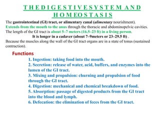
  • 1. T H E D I G E S T I V E S Y S T E M A N D H O M EO S T A S I S The gastrointestinal (GI) tract, or alimentary canal (alimentary nourishment). Extends from the mouth to the anus through the thoracic and abdominopelvic cavities. The length of the GI tract is about 5–7 meters (16.5–23 ft) in a living person. It is longer in a cadaver (about 7–9meters or 23–29.5 ft) . Because the muscles along the wall of the GI tract organs are in a state of tonus (sustained contraction). Functions 1. Ingestion: taking food into the mouth. 2. Secretion: release of water, acid, buffers, and enzymes into the lumen of the GI tract. 3. Mixing and propulsion: churning and propulsion of food through the GI tract. 4. Digestion: mechanical and chemical breakdown of food. 5. Absorption: passage of digested products from the GI tract into the blood and lymph. 6. Defecation: the elimination of feces from the GI tract.
  • 2. Organs of the digestive system
  • 3. Layers of the gastrointestinal tract.
  • 4. Mucosa Layers of the gastrointestinal tract. A.The mucosa, or inner lining of the GI tract, is a mucous membrane. It is composed of: (1) Epithelium in direct contact with the contents of the GI tract. (2) A layer of connective tissue called the lamina propria.88888 (3) A thin layer of smooth muscle (muscularis mucosae). 1. The epithelium: In the mouth, pharynx, esophagus, and anal canal. Nonkeratinized stratified squamous epithelium that serves a protective function. Simple columnar epithelium, which functions in secretion and absorption. Lines the stomach and intestines. The rate of renewal of GI tract epithelial cells is rapid: Every 5 to 7 days they slough off and are replaced by new cells. Enteroendocrine cells, that secrete hormones. 2. The lamina propria : Areolar connective tissue containing many blood and lymphatic vessels. Which are the routes by which nutrients absorbed into the GI tract reach the other tissues of the body. The lamina propria also contains the majority of the cells of the mucosa- associated lymphatic tissue (MALT). These prominent lymphatic nodules contain immune system cells that protect against disease .
  • 5. 3. A thin layer of smooth muscle fibers called the muscularis: Mucosae throws the mucous membrane of the stomach and small intestine into many small folds. Which increase the surface area for digestion and absorption. Movements of the muscularis mucosae ensure that all absorptive cells are fully exposed to the contents of the GI tract. B. Submucosa Consists of areolar connective tissue that binds the mucosa to the muscularis. It contains many blood and lymphatic vessels that receive absorbed food molecules. Also located in the submucosa is an extensive network of neurons known as the submucosal plexus . The submucosa may also contain glands and lymphatic tissue. Muscularis The muscularis of the mouth, pharynx, and superior and middle parts of the esophagus contains skeletal muscle that produces voluntary swallowing. Skeletal muscle also forms the external anal sphincter, which permits voluntary control of defecation. The muscularis consists of smooth muscle that is generally found in two sheets: Inner sheet of circular fibers. Outer sheet of longitudinal fibers. Between the layers of the muscularis is a second plexus of neurons—the myenteric plexus.
  • 6. C. Serosa Those portions of the GI tract that are suspended in the abdominopelvic cavity have a superficial layer called the serosa. The serosa is a serous membrane composed of areolar connective tissue and simple squamous epithelium (mesothelium). The esophagus lacks a serosa.
  • 7. Autonomic Nervous System The vagus (X) nerves supply parasympathetic fibers to most parts of the GI tract. The exception of the last half of the large intestine. Which is supplied with parasympathetic fibers from the sacral spinal cord. The parasympathetic nerves that supply the GI tract form neural connections with the ENS. Parasympathetic preganglionic neurons of the vagus or pelvic splanchnic nerves synapse with parasympathetic postganglionic neurons located in the myenteric and submucosal plexuses. Some of the parasympathetic postganglionic neurons in turn synapse with neurons in the ENS; Others directly innervate smooth muscle and glands within the wall of the GI tract. Stimulation of theparasympathetic nerves that innervate the GI tract causes an increase in GI secretion and motility by increasing the activity of ENS neurons. Sympathetic nerves that supply the GI tract arise from the thoracic and upper lumbar regions of the spinal cord. Sympathetic nerves form neural connections with the ENS. Sympathetic postganglionic neurons synapse with neurons located in the myenteric plexus and the submucosal plexus. The sympathetic nerves that supply the GI tract cause a decrease in GI secretion and motility by inhibiting the neurons of the ENS. Emotions such as anger, fear, and anxiety may slow digestion because they stimulate the sympathetic nerves that supply the GI tract.
  • 8. Organization of the enteric nervous system.
  • 9. The peritoneum Largest serous membrane of the body; It consists of a layer of ; Simple squamous epithelium (mesothelium) with an underlying supporting layer of areolar connective tissue. The peritoneum is divided into the parietal peritoneum: Which lines the wall of the abdomino pelvic cavity. The visceral peritoneum: Which covers some of the organs in the cavity and is their serosa The slim space containing lubricating serous fluid that is between the parietal and visceral portions of the peritoneum is called the peritoneal cavity. In certain diseases the peritoneal cavity may become distended by the accu- mulation of several liters of fluid, a condition called ascites. Some organs are not lie in the peritoneal cavity. Such organs, including the kidneys, ascending and descending colons of the large intestine, duodenum of the small intestine, and pancreas, are said to be retroperitoneal (retro- behind).
  • 10. The peritoneum contains large folds that weave between the viscera. The folds bind the organs to one another and to the walls of the abdominal cavity. They also contain blood vessels, lymphatic vessels, and nerves that supply the abdominal organs. There are five major peritoneal folds: The greater omentum, Falciform ligament, Lesser omentum, Mesentery, Mesocolon.
  • 11. 1. The greater omentum (o¯-MEN-tum fat skin), the largest peritoneal fold. The greater omentum is a double sheet that folds back on itself, giving it a total of four layers. 2. The falciform ligament (FAL-si-form; falc- sickleshaped) attaches the liver to the anterior abdominal wall and diaphragm . The liver is the only digestive organ that is attached to the anterior abdominal wall. 3. The lesser omentum arises as an anterior fold in the serosa of the stomach and duodenum, and it suspends the stomach and duodenum from the liver . It is the pathway for blood vessels entering the liver and contains the hepatic portal vein, common hepatic artery, and common bile duct, along with some lymph nodes. 4. A fan-shaped fold of the peritoneum, called the mesentery binds the jejunum and ileum of the small intestine to the posterior abdominal wall . 5. Two separate folds of peritoneum, called the mesocolon bind the transverse colon (transverse mesocolon) and sigmoid colon (sigmoid mesocolon) of the large intestine to the posterior abdominal wall .
  • 12. Structures of the mouth (oral cavity).
  • 13. The mouth Also referred to as the oral or buccal cavity (BUK-al; bucca cheeks): is formed by the cheeks, hard and soft palates, and tongue . The cheeks form the lateral walls of the oral cavity. They are covered externally by skin and internally by a mucous membrane. Which consists of nonkeratinized stratified squamous epithelium. Buccinator muscles and connective tissue lie between the skin and mucous membranes of the cheeks. The anterior portions of the cheeks end at the lips. The lips or labia ( fleshy borders) are fleshy folds surrounding the opening of the mouth. They contain the orbicularis oris muscle and are covered externally by skin and internally by a mucous membrane. The inner surface of each lip is attached to its corresponding gum by a midline fold of mucous membrane called the labial frenulum . During chewing, contraction of the buccinator muscles in the cheeks and orbicularis oris muscle in the lips helps keep food between the upper and lower teeth. These muscles also assist in speech.
  • 14. The oral vestibule ( entrance to a canal) of the oral cavity is a space bounded externally by the cheeks and lips and internally by the gums and teeth. The palate is a wall or septum that separates the oral cavity from the nasal cavity, forming the roof of the mouth. This important structure makes it possible to chew and breathe at the same time. The hard palate—the anterior portion of the roof of the mouth—is formed by the maxillae and palatine bones and is covered by a mucous membrane; it forms a bony partition between the oral and nasal cavities. The soft palate, which forms the posterior portion of the roof of the mouth, is an arch-shaped muscular partition between the oropharynx and nasopharynx that is lined with mucous membrane. Hanging from the free border of the soft palate is a conical muscular process called the uvula (U¯ -vu¯ -la little grape). During swallowing, the soft palate and uvula are drawn superiorly, closing off the nasopharynx and preventing swallowed foods and liquids from entering the nasal cavity
  • 15. The three major salivary glands—parotid, sublingual, and submandibular
  • 16. A salivary gland is a gland that releases a secretion called saliva into the oral cavity. When food enters the mouth, however, secretion of saliva increases, and it lubricates, dissolves, and begins the chemical breakdown of the food. There are three pairs of major salivary glands: the parotid, submandibular, and sublingual glands The parotid glands (par- near; to- ear) are located inferior and anterior to the ears, between the skin and the masseter muscle. Each secretes saliva into the oral cavity via a parotid duct that pierces the buccinator muscle to open into the vestibule opposite the second maxillary (upper) molar tooth. The submandibular glands are found in the floor of the mouth; They are medial and partly inferior to the body of the mandible. Their ducts, the submandibular ducts, run under the mucosa on either side of the midline of the floor of the mouth and enter the oral cavity proper lateral to the lingual frenulum. The sublingual glands are beneath the tongue and superior to the submandibular glands. Their ducts, the lesser sublingual ducts, open into the floor of the mouth in the oral cavity proper.
  • 17. Composition and Functions of Saliva Chemically, saliva is 99.5% water and 0.5% solutes. Among the solutes are ions, including sodium, potassium, chloride, bicarbonate, and phosphate. Also present are some dissolved gases and various organic substances, including urea and uric acid, mucus, immunoglobulin A, the bacteriolytic enzyme lysozyme, and salivary amylase, a digestive enzyme that acts on starch. The parotid glands secrete a watery (serous) liquid containing salivary amylase. The submandibular glands secrete a fluid that contains amylase but is thickened with mucus. The sublingual glands contain mostly mucous cells, so they secrete a much thicker fluid that contributes only a small amount of salivary amylase. The water in saliva provides a medium for dissolving foods so that they can be tasted by gustatory receptors. Chloride ions in the saliva activate salivary amylase, an enzyme that starts the breakdown of starch. Bicarbonate and phosphate ions buffer acidic foods that enter the mouth, so saliva is only slightly acidic (Ph 6.35–6.85). Immunoglobulin A (IgA) prevents attachment of microbes so they cannot penetrate the epithelium, and the enzyme lysozyme kills bacteria;
  • 18. Salivation The secretion of saliva, called salivation (sal-i-VA¯ -shun). Controlled by the autonomic nervous system. Amounts of saliva secreted daily vary considerably but average 1000–1500 mL (1–1.6 qt). Parasympathetic stimulation promotes continuous secretion of a moderate amount of saliva. Sympathetic stimulation dominates during stress, resulting in dryness of the mouth.
  • 19. The tongue An accessory digestive organ composed of skeletal muscle covered with mucous membrane. It forms the floor of the oral cavity. The tongue is divided into symmetrical lateral halves by a median septum that extends its entire length, and it is attached inferiorly to the hyoid bone, styloid process of the temporal bone, and mandible. Each half of the tongue consists of an identical complement of extrinsic and intrinsic muscles. The extrinsic muscles of the tongue, which originate outside the tongue (attach to bones in the area) and insert into connective tissues in the tongue, include the hyoglossus, genioglossus, and styloglossus muscles . The extrinsic muscles move the tongue from side to side and in and out to maneuver food for chewing, shape the food into a rounded mass, and force the food to the back of the mouth for swallowing. The intrinsic muscles originate in and insert into connective tissue within the tongue. They alter the shape and size of the tongue for speech and swallowing. The intrinsic muscles include the longitudinalis superior, longitudinalis inferior, transversus linguae, and verticalis linguae muscles. The dorsum (upper surface) and lateral surfaces of the tongue are covered with papillae (pa-PIL-e¯ nipple-shaped projections), projections of the lamina propria covered with stratified squamous epithelium . Many papillae contain taste buds, the receptors for gustation (taste).
  • 20. The teeth, or dentes Organs located in sockets of the alveolar processes of the mandible and maxillae. The alveolar processes are covered by the gingivae (JIN-ji-ve¯ ), or gums. Which extend slightly into each socket. The sockets are lined by the periodontal ligament or membrane (odont- tooth). Which consists of dense fibrous connective tissue that anchors the teeth to the socket walls. A typical tooth has three major external regions: Crown Root Neck. The crown is the visible portion above the level of the gums. Embedded in the socket are one to three roots. The neck is the constricted junction of the crown and root near the gum line. Internally, dentin forms the majority of the tooth. Dentin consists of a calcified connective tissue that gives the tooth its basic shape and rigidity. It is harder than bone because of its higher content of calcium salts (70% of dry weight). The dentin of the crown is covered by enamel. Which consists primarily of calcium phosphate and calcium carbonate. Enamel is also harder than bone because of its even higher content of calcium salts (about 95% of dry weight).
  • 21. A typical tooth and surrounding structures.
  • 22.
  • 23. Humans have two dentitions, or sets of teeth: Deciduous and permanent. The first of these—the deciduous teeth (decidu- falling out), also called primary teeth, milk teeth, or baby teeth—begin to erupt at about 6 months of age, and approximately two teeth appear each month thereafter, until all 20 . The incisors : which are closest to the midline, are chisel-shaped and adapted for cutting into food. They are referred to as either central or lateral incisors based on their position. The cuspids (canines): which have a pointed surface called a cusp. Cuspids are used to tear and shred food. Incisors and cuspids have only one root apiece. The first and second molars: which have four cusps. Maxillary (upper) molars have three roots; Mandibular (lower) molars have two roots. The molars crush and grind food to prepare it for swallowing. All the deciduous teeth are lost—generally between ages 6 and 12 years.
  • 24. The permanent dentition: Contains 32 teeth that erupt between age 6 and adulthood. The deciduous molars are replaced by the first and second premolars (bicuspids). Which have two cusps and one root (upper first premolars have two roots) and are used for crushing and grinding. The first molars at age 6 (six-year molars), The second molars at age 12 (twelve-year molars), The third molars (wisdom teeth) after age 17 or not at all.
  • 25.
  • 26. Pharynx ( throat) When food is first swallowed, it passes from the mouth into the pharynx ( throat). a funnel-shaped tube that extends from the internal nares to the esophagus. The pharynx is composed of skeletal muscle and lined by mucous membrane. Divided into three parts: The nasopharynx. The oropharynx. The laryngopharynx. The nasopharynx functions only in respiration. Both the oropharynx and laryngopharynx have digestive as well as respiratory functions. Swallowed food passes from the mouth into the oropharynx and laryngopharynx; The muscular contractions of these areas help propel food into the esophagus and then into the stomach.
  • 27. The esophagus; Collapsible muscular tube, about 25 cm (10 in.) long, that lies posterior to the trachea. The esophagus begins at the inferior end of the laryngopharynx and passes through the mediastinum anterior to the vertebral column. Then it pierces the diaphragm through an opening called the esophageal hiatus, and ends in the superior portion of the stomach . Sometimes, part of the stomach protrudes above the diaphragm through the esophageal hiatus. This condition, termed a hiatus hernia. The esophagus secretes mucus and transports food into the stomach. It does not produce digestive enzymes, and it does not carry on absorption.
  • 28. DEGLUTITION The movement of food from the mouth into the stomach is achieved by the act of swallowing, or deglutition . Deglutition is facilitated by the secretion of saliva and mucus and involves the mouth, pharynx, and esophagus. Swallowing occurs in three stages: (1) the voluntary stage, in which the bolus is passed into the oropharynx; (2) the pharyngeal stage, the involuntary passage of the bolus through the pharynx into the esophagus; (3) the esophageal stage, the involuntary passage of the bolus through the esophagus into the stomach. Swallowing starts when the bolus is forced to the back of the oral cavity and into the oropharynx by the movement of the tongue upward and backward against the palate; these actions
  • 29.
  • 30.
  • 31. The stomach J-shaped enlargement of the GI tract directly inferior to the diaphragm in the epigastric, umbilical, and left hypochondriac regions of the abdomen. The stomach connects the esophagus to the duodenum, the first part of the small intestine. converted to a liquid, and certain substances are absorbed. Anatomy of the Stomach The stomach has four main regions: the cardia, fundus, body, and pylorus . The cardia (CAR-de¯-a) surrounds the superior opening of the stomach. The rounded portion superior to and to the left of the cardia is the fundus (FUN-dus). Inferior to the fundus is the large central portion of the stomach, called the body. The region of the stomach that connects to the duodenum is the pylorus ; It has two parts, the pyloric antrum (AN-trum cave), which connects to the body of the stomach, and the pyloric canal, which leads into the duodenum. When the stomach is empty, the mucosa lies in large folds, called rugae . The pylorus communicates with the duodenum of the small intestine via a smooth muscle sphincter called the pyloric sphincter. The concave medial border of the stomach is called the lesser curvature, and the convex lateral border is called the greater curvature.
  • 32.
  • 33. Functions of the Stomach 1. Mixes saliva, food, and gastric juice to form chyme. 2. Serves as a reservoir for food before release into small intestine. 3. Secretes gastric juice, which contains HCl (kills bacteria and denatures protein), pepsin (begins the digestion of proteins), intrinsic factor (aids absorption of vitamin B12), and gastric lipase (aids digestion of triglycerides). 4. Secretes gastrin into blood.
  • 34. Histology of the stomach.
  • 35. The stomach mucosa showing gastric glands and cell types
  • 36.
  • 37.
  • 38.
  • 39. PANCREAS Anatomy of the Pancreas The pancreas a retroperitoneal gland that is about 12–15 cm (5–6 in.) long and 2.5 cm (1 in.) thick. Lies posterior to the greater curvature of the stomach. The pancreas consists of a head, a body, and a tail and is usually connected to the duodenum by two ducts . The head is the expanded portion of the organ near the curve of the duodenum; Superior to and to the left of the head are the central body and the tapering tail. Pancreatic juices are secreted by exocrine cells into small ducts that ultimately unite to form two larger ducts, the pancreatic duct and the accessory duct. The pancreatic duct (duct of Wirsung) is the larger of the two ducts. These in turn convey the secretions into the small intestine. Pancreatic duct joins the common bile duct from the liver and gallbladder and enters the duodenum as a dilated common duct called the hepatopancreatic ampulla (ampulla of Vater). The ampulla opens on an elevation of the duodenal mucosa known as the major duodenal papilla, which lies about 10 cm (4 in.) inferior to the pyloric sphincter of the stomach.
  • 40. The passage of pancreatic juice and bile through the hepatopancreatic ampulla into the small intestine is regulated by a mass of smooth muscle known as the sphincter of the hepatopancreatic ampulla (sphincter of Oddi). The other major duct of the pancreas, the accessory duct (duct of Santorini), leads from the pancreas and empties into the duodenum about 2.5 cm (1 in.) uperior to the hepatopancreatic ampulla. The pancreas is made up of small clusters of glandular epithelial cells. About 99% of the clusters, called acini (AS-i-ne¯ ), constitute the exocrine portion of the organ . The cells within acini secrete a mixture of fluid and digestive enzymes called pancreatic juice. The remaining 1% of the clusters, called pancreatic islets (islets of Langerhans), form the endocrine portion of the pancreas. These cells secrete the hormones glucagon, insulin, somatostatin, and pancreatic polypeptide.
  • 41. Relation of the pancreas to the liver, gallbladder, and duodenum.
  • 43. Ducts carrying bile from liver and gallbladder and pancreatic juice from pancreas to the duodenum
  • 44. Composition and Functions of Pancreatic Juice Each day the pancreas produces 1200–1500 mL (about 1.2– 1.5 qt) of pancreatic juice, a clear, colorless liquid consisting mostly of water, some salts, sodium bicarbonate, and several enzymes. The sodium bicarbonate gives pancreatic juice a slightly alkaline pH (7.1–8.2) that buffers acidic gastric juice in chyme, stops the action of pepsin from the stomach, and creates the proper pH for the action of digestive enzymes in the small intestine. Starch digesting enzyme called pancreatic amylase; Protein digesting enzymes called trypsin ,chymotrypsin ,carboxypeptidase, and elastase; Triglyceride-digesting enzyme in adults, called pancreatic lipase; Nucleic acid– digesting enzymes called ribonuclease and deoxyribonuclease.
  • 45. LIVER AND GALLBLADDER The liver is the heaviest gland of the body, weighing about 1.4 kg (about 3 lb) in an average adult. Of all of the organs of the body, it is second only to the skin in size. The liver is inferior to the diaphragm and occupies most of the right hypochondriac and part of the epigastric regions of the abdominopelvic cavity The gallbladder (gall- bile) is a pear-shaped sac that is located in a depression of the posterior surface of the liver. It is 7–10 cm (3–4 in.) long and typically hangs from the anterior inferior margin of the liver. Anatomy of the Liver and Gallbladder The liver is divided into two principal lobes: A large right lobe and a smaller left lobe— by the falciform ligament, a fold of the mesentery. The right lobe include: An inferior quadrate lobe . Posterior caudate lobe. The falciform ligament extends from the undersurface of the diaphragm between the two principal lobes of the liver to the superior surface of the liver, helping to suspend the liver in the abdominal cavity. In the free border of the falciform ligament is the ligamentum teres round ligament), a remnant of the umbilical vein of the fetus this fibrous cord extends from the liver to the umbilicus.
  • 46. The right and left coronary ligaments are narrow extensions of the parietal peritoneum that suspend the liver from the diaphragm. The parts of the gallbladder include the broad fundus, which projects inferiorly beyond the inferior border of the liver; the body, the central portion; and the neck, the tapered portion. The body and neck project superiorly. Histology of the Liver and Gallbladder Histologically, the liver is composed of several compon
  • 48. Hepatic blood flow: sources, path through the liver, and return to the heart.
  • 49. Functions of liver: • Carbohydrate metabolism: The liver is especially important in maintaining a normal blood glucose level. When blood glucose is low: The liver can break down glycogen to glucose and release the glucose into the bloodstream. When blood glucose is high: as occurs just after eating a meal, the liver converts glucose to glycogen and triglycerides for storage. • Lipid metabolism: Hepatocytes store some triglycerides; break down fatty acids to generate ATP; • Protein metabolism: Hepatocytes deaminate (remove the amino group, NH2, from) amino acids so that the amino acids can be used for ATP production or converted to carbohydrates or fats. • Processing of drugs and hormones: The liver can detoxify substances such as alcohol and excrete drugs such as penicillin, erythromycin, and sulfonamides into bile. It can also chemically alter or excrete thyroid hormones and steroid hormones such as estrogens and aldosterone. • Excretion of bilirubin: Bilirubin, derived from the heme of aged red blood cells, is absorbed by the liver from the blood and secreted into bile. • Storage. In addition to glycogen, the liver is a prime storage • Phagocytosis. The stellate reticuloendothelial (Kupffer) • Activation of vitamin D. The skin, liver, and kidneys participate in synthesizing the active form of vitamin D.
  • 50. The small intestine. Most digestion and absorption of nutrients occur in a long tube called the small intestine. Because of this, its structure is specially Its length alone provides a large surface area for digestion and absorption, and that area is further increased by circular folds, villi, and microvilli. 2.5 cm (1 in.) in diameter; its length is about 3 m (10 ft) in a living person 6.5 m (21 ft) in a cadaver due to the loss of smooth muscle tone after death. The small intestine is divided into three regions . The duodenum : the shortest region, is retroperitoneal. It starts at the pyloric sphincter of the stomach and extends about 25 cm (10 in.) until it merges with the jejunum. Duodenum means “12”; it is so named because it is about as long as the width of 12 fingers. The jejunum : is about 1 m (3 ft) long and extends to the ileum. The final and longest region of the small intestine the ileum : twisted), measures about 2 m (6 ft) and joins the large intestine at a smooth muscle sphincter called the ileocecal sphincter
  • 51. Anatomy of the small intestine.
  • 52. Histology of the small intestine
  • 53. Enlarged villus showing lacteal, capillaries, intestinal glands, and cell types
  • 54.
  • 55. Absorption of digested nutrients in the small intestine
  • 56. Daily volumes of fluid ingested, secreted, absorbed, and excreted from the GI tract.
  • 57. Anatomy of the large intestine
  • 58. PHASES OF DIGESTION Digestive activities occur in three overlapping phases: The cephalic phase, the gastric phase, and the intestinal phase. Cephalic Phase: During the cephalic phase of digestion, the smell, sight, thought, or initial taste of food activates neural centers in the cerebral cortex, hypothalamus, and brain stem. The brain stem then activates the facial (VII), glossopharyngeal (IX), and vagus (X) nerves. The facial and glossopharyngeal nerves stimulate the salivary glands to secrete saliva, while the vagus nerves stimulate the gastric glands to secrete gastric juice. The purpose of the cephalic phase of digestion is to prepare the mouth and stomach for food that is about to be eaten. Gastric Phase : Once food reaches the stomach, the gastric phase of digestion begins. Neural and hormonal mechanisms regulate the gastric phase of digestion to promote gastric secretion and gastric motility. • Neural regulation. Food of any kind distends the stomach and stimulates stretch receptors in its walls. Chemoreceptors in the stomach monitor the pH of the stomach chyme. When the stomach walls are distended or pH increases because proteins have entered the stomach and buffered some of the stomach acid, the stretch receptors and chemoreceptors are activated, and a neural negative feedback loop is set in motion (Figure 24.24). From the stretch receptors and chemoreceptors, nerve
  • 59. Hormonal regulation. Gastric secretion during the gastric phase is also regulated by the hormone gastrin. Gastrin is released from the G cells of the gastric glands in response to several stimuli: distension of the stomach by chyme, partially digested proteins in chyme, the high pH of chyme due to the presence of food in the stomach, caffeine in gastric chyme, and acetycholine released from parasympathetic neurons. Once it is released, gastrin enters the bloodstream, makes a round-trip through the body, and finally reaches its target organs in the digestive system. Gastrin stimulates gastric glands to secrete large amounts of gastric juice. It also strengthens the contraction of the lower esophageal sphincter to prevent reflux of acid chyme into the esophagus, increases motility of the stomach, and relaxes the pyloric sphincter, which promotes gastric emptying. Gastrin secretion is inhibited when the pH of gastric juice drops below 2.0 and is stimulated when the pH rises. This negative feedback mechanism helps provide an optimal low pH for the functioning of pepsin, the killing of microbes, and the denaturing of proteins in the stomach.
  • 60. Intestinal Phase The intestinal phase of digestion begins once food enters the small intestine. In contrast to reflexes initiated during the cephalic and gastric phases, which stimulate stomach secretory activity and motility, those occurring during the intestinal phase have inhibitory effects that slow the exit of chyme from the stomach. This prevents the duodenum from being overloaded with more chyme than it can handle. In addition, responses occurring during the intestinal phase promote the continued digestion of foods that have reached the small intestine. These activities of the intestinal phase of digestion are regulated by neural and hormonal mechanisms. • Neural regulation. Distension of the duodenum by the presence of chyme causes the enterogastric reflex. Stretch receptors in the duodenal wall send nerve impulses to the medulla oblongata, where they inhibit parasympathetic stimulation and stimulate the sympathetic nerves to the stomach. As a result, gastric motility is inhibited and there is an increase in the contraction of the pyloric sphincter, which decreases gastric emptying. • Hormonal regulation. The intestinal phase of digestion is mediated by two major hormones secreted by the small intestine: cholecystokinin and secretin. Cholecystokinin (CCK) is secreted by the CCK cells of the small intestinal