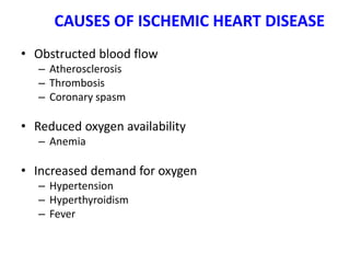
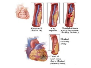
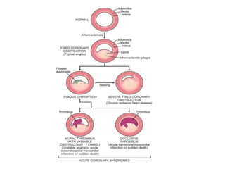
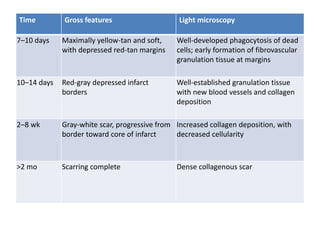
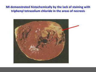
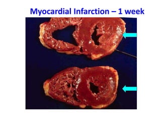
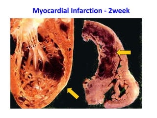
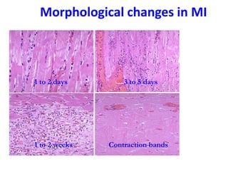
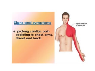
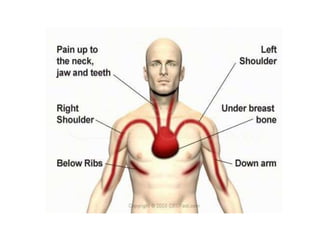
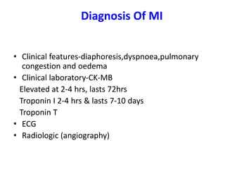
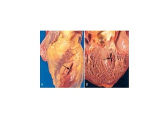
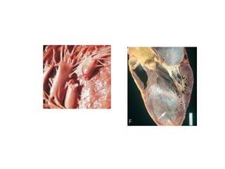
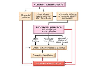
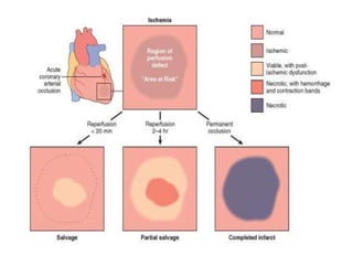
MYOCARIAL INFARCTION
DR. SHANMUGASAMY. K
ASSISTANT PROFESSOR
Case history:
• 50 yrs old hypertensive male came with
complaints of severe chest pain, dyspnoea
and sweating. On examination, pulse was
rapid and feeble. ECG- shows------------,
biochemical investigation shows elevated
cardiac markers like troponin and CK-MB .
• 1. what is your diagnosis
• 2. morphology of this disease
• 3. lab diagnosis
ECG
CONTENT:
• Introduction
• Causes and pathogenesis of IHD
• Angina pectoris
• Myocardial infarction
- Etiopathogenesis
- Morphology
- Lab diagnosis
- Complications
Introduction
• Leading cause of death.
• Myocardial ischemia.
• Ischemia versus hypoxia.
• Coronary atheroscelerosis- 90%
IHD presenting clinical syndromes:
• Myocardial infarction.
• Angina pectoris.
• Chronic IHD with heart failure.
• Sudden cardiac death
Ischemic Heart Disease (IHD)
Asymptomatic
Disease
Angina
pectoris
Myocardial
Infarction
Heart
Failure
Sudden
Death
Etiology:
1. Reduced coronary blood flow.
2. Increased myocardial metabolism.
3. Decreased oxygen transport
CAUSES OF ISCHEMIC HEART DISEASE
• Obstructed blood flow
– Atherosclerosis
– Thrombosis
– Coronary spasm
• Reduced oxygen availability
– Anemia
• Increased demand for oxygen
– Hypertension
– Hyperthyroidism
– Fever
Pathogenesis :
• Chronic atheroscelerosis.
• Acute plaque change.
• Consequences of myocardial ischemia.
Chronic atheroscelerosis:
• More than 90% of patients with IHD have
atherosclerosis of one or more of the
epicardial coronary arteries.
• stenosis (“fixed” obstructions) , acute plaque
disruption with thrombosis.
• single major coronary epicardial trunk may be
affected, two or all three. 1. LAD, 2.LCX, and 3.
RCA.
Acute Plaque Change:
• Number, Distribution, Structure, and Degree
of obstruction of atheromatous plaques.
• Stable atherosclerotic plaque to an unstable
atherothrombotic lesion through rupture,
superficial erosion, ulceration, fissuring, or
deep hemorrhage.
• Formation of a superimposed thrombus that
partially or completely occludes the affected
artery.
Angina pectoris
• A symptom complex of IHD characterized
by paroxysmal attacks of chest pain,
usually substernal or precordial, caused
by myocardial ischemia that falls short of
inducing infarction. There are several
patterns
Variants of Angina
• 1. Stable angina (typical) - paroxysms of pain
related to exertion and relieved by rest or
vasodilators. subendocardial ischemia with ST-
segment depression
• 2. Variant or Prinzmetal's angina - angina that
classically occurs at rest and is caused by reversible
spasm in normal to severely atherosclerotic
coronary arteries. ST-segment elevation or
depression maybe seen during attacks.
Variants of Angina
• 3. Unstable angina - prolonged pain, pain at rest in
a person with stable angina, or worsening of pain in
stable angina. ST-segment depression (usually) and
ST-segment elevation.
• .
Myocardial infarction
• Is the death of heart muscle following
ischaemic heart disease
Pathogenesis-MI
• Coronary artery occlusion-
• Sudden change in the morphology of the plaque
• Adhesion,aggregation, activation and release of
potent platelet aggregators including thromboxane
A2,serotonin,platelet factor 3 & 4
• Vasospasm
• Activation of extrinsic pathway of coagulation
• Thrombus completely occludes vessel lumen
Pathogenesis (contd)
• Other mechanisms-
• Emboli-from lt. atrium,paradoxical emboli
• Unexplained-diseases of small intra-mural
coronary vessels like
vasculitis,haemoglobinopathies,amyloid
deposition in vessel walls,other unusual
disorders like vascular dissection and
inadequate protection during cardiac surgery.
Pathogenesis -contd
• Myocardial response-Coronary artery
occlusion leads to loss of critical blood supply
to the myocardium,producing profound
functional,biologic and morphologic
consequences.
• Results in cell death in the area of supply,seen
in sub-endocardium.Outcome depends on
duration and severity of flow deprivation
Pathogenesis -contd
• Early biochemical change-switch from aerobic to anaerobic
glycolysis within seconds→inadequate production of high
energy phosphates(creatine phosphate and adenosine
triphosphate)
• Accumulation of lactic acid
• Within 60 secs of onset of ischaemia-loss of contractility
occurs→acute heart failure(myofibrillar relaxation,glycogen
deposition,cell and mitochondrial swelling)
• These changes are reversible and cell death is not immediate
Pathogenesis -contd
• Severe ischaemia lasting 20-40 mins leads to
irreversible necrosis of some cardiomyocytes(primary
structural defects in sarcolemmal membrane)
• With prolonged ischemia injury to micro-vasculature
follows
• Arrhythmias occur through poorly understood
mechanism
• Sudden cardiac death
• “Wavefront”of myocardial ischaemia
Gross morphology -MI
Patterns include:
• Transmural infarct - involving the entire thickness
of the left ventricular wall from endocardium to
epicardium, usually the anterior free wall and
posterior free wall and septum with extension into
the RV wall in 15-30%. Isolated infarcts of RV and
right atrium are extremely rare.
• Subendocardial infarct - multifocal areas of
necrosis confined to the inner 1/3-1/2 of the left
ventricular wall. These do not show the same
evolution of changes seen in a transmural MI.
Global hypotension-sub-endocardial infarcts are usually circumferential.
Transmural infarction
Irreversible injury
Time Gross features Light microscopy
½–4 hr None Usually none; variable waviness of fibers at border
4-12 hrs Dark mottling
(occasional)
Early coagulation necrosis; edema; hemorrhage
12–24 hr Dark mottling Ongoing coagulation necrosis; pyknosis of nuclei;
myocyte hypereosinophilia; marginal contraction band
necrosis; early neutrophilic infiltrate
1–3 days Mottling with yellow-tan
infarct center
Coagulation necrosis, with loss of nuclei and
striations; brisk interstitial infiltrate of neutrophils
3–7 days Hyperemic border;
central yellow-tan
softening
Beginning disintegration of dead myofibers, with dying
neutrophils; early phagocytosis of dead cells by
macrophages at infarct border
Time Gross features Light microscopy
7–10 days Maximally yellow-tan and soft,
with depressed red-tan margins
Well-developed phagocytosis of dead
cells; early formation of fibrovascular
granulation tissue at margins
10–14 days Red-gray depressed infarct
borders
Well-established granulation tissue
with new blood vessels and collagen
deposition
2–8 wk Gray-white scar, progressive from
border toward core of infarct
Increased collagen deposition, with
decreased cellularity
>2 mo Scarring complete Dense collagenous scar
MI demonstrated histochemically by the lack of staining with
triphenyl tetrazolium chloride in the areas of necrosis
Myocardial Infarction – 1 week
Myocardial Infarction - 2week
Morphological changes in MI
1 to 2 days 3 to 5 days
1 to 2 weeks Contraction bands
Diagnosis Of MI
• Clinical features-diaphoresis,dyspnoea,pulmonary
congestion and oedema
• Clinical laboratory-CK-MB
Elevated at 2-4 hrs, lasts 72hrs
Troponin I 2-4 hrs & lasts 7-10 days
Troponin T
• ECG
• Radiologic (angiography)
Creatine Kinase -Total
• The total CK is a simple and inexpensive test
that is readily available using many laboratory
instruments.
• However, an elevation in total CK is not
specific for myocardial injury,
• because most CK is located in skeletal muscle,
and elevations are possible from a variety of
non-cardiac conditions.
Creatine Kinase
• Three isoenzymes
– CK – MM (heart & skeletal muscle)
– CK – BB (brain, lung & other tissues)
– CK – MB (Principally myocardium, variable
amounts in skeletal muscle)
• CK – MB rises within 2 – 8 hours of MI, peaks
at 18 hours and disappears by 48 to 72 hours
• An absence of a change in levels in CK – MB
during the first 2 days of chest pain essentially
excludes MI
CK-MB
• Is a very good marker for acute myocardial injury, because
of its excellent specificity, and it rises in serum within 2 to 8
hours of onset of acute myocardial infarction.
• Serial measurements every 2 to 4 hours for a period of 9 to
12 hours after the patient is first seen will provide a pattern
to determine whether the CK-MB is rising, indicative of
myocardial injury.
• The CK-MB is also useful for diagnosis of reinfarction or
extension of an MI because it begins to fall after a day,
dissipating in 1 to 3 days, so subsequent elevations are
indicative of another event.
Troponin I and T
• Troponin I and T are structural components of cardiac muscle.
• They are released into the bloodstream with myocardial
injury.
• They are highly specific for myocardial injury – more so than
CK-MB – and help to exclude elevations of CK with skeletal
muscle trauma.
• Troponins will begin to increase following MI within 2 to 12
hours, about the same time frame as CK-MB. However, the
rate of rise for early infarction may not be as dramatic as for
CK-MB.
Troponins
• Troponins will remain elevated longer than CK--up to 5 to
9 days for troponin I and up to 2 weeks for troponin T.
• This makes troponins a superior marker for diagnosing
myocardial infarction in the recent past--better than
lactate dehydrogenase (LDH).
• However, this continued elevation has the disadvantage
of making it more difficult to diagnose reinfarction or
extension of infarction in a patient who has already
suffered an initial MI.
• Troponin T lacks some specificity because elevations can
appear with skeletal myopathies and with renal failure.
Myoglobin
• Is a protein found in skeletal and cardiac muscle
which binds oxygen.
• It is a very sensitive indicator of muscle injury.
The rise in myoglobin can help to determine the
size of an infarction.
• A negative myoglobin can help to rule out
myocardial infarction. It is elevated even before
CK-MB.
• However, it is not specific for cardiac muscle, and
can be elevated with any form of injury to skeletal
muscle.
LDH
• The LDH has been supplanted by other tests. It begins to
rise in 12 to 24 hours following MI, and peaks in 2 to 3
days, gradually dissipating in 5 to 14 days.
• Measurement of LDH isoenzymes is necessary for
greater specificity for cardiac injury.
• There are 5 isoenzymes (1 through 5). Ordinarily,
isoenzyme 2 is greater than 1, but with myocardial
injury, this pattern is "flipped" and 1 is higher than 2.
• LDH-5 from liver may be increased with centrilobular
necrosis from passive congestion with congestive heart
failure following ischemic myocardial injury.
Infarct modification by reperfusion
• Restoration of coronary flow→salvage ischaemic
myocardium.To be established in 3-4 hrs.Within 15-
20 mins-prevents all necrosis
• Contraction bands-occurs in lethally injured cells on
reperfusion
• Reperfusion injury-Leukocytes in the reperfused
blood→apoptosis of myocytes and microvascular
injury→haemorrhage,endothelial
swelling→occludes capillaries→prevents local
reperfusion-no reflow
Complications
Complicated(80%)
• Contractile dysfunction,pump failure,cardiogenic shock(15%)
• >40% ventricular wall involved
• Arrhythmias and conduction defects, with possible "sudden
death" (85%)
• Myocardial rupture-3-7 days,mid-ventricular wall
• Risk factors-female gender,older than 60 yrs,pre-existing
hypertension,lack of lt ventr hypertrophy
• Myocardial wall rupture, with possible tamponade (1-2%)
• Papillary muscle rupture, with possible valvular insufficiency
• Rupture of Ventricular septum-L→R shunt(1-2%)
• Extension of infarction, or re-infarction
• Congestive heart failure (pulmonary edema) (60%)
• Pericarditis-2-3 days
• Right ventricular infarction
• Infarct expansion-Stretching,thinning and dilatation
of infarct region as with mural thrombus
• Mural thrombosis, with possible embolization (30%)
• Ventricular aneurysm formation-late
complication,rarely undergoes rupture
• Papillary muscle dysfunction
• Progressive late heart failure
• Uncomplicated (20%)
THANK YOU

4 4-2016 myocardial infarction

  • 1.
  • 2.
    Case history: • 50yrs old hypertensive male came with complaints of severe chest pain, dyspnoea and sweating. On examination, pulse was rapid and feeble. ECG- shows------------, biochemical investigation shows elevated cardiac markers like troponin and CK-MB . • 1. what is your diagnosis • 2. morphology of this disease • 3. lab diagnosis
  • 3.
  • 4.
    CONTENT: • Introduction • Causesand pathogenesis of IHD • Angina pectoris • Myocardial infarction - Etiopathogenesis - Morphology - Lab diagnosis - Complications
  • 5.
    Introduction • Leading causeof death. • Myocardial ischemia. • Ischemia versus hypoxia. • Coronary atheroscelerosis- 90%
  • 6.
    IHD presenting clinicalsyndromes: • Myocardial infarction. • Angina pectoris. • Chronic IHD with heart failure. • Sudden cardiac death
  • 7.
    Ischemic Heart Disease(IHD) Asymptomatic Disease Angina pectoris Myocardial Infarction Heart Failure Sudden Death
  • 8.
    Etiology: 1. Reduced coronaryblood flow. 2. Increased myocardial metabolism. 3. Decreased oxygen transport
  • 9.
    CAUSES OF ISCHEMICHEART DISEASE • Obstructed blood flow – Atherosclerosis – Thrombosis – Coronary spasm • Reduced oxygen availability – Anemia • Increased demand for oxygen – Hypertension – Hyperthyroidism – Fever
  • 10.
    Pathogenesis : • Chronicatheroscelerosis. • Acute plaque change. • Consequences of myocardial ischemia.
  • 11.
    Chronic atheroscelerosis: • Morethan 90% of patients with IHD have atherosclerosis of one or more of the epicardial coronary arteries. • stenosis (“fixed” obstructions) , acute plaque disruption with thrombosis. • single major coronary epicardial trunk may be affected, two or all three. 1. LAD, 2.LCX, and 3. RCA.
  • 12.
    Acute Plaque Change: •Number, Distribution, Structure, and Degree of obstruction of atheromatous plaques. • Stable atherosclerotic plaque to an unstable atherothrombotic lesion through rupture, superficial erosion, ulceration, fissuring, or deep hemorrhage. • Formation of a superimposed thrombus that partially or completely occludes the affected artery.
  • 15.
    Angina pectoris • Asymptom complex of IHD characterized by paroxysmal attacks of chest pain, usually substernal or precordial, caused by myocardial ischemia that falls short of inducing infarction. There are several patterns
  • 16.
    Variants of Angina •1. Stable angina (typical) - paroxysms of pain related to exertion and relieved by rest or vasodilators. subendocardial ischemia with ST- segment depression • 2. Variant or Prinzmetal's angina - angina that classically occurs at rest and is caused by reversible spasm in normal to severely atherosclerotic coronary arteries. ST-segment elevation or depression maybe seen during attacks.
  • 17.
    Variants of Angina •3. Unstable angina - prolonged pain, pain at rest in a person with stable angina, or worsening of pain in stable angina. ST-segment depression (usually) and ST-segment elevation. • .
  • 18.
    Myocardial infarction • Isthe death of heart muscle following ischaemic heart disease
  • 20.
    Pathogenesis-MI • Coronary arteryocclusion- • Sudden change in the morphology of the plaque • Adhesion,aggregation, activation and release of potent platelet aggregators including thromboxane A2,serotonin,platelet factor 3 & 4 • Vasospasm • Activation of extrinsic pathway of coagulation • Thrombus completely occludes vessel lumen
  • 21.
    Pathogenesis (contd) • Othermechanisms- • Emboli-from lt. atrium,paradoxical emboli • Unexplained-diseases of small intra-mural coronary vessels like vasculitis,haemoglobinopathies,amyloid deposition in vessel walls,other unusual disorders like vascular dissection and inadequate protection during cardiac surgery.
  • 22.
    Pathogenesis -contd • Myocardialresponse-Coronary artery occlusion leads to loss of critical blood supply to the myocardium,producing profound functional,biologic and morphologic consequences. • Results in cell death in the area of supply,seen in sub-endocardium.Outcome depends on duration and severity of flow deprivation
  • 23.
    Pathogenesis -contd • Earlybiochemical change-switch from aerobic to anaerobic glycolysis within seconds→inadequate production of high energy phosphates(creatine phosphate and adenosine triphosphate) • Accumulation of lactic acid • Within 60 secs of onset of ischaemia-loss of contractility occurs→acute heart failure(myofibrillar relaxation,glycogen deposition,cell and mitochondrial swelling) • These changes are reversible and cell death is not immediate
  • 24.
    Pathogenesis -contd • Severeischaemia lasting 20-40 mins leads to irreversible necrosis of some cardiomyocytes(primary structural defects in sarcolemmal membrane) • With prolonged ischemia injury to micro-vasculature follows • Arrhythmias occur through poorly understood mechanism • Sudden cardiac death • “Wavefront”of myocardial ischaemia
  • 25.
    Gross morphology -MI Patternsinclude: • Transmural infarct - involving the entire thickness of the left ventricular wall from endocardium to epicardium, usually the anterior free wall and posterior free wall and septum with extension into the RV wall in 15-30%. Isolated infarcts of RV and right atrium are extremely rare. • Subendocardial infarct - multifocal areas of necrosis confined to the inner 1/3-1/2 of the left ventricular wall. These do not show the same evolution of changes seen in a transmural MI. Global hypotension-sub-endocardial infarcts are usually circumferential.
  • 27.
  • 29.
    Irreversible injury Time Grossfeatures Light microscopy ½–4 hr None Usually none; variable waviness of fibers at border 4-12 hrs Dark mottling (occasional) Early coagulation necrosis; edema; hemorrhage 12–24 hr Dark mottling Ongoing coagulation necrosis; pyknosis of nuclei; myocyte hypereosinophilia; marginal contraction band necrosis; early neutrophilic infiltrate 1–3 days Mottling with yellow-tan infarct center Coagulation necrosis, with loss of nuclei and striations; brisk interstitial infiltrate of neutrophils 3–7 days Hyperemic border; central yellow-tan softening Beginning disintegration of dead myofibers, with dying neutrophils; early phagocytosis of dead cells by macrophages at infarct border
  • 30.
    Time Gross featuresLight microscopy 7–10 days Maximally yellow-tan and soft, with depressed red-tan margins Well-developed phagocytosis of dead cells; early formation of fibrovascular granulation tissue at margins 10–14 days Red-gray depressed infarct borders Well-established granulation tissue with new blood vessels and collagen deposition 2–8 wk Gray-white scar, progressive from border toward core of infarct Increased collagen deposition, with decreased cellularity >2 mo Scarring complete Dense collagenous scar
  • 31.
    MI demonstrated histochemicallyby the lack of staining with triphenyl tetrazolium chloride in the areas of necrosis
  • 32.
  • 33.
  • 34.
    Morphological changes inMI 1 to 2 days 3 to 5 days 1 to 2 weeks Contraction bands
  • 37.
    Diagnosis Of MI •Clinical features-diaphoresis,dyspnoea,pulmonary congestion and oedema • Clinical laboratory-CK-MB Elevated at 2-4 hrs, lasts 72hrs Troponin I 2-4 hrs & lasts 7-10 days Troponin T • ECG • Radiologic (angiography)
  • 38.
    Creatine Kinase -Total •The total CK is a simple and inexpensive test that is readily available using many laboratory instruments. • However, an elevation in total CK is not specific for myocardial injury, • because most CK is located in skeletal muscle, and elevations are possible from a variety of non-cardiac conditions.
  • 39.
    Creatine Kinase • Threeisoenzymes – CK – MM (heart & skeletal muscle) – CK – BB (brain, lung & other tissues) – CK – MB (Principally myocardium, variable amounts in skeletal muscle) • CK – MB rises within 2 – 8 hours of MI, peaks at 18 hours and disappears by 48 to 72 hours • An absence of a change in levels in CK – MB during the first 2 days of chest pain essentially excludes MI
  • 40.
    CK-MB • Is avery good marker for acute myocardial injury, because of its excellent specificity, and it rises in serum within 2 to 8 hours of onset of acute myocardial infarction. • Serial measurements every 2 to 4 hours for a period of 9 to 12 hours after the patient is first seen will provide a pattern to determine whether the CK-MB is rising, indicative of myocardial injury. • The CK-MB is also useful for diagnosis of reinfarction or extension of an MI because it begins to fall after a day, dissipating in 1 to 3 days, so subsequent elevations are indicative of another event.
  • 41.
    Troponin I andT • Troponin I and T are structural components of cardiac muscle. • They are released into the bloodstream with myocardial injury. • They are highly specific for myocardial injury – more so than CK-MB – and help to exclude elevations of CK with skeletal muscle trauma. • Troponins will begin to increase following MI within 2 to 12 hours, about the same time frame as CK-MB. However, the rate of rise for early infarction may not be as dramatic as for CK-MB.
  • 42.
    Troponins • Troponins willremain elevated longer than CK--up to 5 to 9 days for troponin I and up to 2 weeks for troponin T. • This makes troponins a superior marker for diagnosing myocardial infarction in the recent past--better than lactate dehydrogenase (LDH). • However, this continued elevation has the disadvantage of making it more difficult to diagnose reinfarction or extension of infarction in a patient who has already suffered an initial MI. • Troponin T lacks some specificity because elevations can appear with skeletal myopathies and with renal failure.
  • 43.
    Myoglobin • Is aprotein found in skeletal and cardiac muscle which binds oxygen. • It is a very sensitive indicator of muscle injury. The rise in myoglobin can help to determine the size of an infarction. • A negative myoglobin can help to rule out myocardial infarction. It is elevated even before CK-MB. • However, it is not specific for cardiac muscle, and can be elevated with any form of injury to skeletal muscle.
  • 44.
    LDH • The LDHhas been supplanted by other tests. It begins to rise in 12 to 24 hours following MI, and peaks in 2 to 3 days, gradually dissipating in 5 to 14 days. • Measurement of LDH isoenzymes is necessary for greater specificity for cardiac injury. • There are 5 isoenzymes (1 through 5). Ordinarily, isoenzyme 2 is greater than 1, but with myocardial injury, this pattern is "flipped" and 1 is higher than 2. • LDH-5 from liver may be increased with centrilobular necrosis from passive congestion with congestive heart failure following ischemic myocardial injury.
  • 45.
    Infarct modification byreperfusion • Restoration of coronary flow→salvage ischaemic myocardium.To be established in 3-4 hrs.Within 15- 20 mins-prevents all necrosis • Contraction bands-occurs in lethally injured cells on reperfusion • Reperfusion injury-Leukocytes in the reperfused blood→apoptosis of myocytes and microvascular injury→haemorrhage,endothelial swelling→occludes capillaries→prevents local reperfusion-no reflow
  • 46.
    Complications Complicated(80%) • Contractile dysfunction,pumpfailure,cardiogenic shock(15%) • >40% ventricular wall involved • Arrhythmias and conduction defects, with possible "sudden death" (85%) • Myocardial rupture-3-7 days,mid-ventricular wall • Risk factors-female gender,older than 60 yrs,pre-existing hypertension,lack of lt ventr hypertrophy • Myocardial wall rupture, with possible tamponade (1-2%) • Papillary muscle rupture, with possible valvular insufficiency • Rupture of Ventricular septum-L→R shunt(1-2%)
  • 47.
    • Extension ofinfarction, or re-infarction • Congestive heart failure (pulmonary edema) (60%) • Pericarditis-2-3 days • Right ventricular infarction • Infarct expansion-Stretching,thinning and dilatation of infarct region as with mural thrombus • Mural thrombosis, with possible embolization (30%) • Ventricular aneurysm formation-late complication,rarely undergoes rupture • Papillary muscle dysfunction • Progressive late heart failure • Uncomplicated (20%)
  • 53.