Downloaded 357 times
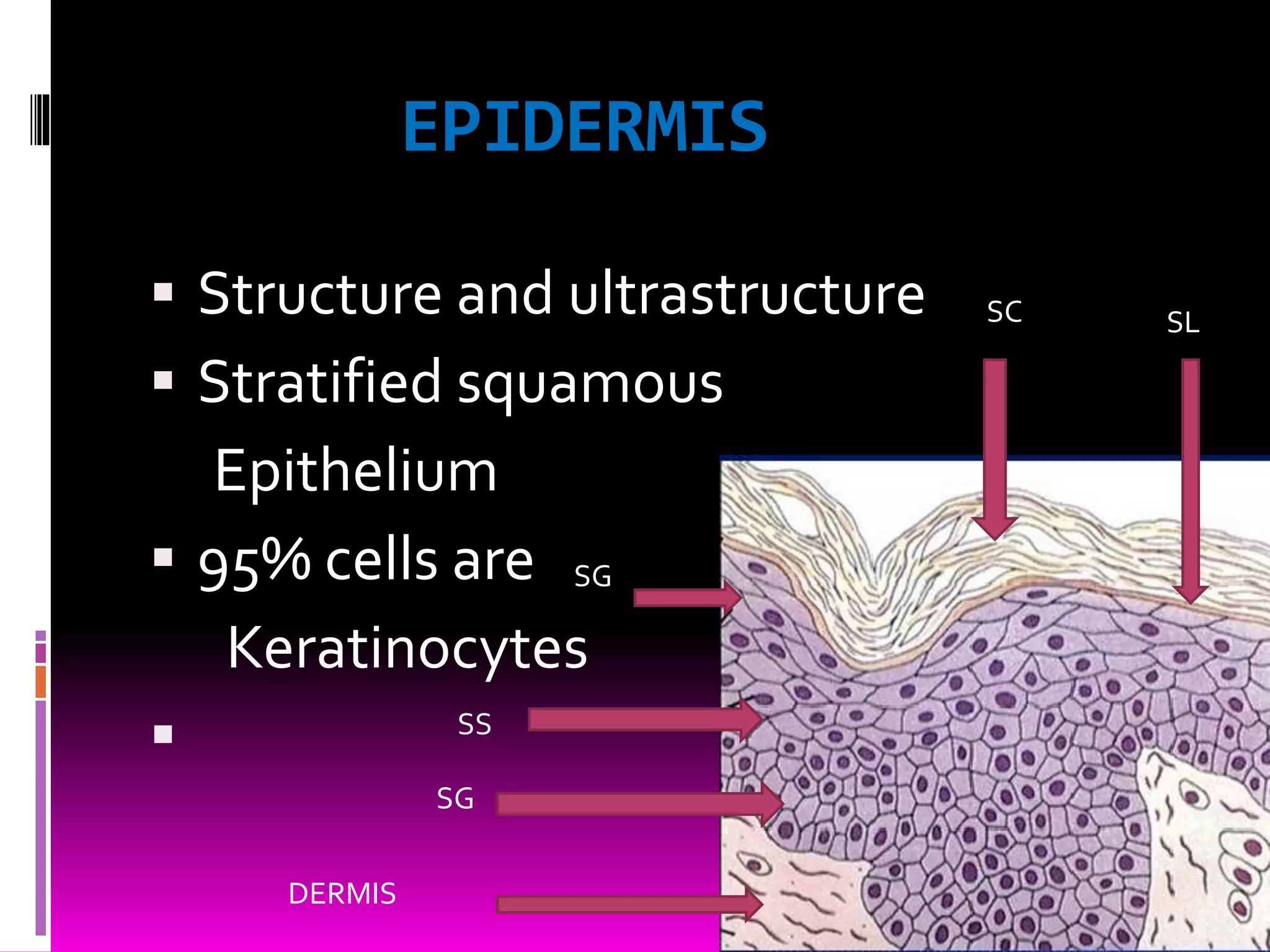
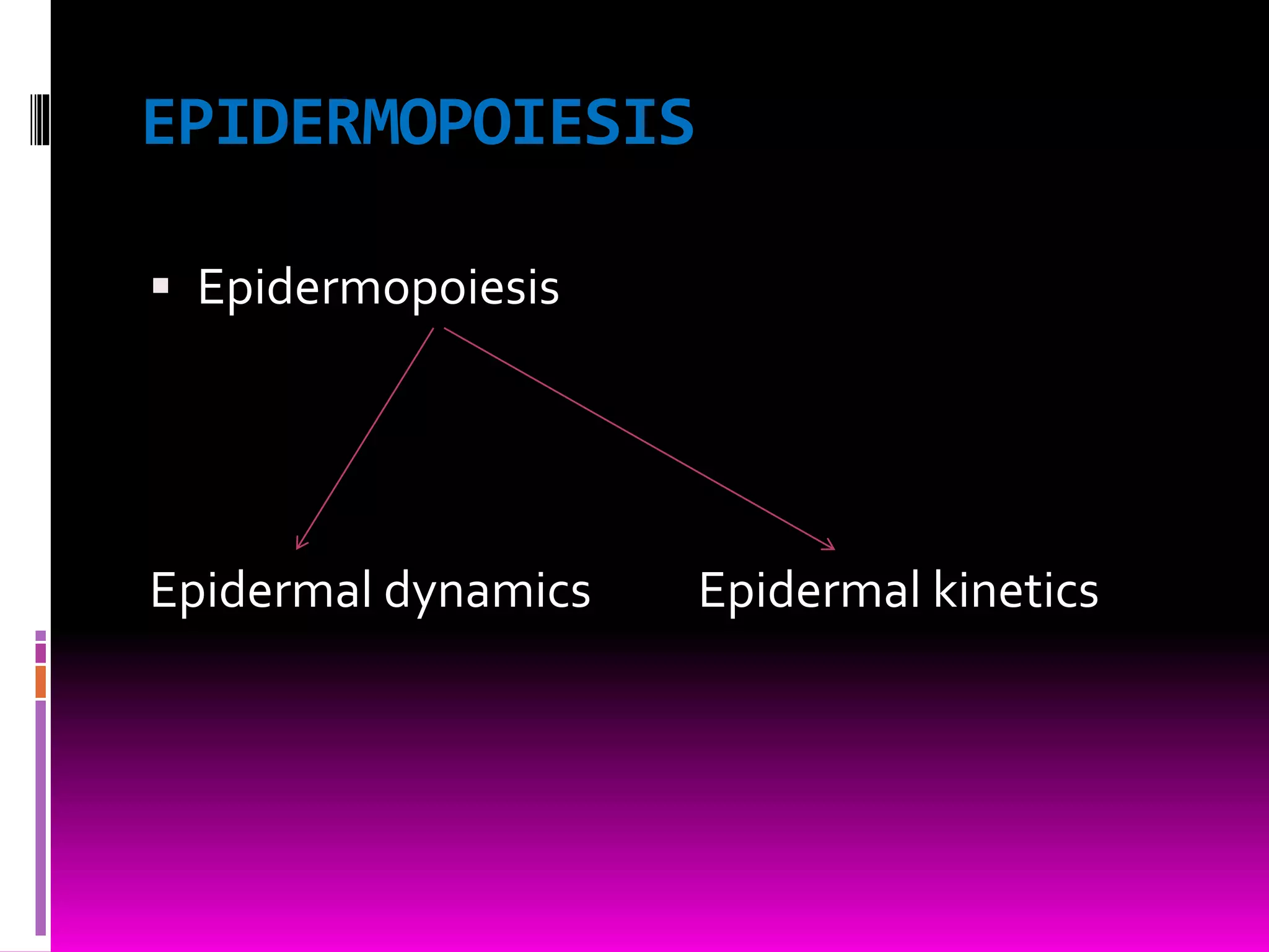
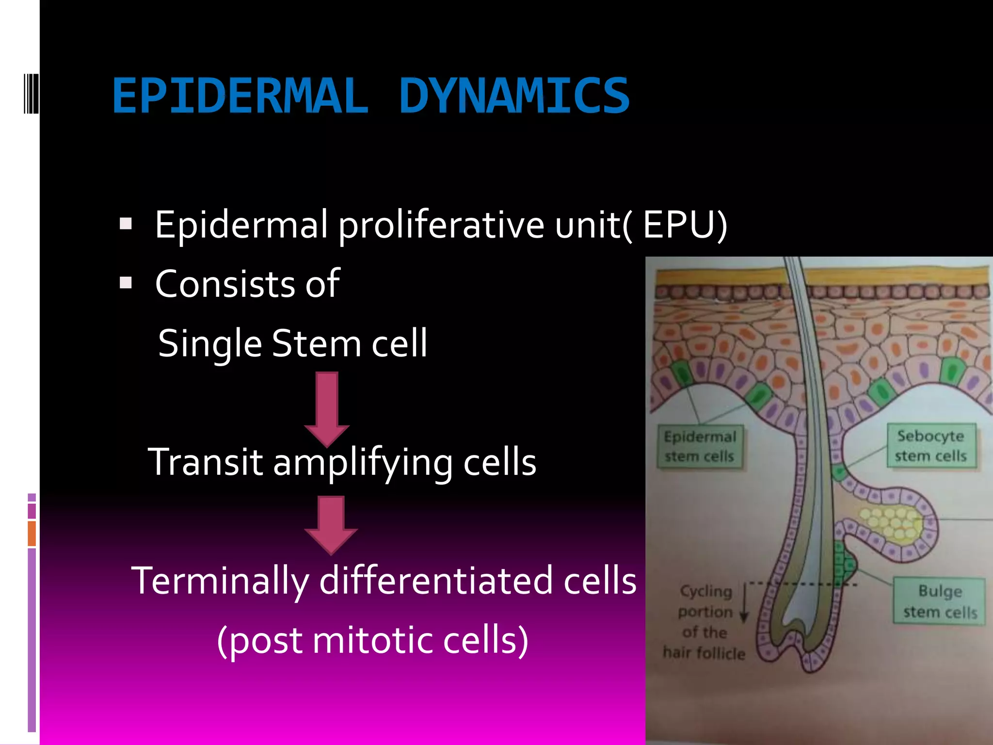
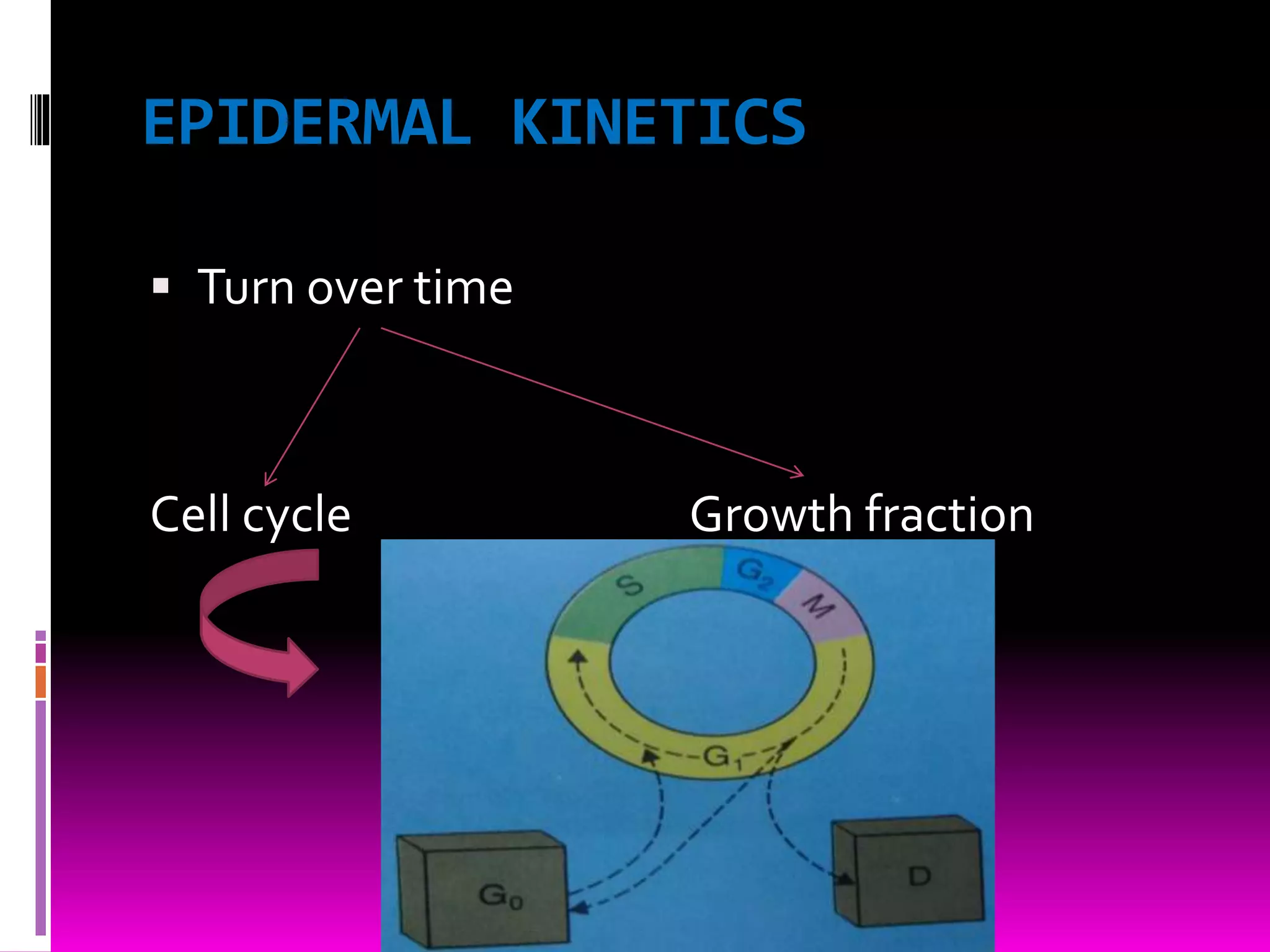
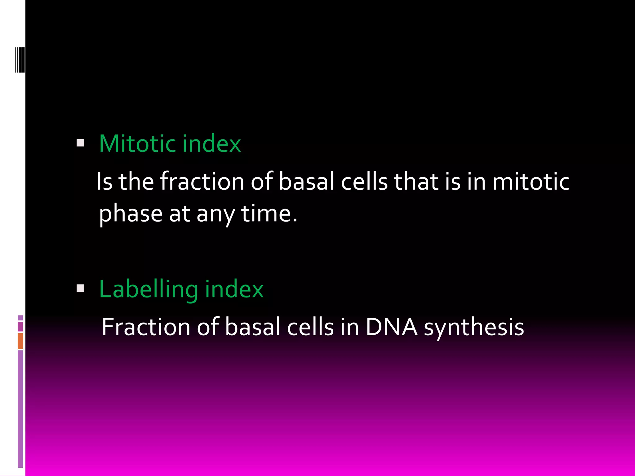
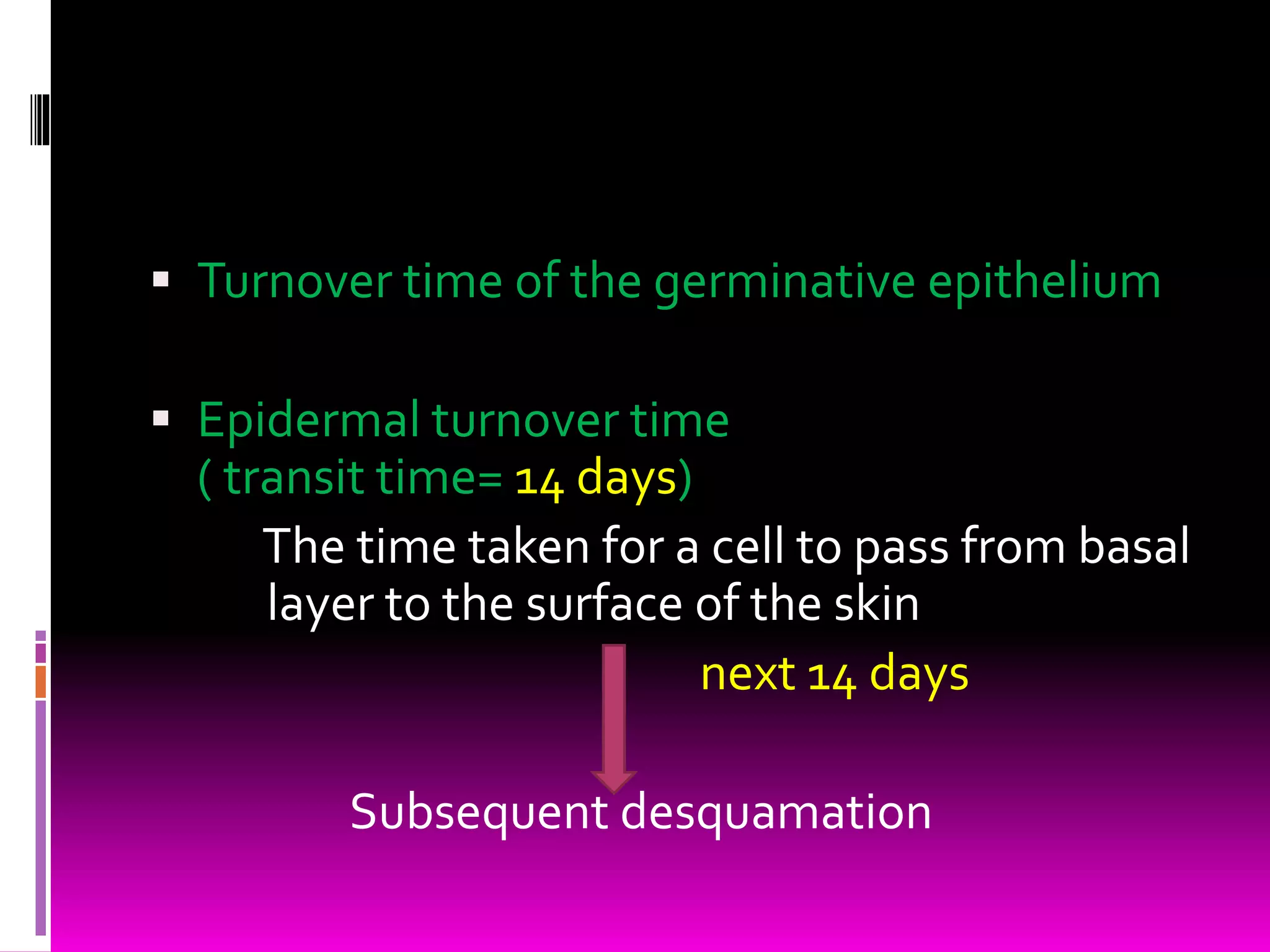
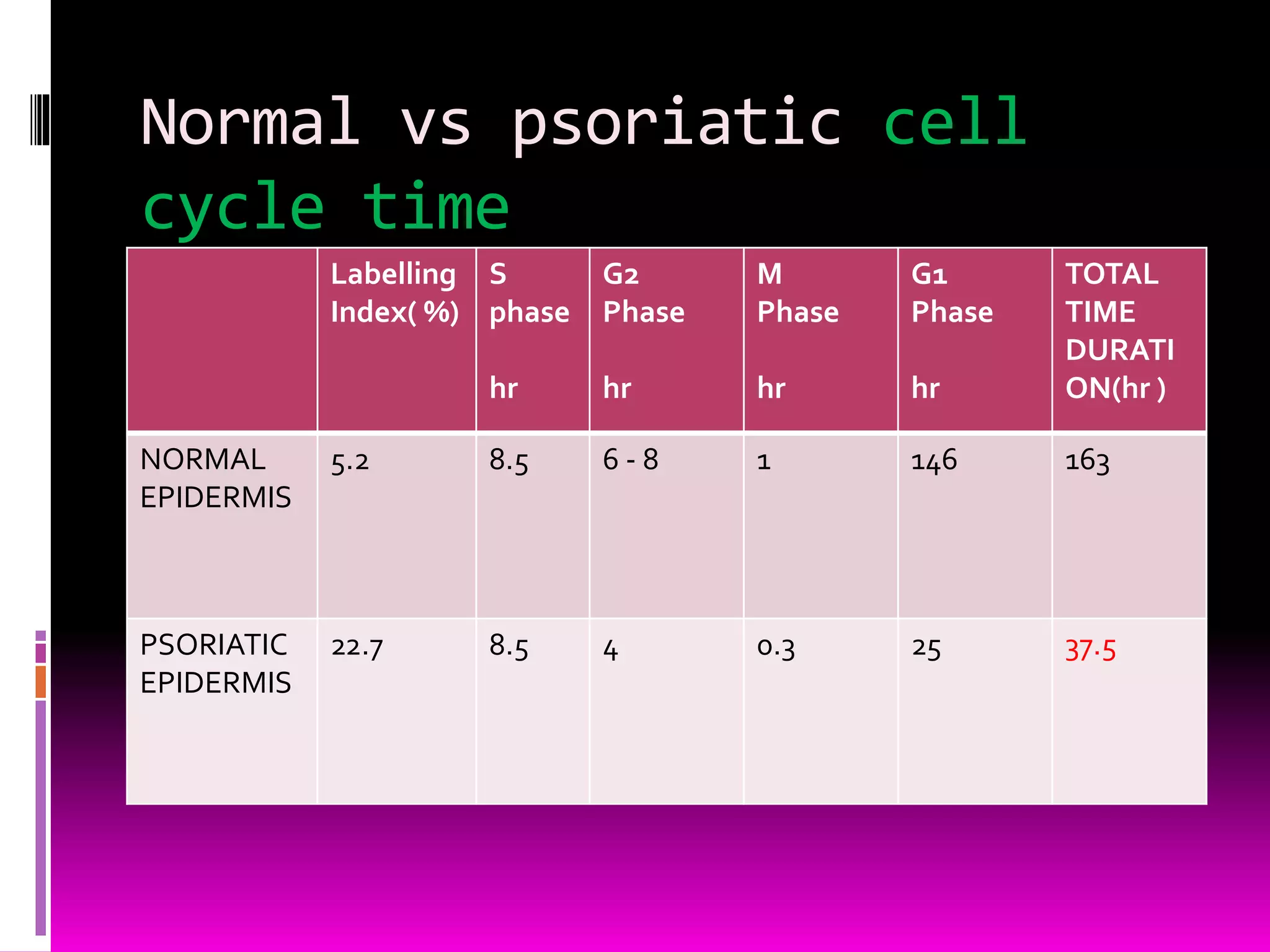
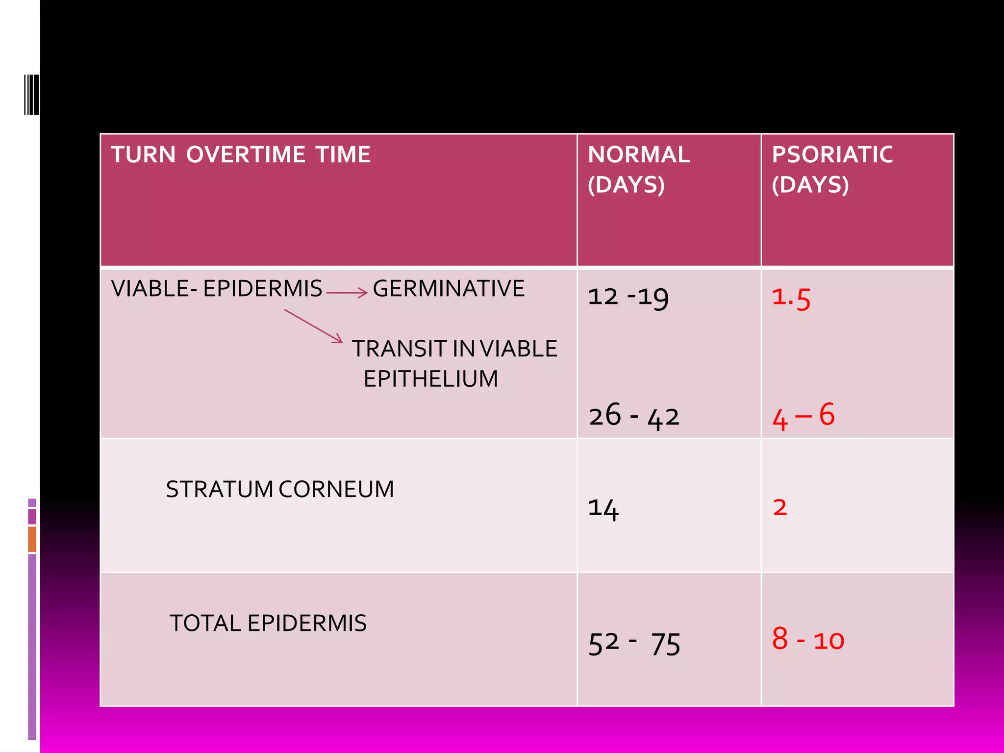
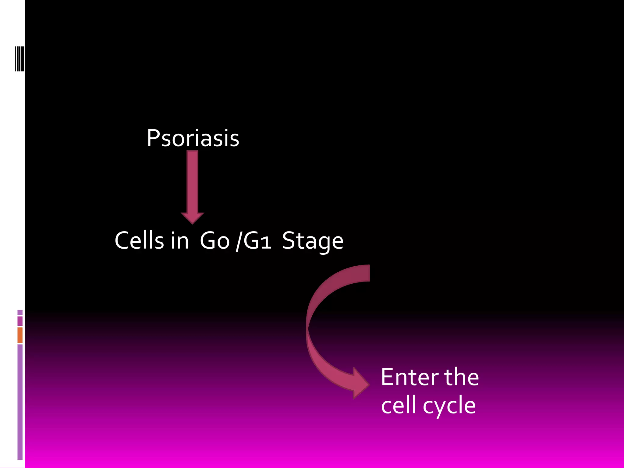
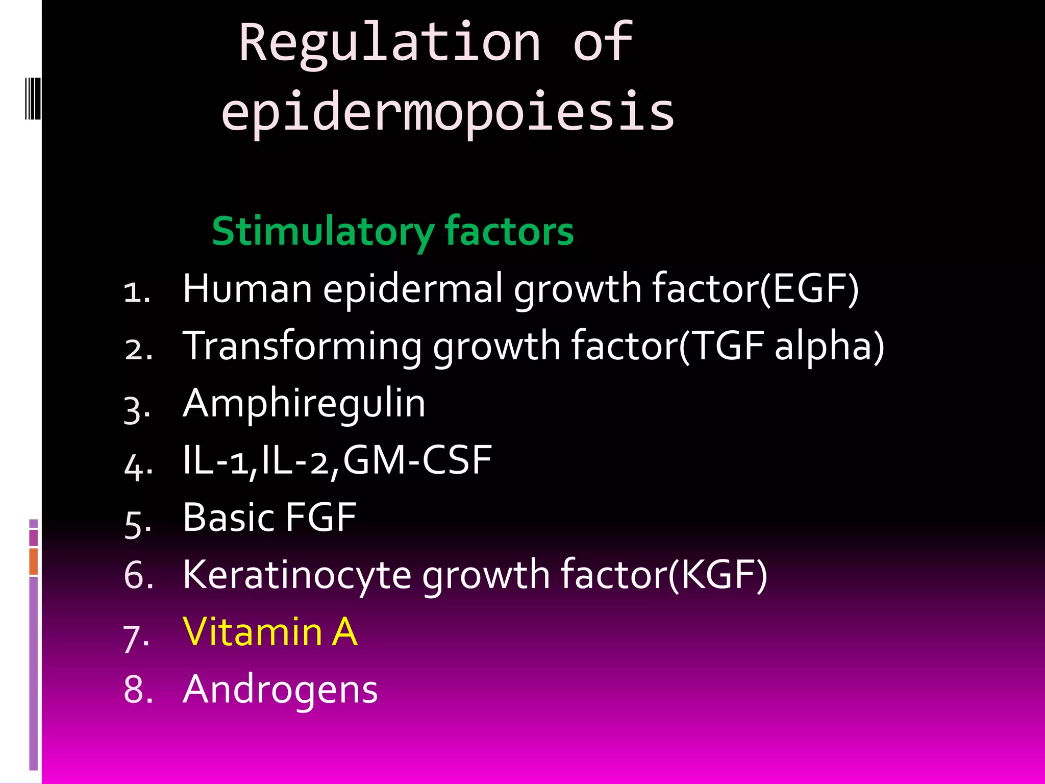
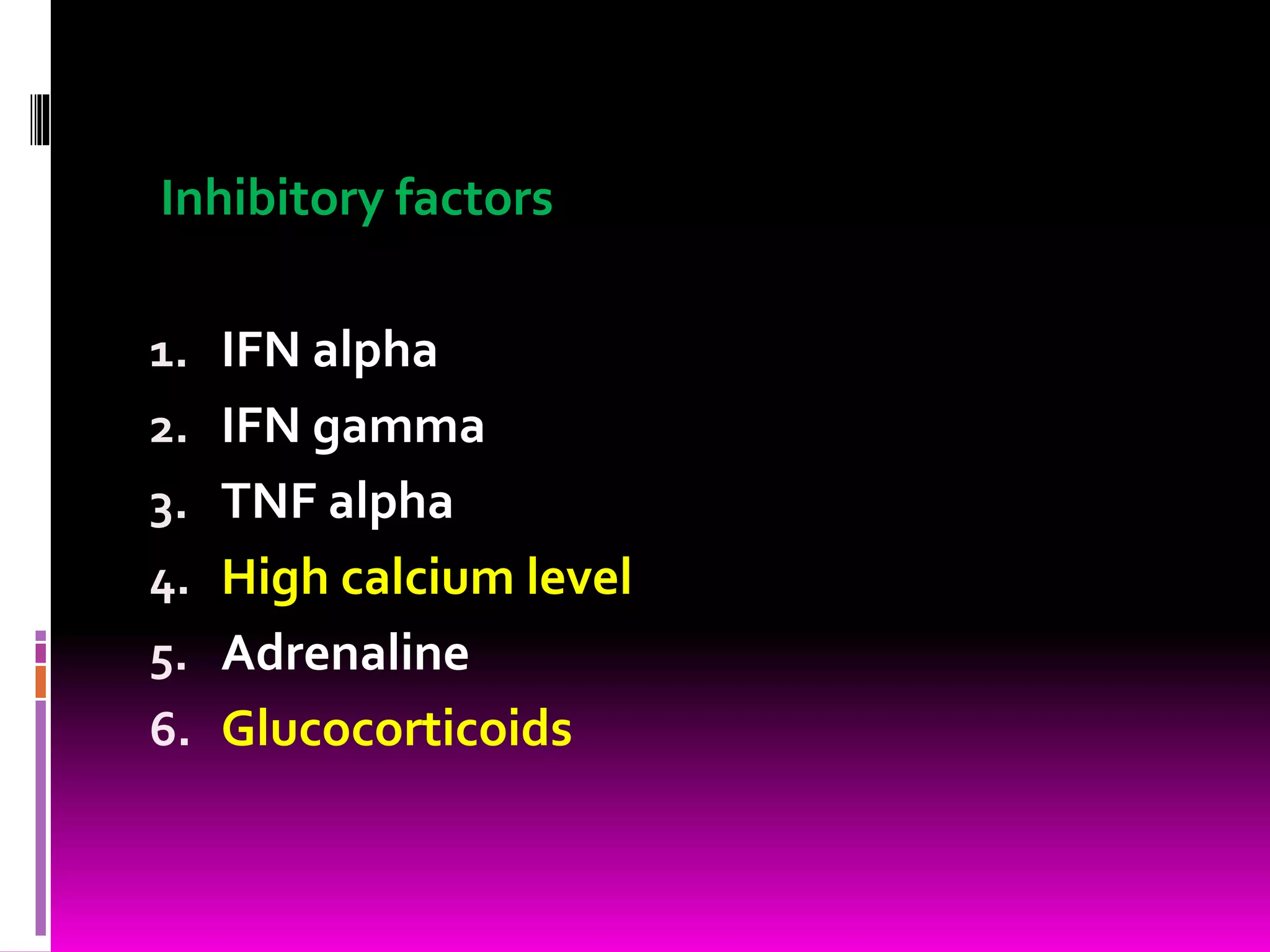
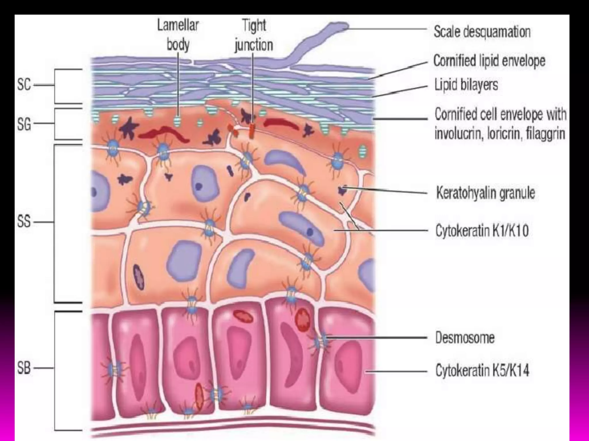
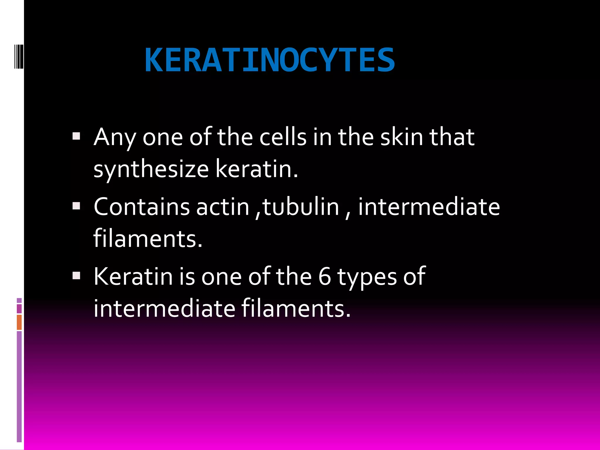
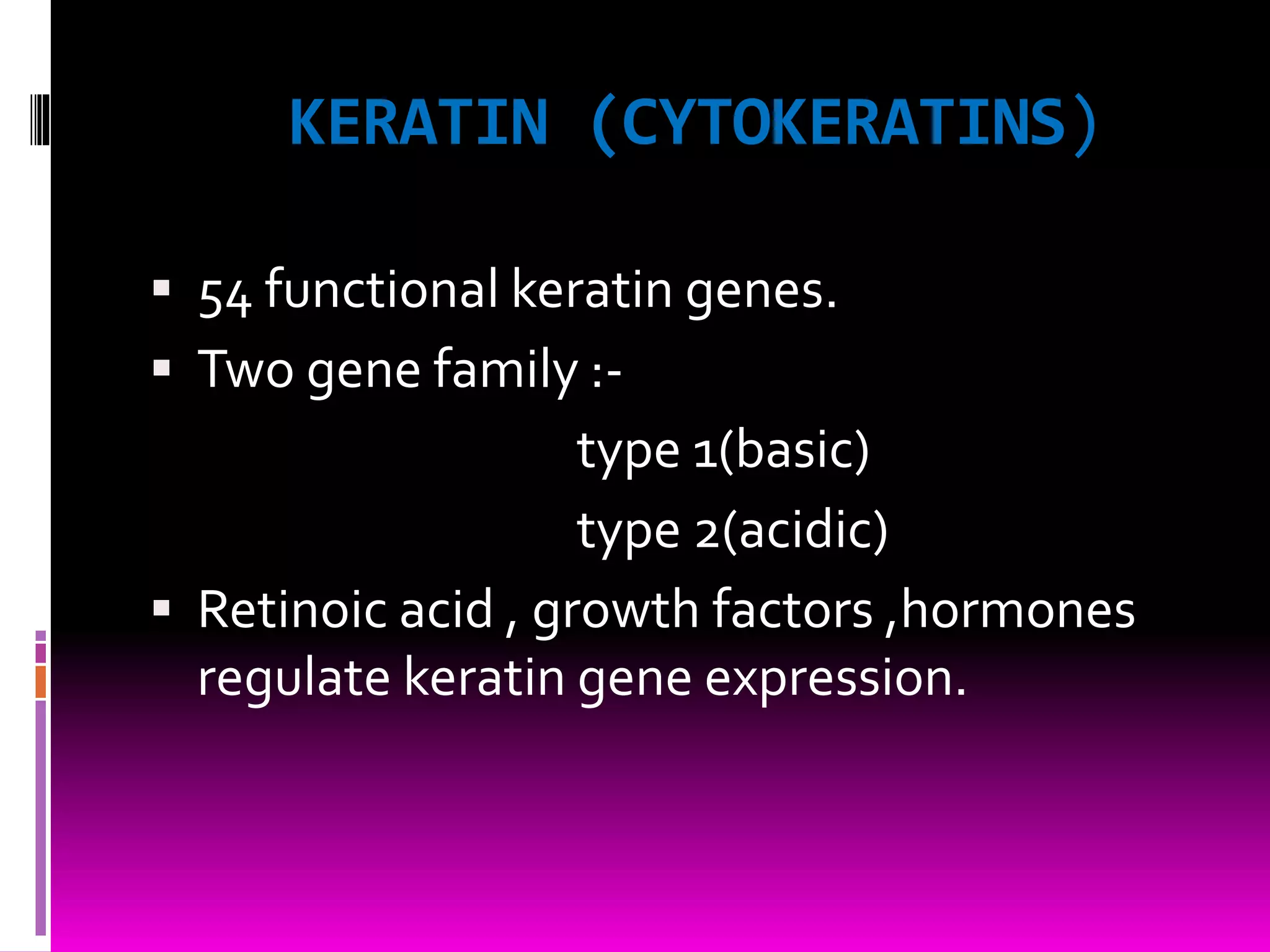
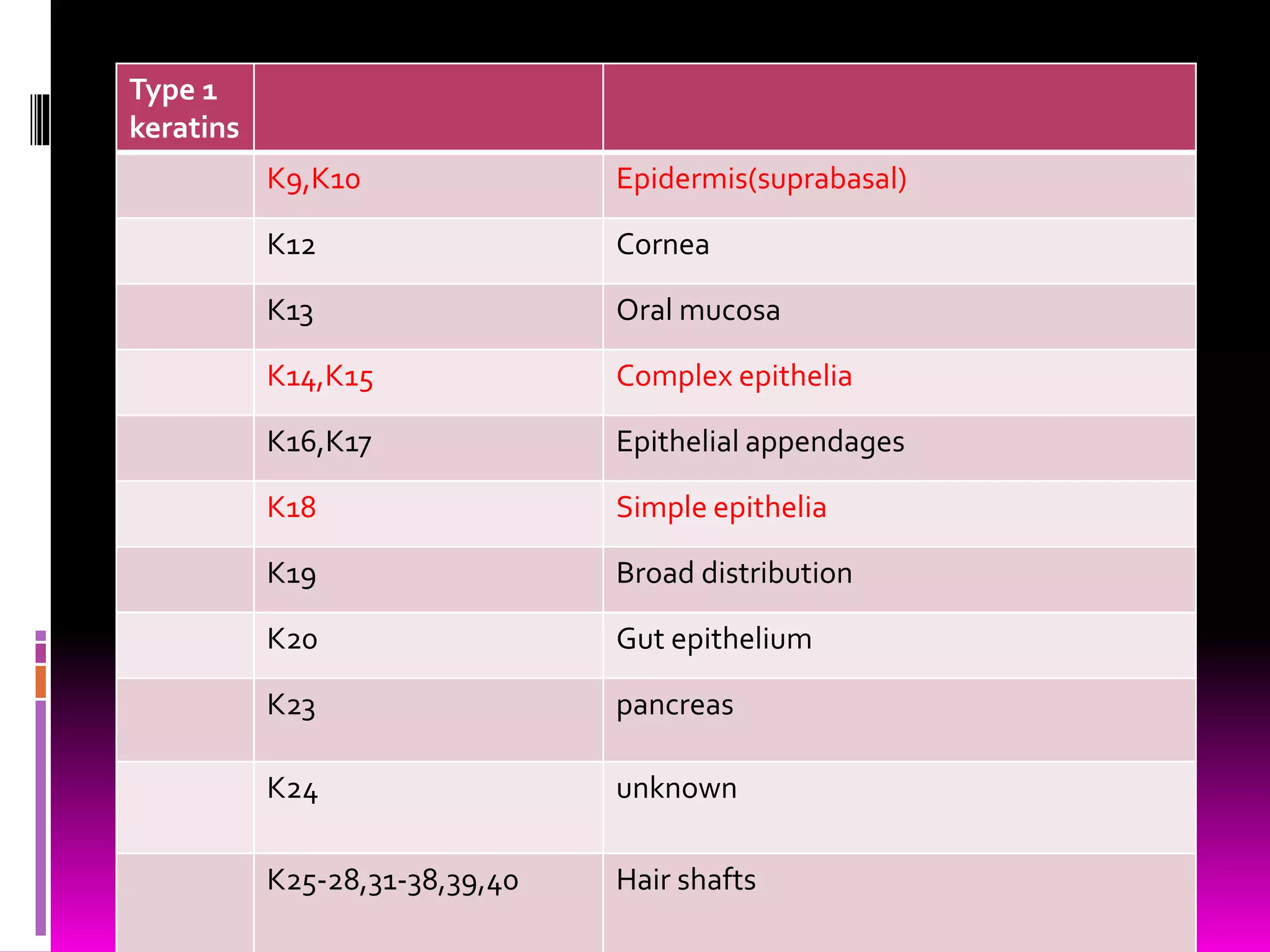
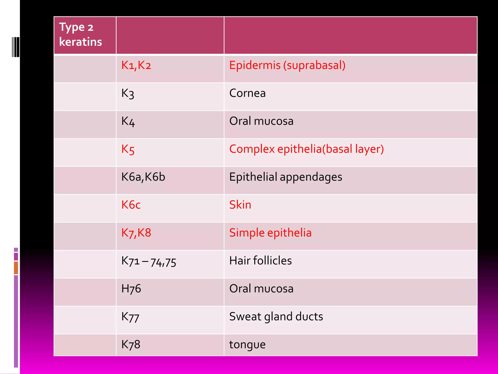
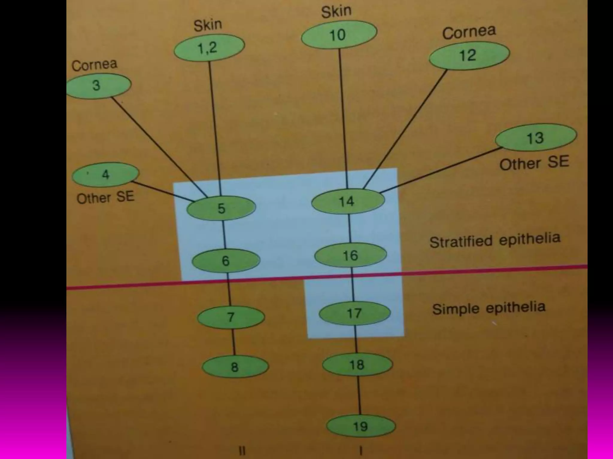
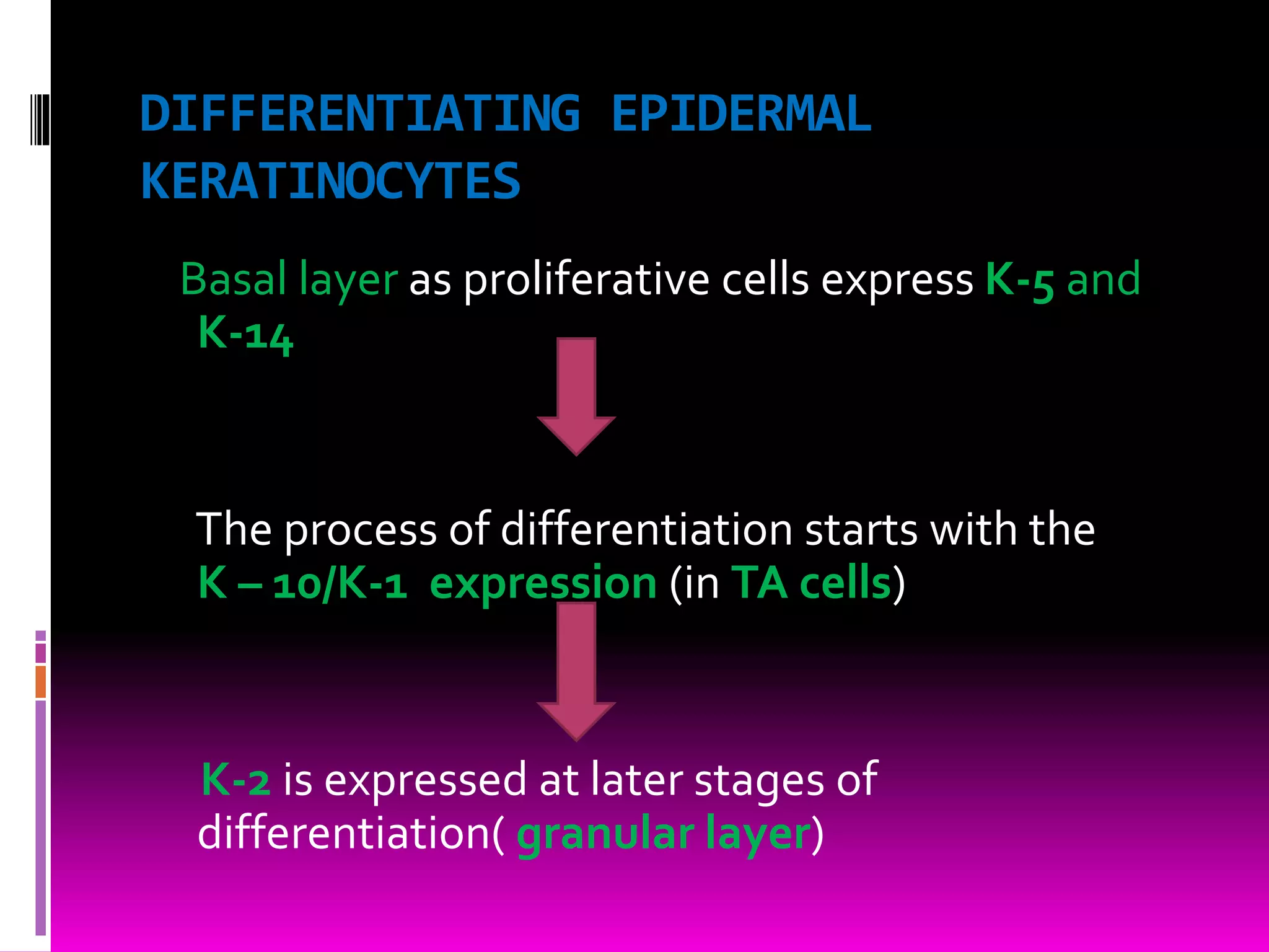
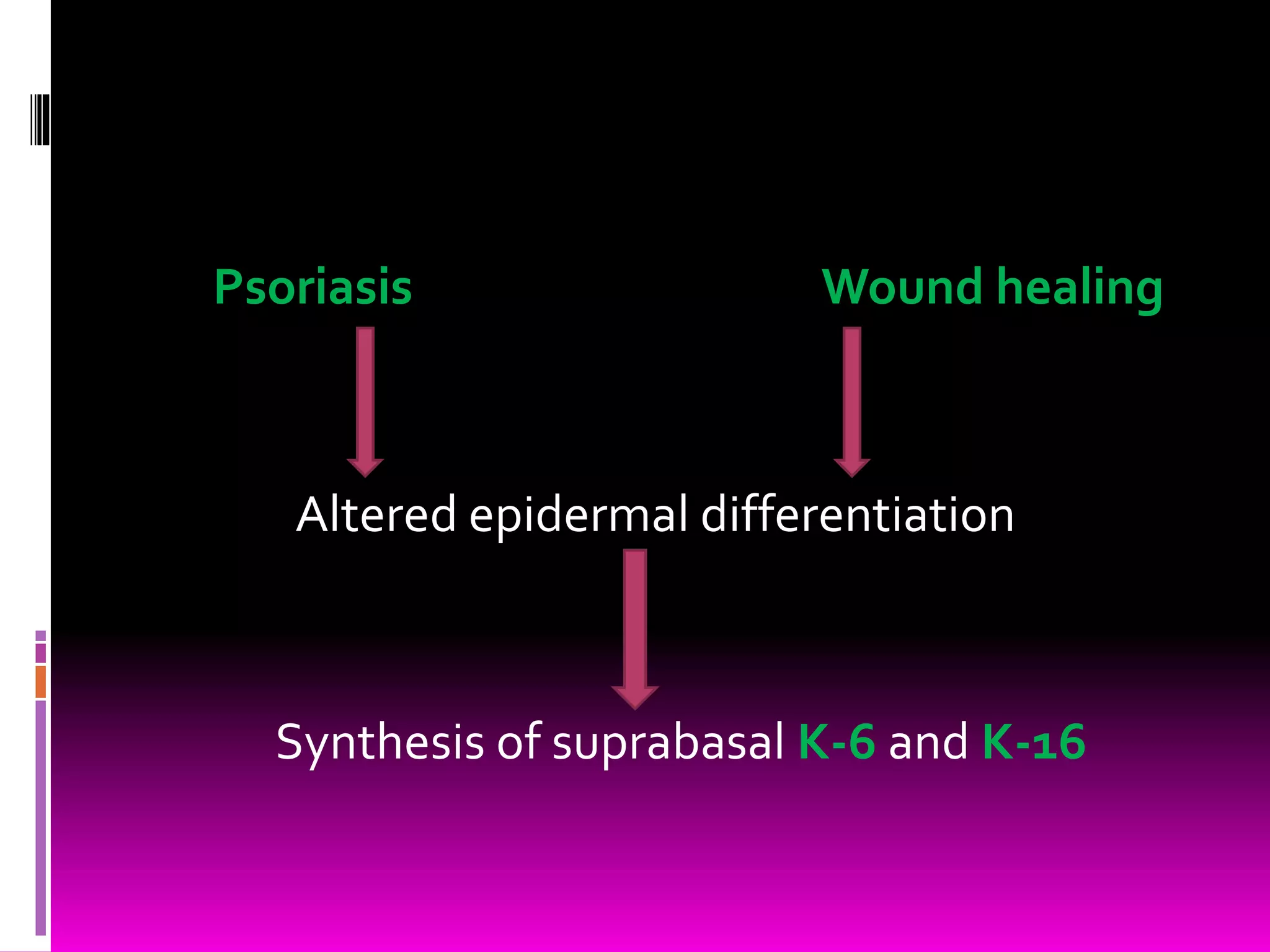
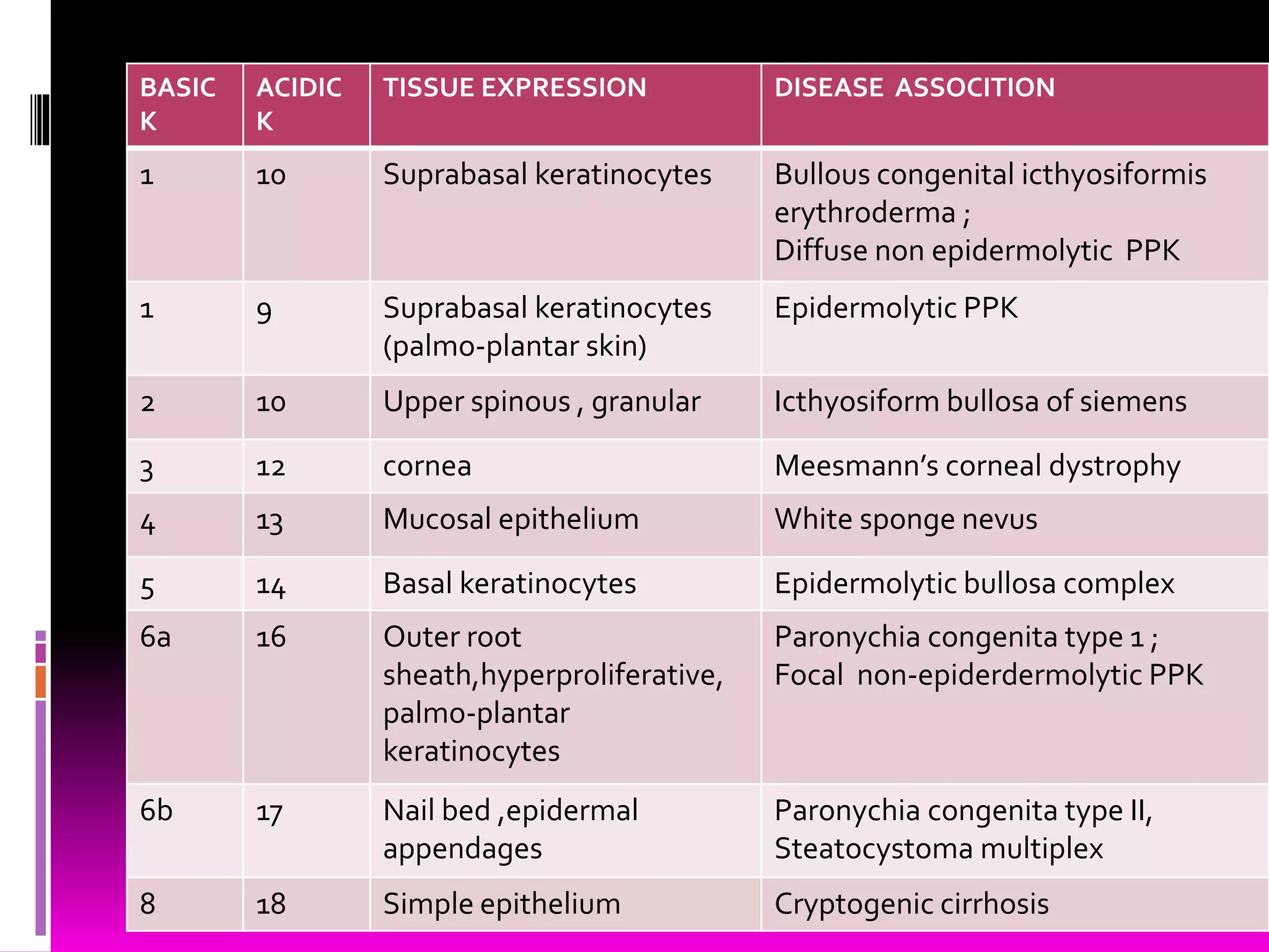

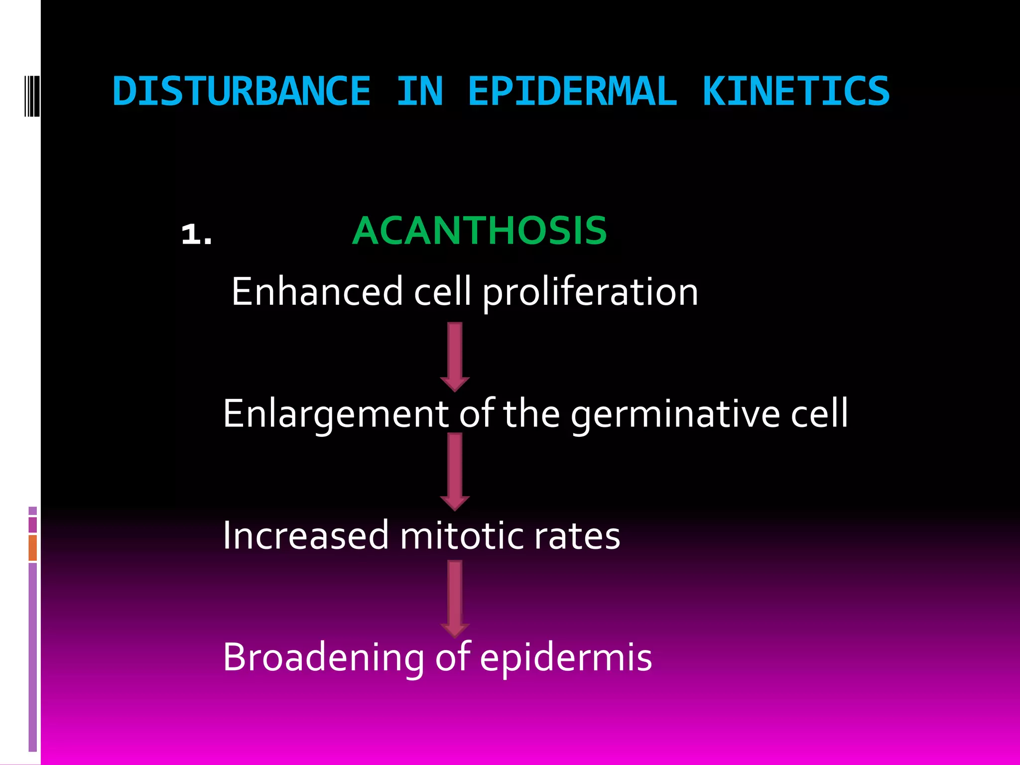
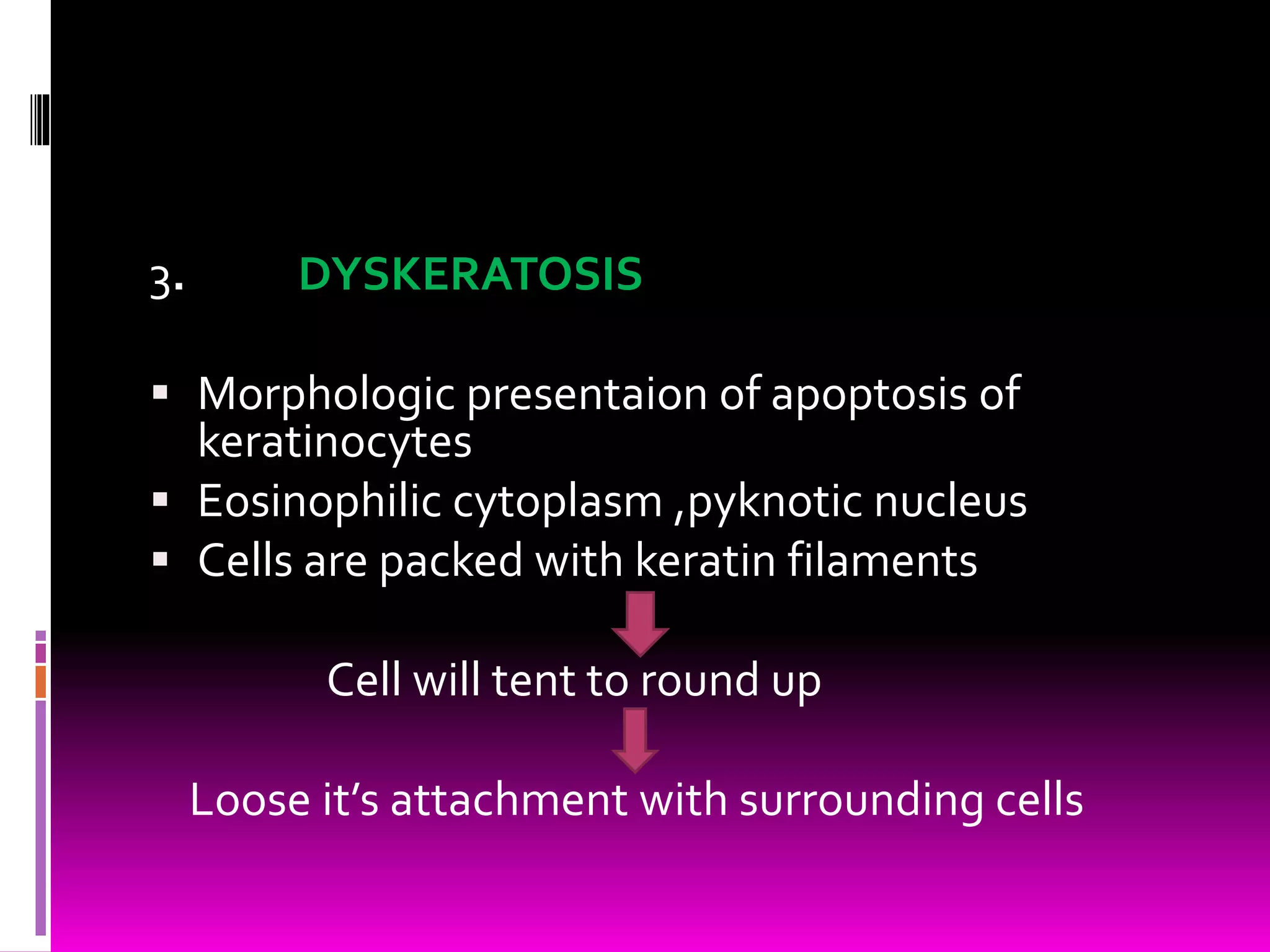
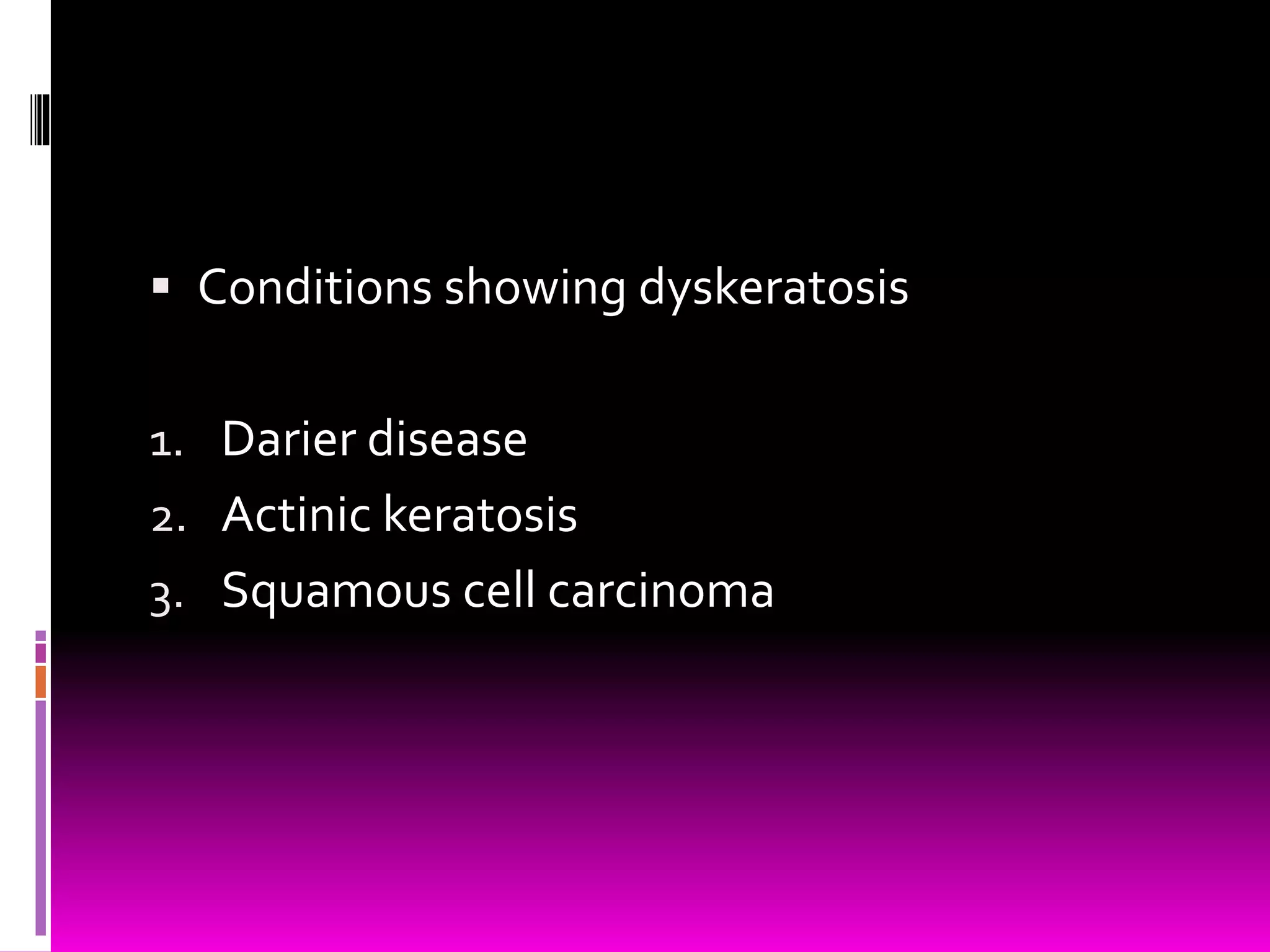
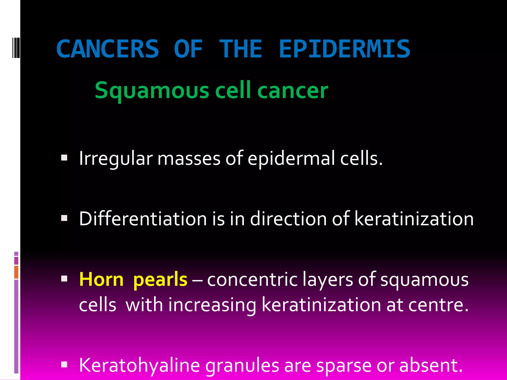
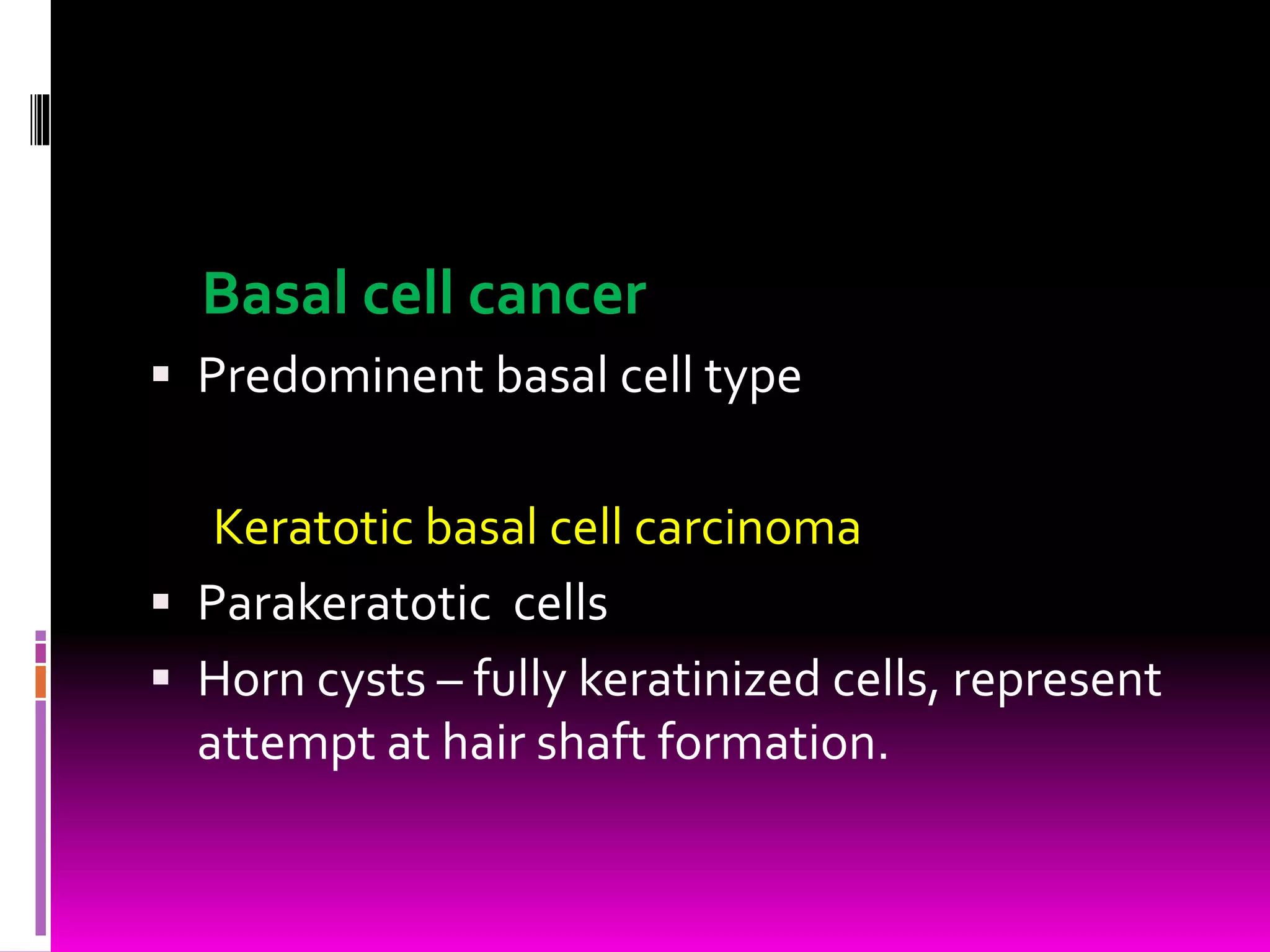

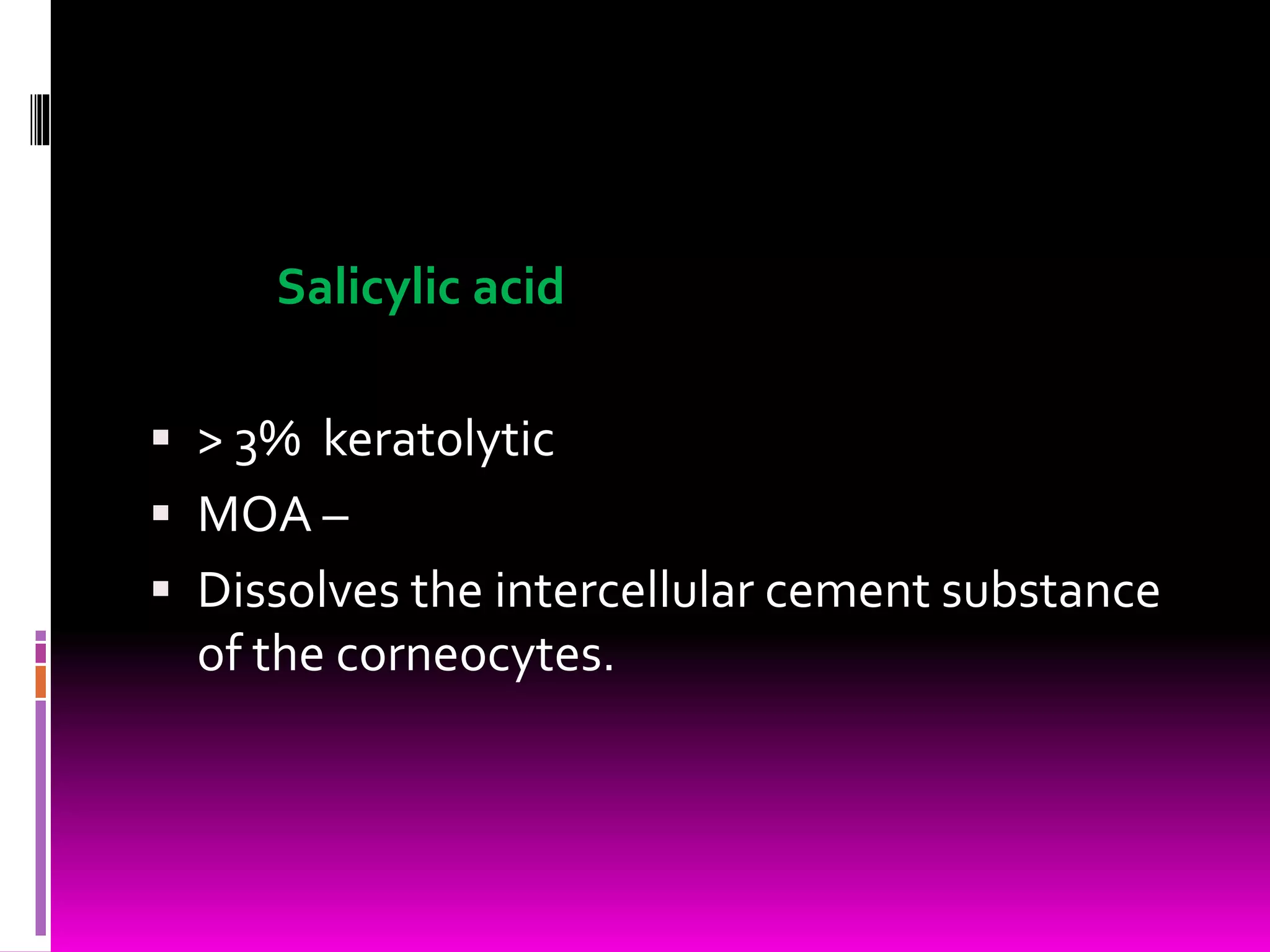


This document summarizes epidermal kinetics and dynamics. It discusses the structure of the epidermis, epidermal proliferation units, cell cycle kinetics like turnover time and labeling index. Disturbances in epidermal kinetics like acanthosis, parakeratosis and dyskeratosis are described. Kinetics in normal skin versus psoriasis are compared. Epidermal differentiation and terminal differentiation involving keratinization are outlined. Drugs acting on epidermal cells like retinoids, vitamin D analogues, and salicylic acid are mentioned. Cancers of the epidermis are briefly described.


