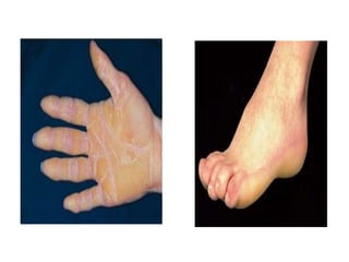
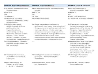
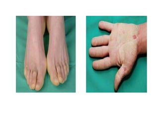
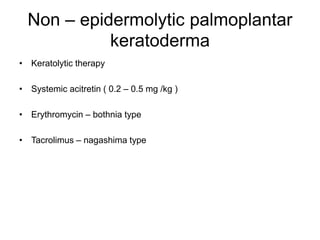
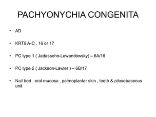
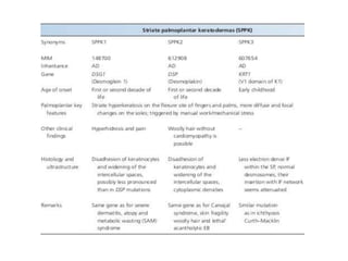
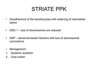
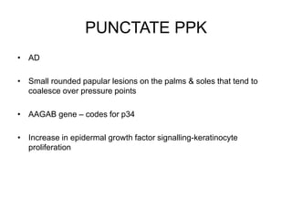
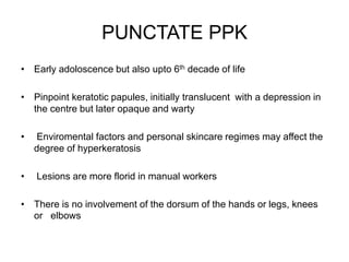
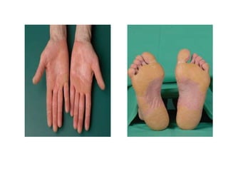
SlideShare a Scribd company logo
PALMOPLANTAR
KERATODERMA
MAJ TEGHVEER SINGH
RESIDENT DERMATOLOGY
INTRODUCTION
• Excessive epidermal thickening of palms & soles
• Acquired or heriditary
• Syndromic or non syndromic
• Genes Implication
1. Structure proteins
2. Cornified envelop
3. Cohesion
4. Cell to cell communications
5. Transmembrane signal transduction
CLINICAL PATTERNS
• Diffuse – complete palmar or planar surface
centripalmar or insteps , uniform thickening
• Focal,areate or annular – pressure points are disproportionately thickened
• Striate keratoderma – focal keratoderma , conspiciously longitudinal lesions on
fingers overlying flexor tendons
• Punctate , papular or disseminated – multiple scattered discrete round lesions
• Transgradient – extends beyond palmo plantar skin contiguously or as
callosities at pressure points
• Confluent – cover whole digits
• Cicatrizing keratoderma (mutilating) – constriction bands appear around digits
EPIDERMOLYTIC PPK
• Voerne’r disease
• AD
• KRT 9 or KRT 1
• Diffuse
• Disruption of intermediate filament integrity – blistering & hyperkeratosis
epidermolysis with tonofilament clumping to minor trauma
• Infants – diffuse kerotoderma
• Adults – confluent , even ,thick yellowish hyperkeratosis with sparing of
dorsal surface, sharp demarcation with erythematous edges
EPIDERMOLYTIC PPK
• Epidermolysis in suprabasal keratinocytes
• Hyperkeratosis , acanthosis & papillomatosis
• Round or ovoid eosinophilic inclusions with large tonofilament
aggregates
• Whorls of keratins containing tubular structure – tono tubules
• MANAGEMENT
1. Mechanical debridement
2. Mild keratolytics
3. Oral retinoids
4. Topical calcipotriol
Non – epidermolytic palmoplantar
keratoderma
• Diffuse keratoderma
• No cytolysis in upper spinous or granular layer
• Bothnia type , kimonis type, nagashima type
• AQP5 gene
Non – epidermolytic palmoplantar
keratoderma
• Present in the 1st few months of life
• Obvious > 2yrs age
• Variably thick , yellow hyperkeratosis occurs on the whole surface of
foot
• Starting on the heel & anterior arch
• Spreading to palms
• Hyperhidrosis ,dermophytic infections & pitted keratolysis is
common
Non – epidermolytic palmoplantar
keratoderma
• Keratolytic therapy
• Systemic acitretin ( 0.2 – 0.5 mg /kg )
• Erythromycin – bothnia type
• Tacrolimus – nagashima type
PACHYONYCHIA CONGENITA
• AD
• KRT6 A-C , 16 or 17
• PC type 1 ( Jadassohn-Lewandowsky) – 6A/16
• PC type 2 ( Jackson-Lawler ) – 6B/17
• Nail bed , oral mucosa , palmoplantar skin , teeth & pilosebaceous
unit
PACHYONYCHIA CONGENITA
• Toe nail dystrophy + plantar kerotoderma + plantar pain in > 3yr old
• Thickened toenail occur within 1st yr to 9yrs of life
• KRT6A mutation carriers manifest in early age
• Not all 20nails are involved
• Plantar keratoderma manifest as calluses , fissures & thickened skin
• Thick yellow keratosis
• Friction blisters
• Plantar pain
PACHYONYCHIA CONGENITA
• Follicular hyperkeratoses on knees & elbows
• Oral leukokeratosis
• Palmoplantar hyperhydrosis
• Natal teeth
• Patchy white thickened areas on tongue & oral mucosa
• Oral lesions resembling candidiasis
• Hoarseness of voice & fatal respiratory obstruction in infancy
• Steatocystoma or pilosebacious cyst
PACHYONYCHIA CONGENITA
• Tonofilament aggregates demonstrate intermediate filament disorder
• Gross hyperkeratosis
• Alternating ortho & parakeratosis
• Acanthosis with patchy hypergranulosis – large & malformed
keratohyaline granules
• Cyst may be keratinous epidermoid cyst, eruptive vellous cyst or
true steatocyst
PACHYONYCHIA CONGENITA
• Mechanical reduction of hyperkeratosis & nails
• Proper footware & orthotics to reduce blistering & callosities
• Cyst – surgical excision
• Emollients & keratolytics
• Treatment of hyperhidrosis
MAL DE MELEDA
• AR
• Transgradient keratoderma
• Croatian island of meleda
• SLURP1 – transmembrane signal transduction , cell activation &
adhesion
MAL DE MELEDA
• Early childhood
• Hyperkeratosis is preceded by erythema
• Patches of waxy ivory-yellow hyperkeratosis extend across the
glove & stocking distribution
• Insular lesions of knees & elbows – key feature
• Eythema persists in central palms & soles
• Hyperhidrotic maceration & maladour – fungal superinfection
MAL DE MELEDA
• Circumferential hyperkeratosis of fingers – sclerodactyly & digital
constriction
• Nail changes – hypercurvature , thickening & koilonychia
• Angular cheilitis
• Ridged tongue
• High arched palate
MAL DE MELEDA
• Greatly thickened corneal layer
• Increased stratum lucidum
• Marked acanthosis
• Papillomatosis is a typical feature
MAL DE MELEDA
• Oral retinoids – hyperkeratosis responds better than erythema
• Excision of keratoses
• Split – thickness skin graft
LORICRIN KERATODERMA
• Diffuse transgradient
• LOR gene – loricrin
• Glycine rich cornefied envelope protein
• Mutant protein is transported to the nucleus
• Interfares with regulation of cornification
LORICRIN KERATODERMA
• Generalised desquamation at birth
• Collodion babies
• Icthyosis is mild
• Rugose keratoderma – childhood
• Confluent honeycomb pattern
• Edges are diffuse & cicatricial bands (psuedo– ainhum)
• Knuckle pad & warty keratoses
LORICRIN KERATODERMA
• Hyperkeratosis with hypergranulosis & parakeratosis
• Intranuclear granules in granular cells and a thin corneified cell
envelope in the lower cornified layers with abnormal extracellular
lamellae
• Immunoelectron microscopy – presence of loricrin in the nucleus
LORICRIN KERATODERMA
• Isotreninoin
• Inhibitors of VEGF receptor 2
STRIATE PPK
• AD
• Keratinocytic disadhesion
• Desmosomal defects
• Haploinsufficiency for desmoglein 1 – mutation in DSG1
• Mutation in desmoplakin
STRIATE PPK
• Linear pattern of thickening on the palms & flexor aspect of fingers
• Soles – confluent
• Mechanical stress
• Pain , hyperhidrosis & mild hyperkeratosis of knees
• Woolly hairs
STRIATE PPK
• Disadherence of the keratinocytes with widening of intercellular
space
• DSG 1 – size of desmosomes are reduced
• DSP – abnormal keratin filament with loss of desmosomal
connections
• Management :
1. Systemic acetretin
2. Urea cream
PUNCTATE PPK
• AD
• Small rounded papular lesions on the palms & soles that tend to
coalesce over pressure points
• AAGAB gene – codes for p34
• Increase in epidermal growth factor signalling-keratinocyte
proliferation
PUNCTATE PPK
• Early adoloscence but also upto 6th decade of life
• Pinpoint keratotic papules, initially translucent with a depression in
the centre but later opaque and warty
• Enviromental factors and personal skincare regimes may affect the
degree of hyperkeratosis
• Lesions are more florid in manual workers
• There is no involvement of the dorsum of the hands or legs, knees
or elbows
PUNCTATE PPK
• Orthohyperkeratotic with compact acanthosis & hypergranulosis with
a depression in the centre
• Cornoid lamella
• Hypogranulosis and focal parakeratosis
• Management :
1. Pumice stone
2. Oral retinoids (0.5 – 1 mg/kg/day)
3. Tyrosine kinase receptor inhibitors
Cole Disease
• Punctate keratoderma & pigmentary anomaly
• Congenital or early onset
• Defective melanosome transfer from melanocytes to keratinocytes
• Relatively mild focal or punctuate keratoderma
• Sharply demarcated irregular macules with varying degrees of
hypopigmentation
• located over the extremities
• There might be an early onset calcific tendinopathy or calcinosis cutis
Cole Disease
• Histology shows hyperorthokeratosis, hypergranulosis and
acanthosis
• Ultrastructure confirms a reduction of the melanin content of
keratinocytes with disproportionately large melanosomes of
melanocytes
SPINY KERATODERMA
• Fine 1–2 mm papules that project from the palmoplantar surface
• filiform, spiked, prickly, minute digitate or music box‐like spines
• start in early childhood and gradually increase in number, i.e. they
appear in the first up to the fifth decade
SPINY
KERATODERA
• Dense columns of parakeratosis above
a hypogranular epidermis
• Management :
• Mechanical debridement -
dermabrasion, topical keratolytics
• Etretinate or actitretin have a temporary
effect
• Topical 5‐fluorouracil
MARGINAL PAPULAR
KERATODERMA
• Acrokeratoelastoidosis with
elastorrhexis
• Focal acral hyperkeratosis
• characterized by papules, plaques and
nodules
• located at the junction between the
palmar and dorsal skin of the hands or
feet along the thenar and hypothenar
eminences
• Focal orthohyperkeratosis
• Massive elastosis
TRANSIENT AQUAGENIC
KERATODERMA
• Mainly palmar, disorder represents a mild keratoderma
• Triggered or exacerbated by contact with water or sweat
• acquired but autosomal recessive or dominant
• cyclo‐oxygenase 2 inhibitor
TRANSIENT AQUAGENIC
KERATODERMA
• 6–45 years, more often women
• Subtle keratoderma appearing after a few minutes of immersion of their
hands in water or after sweating
• Soles
• Characteristic sign might be that patients bring with them a vessel to
immerse their hands in water (‘hands in the bucket’ sign)
• The painful, burning or itching, whitish papular lesions are associated
with dilated acrosyringeal ostia which can be seen by dermoscopy
• Lesions subside shortly after drying the hands, leaving minimal
hyperkeratosis in the centre of the palms
TRANSIENT AQUAGENIC
KERATODERMA
• 20% aluminium chloride hexahydrate followed by urea cream or
botulinum toxin.
NAXOS SYNDROME
• PPK + Arrythmogenic right ventricle cardiomyopathy + woolly hairs
• gene encoding plakoglobin - a cell junction protein found in desmosomes in
the epidermis and cardiac muscle
• Different mutations of JUP show a large phenotypic spectrum
• Expression of altered plakoglobin may show mild skin fragility, keratoderma
and woolly hair only
• Autosomal dominant JUP mutations underlie isolated arrhythmogenic right
ventricular cardiomyopathy and a Naxos variant with leukonychia and
oligodontia
• Complete loss of plakoglobin due to homozygous nonsense mutations may
lead to acantholytic epidermolysis bullosa
NAXOS SYNDROME
• Woolly hair develops from birth while diffuse or striate hyperker-
atoses of the palms and soles appear during the first year of life
when the skin starts to become mechanically stressed
• Cardiomyopathy manifests by adolescence and shows 100%
penetrance
CARVAJAL–HEURTA SYNDROME
• Cardiocutaneous syndrome with NEPPK
• Woolly hair and dilated cardiomyopathy
• Recessive desmoplakin (DSp) mutations producing a premature stop codon
leading to a truncated protein
• Dominant negative effect on desmosomal assembly has been found to be
associated with cardiomyopathy and mild hyperkeratosis of the elbows and
knees
• Heterozygous carriers of the missense mutation p.S299R showed isolated
arrhythmogenic right ventricular cardiomyopathy without cutaneous phenotype
• Complete loss of the tail domain of desmoplakin presents as
acantholytic epidermolysis bullosa
CARVAJAL–HEURTA SYNDROME
• Striate lichenoid keratoses of flexures, follicular keratoses of elbows & knees
• Presents at a younger age with bilateral predominantly left ventricular
involvement leading to early heart failure with cardiac enlargement and
disrupted cardiac contraction
• Large intercellular spaces of supra basal keratinocytes (keratinocytic
disadhesion)
• Reveal clumping of desmosomes or signs for intermediate filament disruption
such as perinuclear localization of keratin in suprabasal keratinocytes
• On resting electrocardiogram, affected patients exhibit repolarization and or
depolarization abnormalities
• structural/functional abnormalities of the ventricles
CARVAJAL–HEURTA SYNDROME
• Investigate cardiac function in patients with striate keratoderma as
early diagnosis and intervention may improve outcome
• Depending on the myocardial symptoms, implantation of an
automatic cardioverter defibrillator with/without antiarrhythmic drugs
• Heart transplantation
• Screening of possibly affected family members should be initiated
considering dominant as well as recessive inheritance
VOHWINKEL SYNDROME
• AD
• Hereditary keratoderma with hearing loss
• Defective connexin function
• Maternal inheritance - rare mitochondrial type of keratoderma
• connexin 26 is expressed in the cochlea where it may permit the
recycling of potassium to endolymph
• In skin, the protein is found in palmoplantar epidermis and sweat
glands
VOHWINKEL SYNDROME
• Palmoplantar keratoderma begins in childhood as shiny or translucent papular
hyperkeratosis, gradually becoming confluent of the hands and feet
• Striate lesions may be seen
• Warty papules on the knuckles and other extensor sites coalesce into the
pathognomonic ‘starfish’ keratoses
• The edge of the keratoderma at the wrists and Achilles tendon consists of spiky
digitate hyperkeratotic projections onto normal skin
• Multiple keratoses on the digits produce circumferential hyperkeratosis, which
predisposes to the formation of cicatricial bands and autoamputation
• The little finger and fifth toe are commonly affected
• A high tone sensorineural hearing loss is probably present from birth
BART –PUMPHREY SYNDROME
• knuckle pads (similar to the focal keratoses of Vohwinkel syndrome)
• Diffuse PPK in childhood
• Leukonychia
• Mixed sensorineural and conductive deafness – at birth
HURIEZ SYNDROME
• AD
• Onset – early infancy
• Accentuated scleroatrophy of palms & soles
• Hyperkeratosis & dry skin
• Absent dermatoglyphics
• Risk of squamous cell carcinoma of the affected skin is increased by
around 100‐fold
• The disease is characterized by a triad of diffuse scleroatrophy of the
hand, mild PPK and hypoplastic nail change
TYLOSIS EOSOPHAGIAL
CANCER/HOWEL-EWANS
SYNDROME
• 42.5‐kb segment of chromosome 17q23
• commonly deleted in oesophageal carcinoma
• Mutant RHBDF2 increase signalling through EGFR resulting in
hyperproliferation and dysregulation of wound repair which might
stimulate the subsequent development of precancerous lesions
• AD focal keratoderma associated with later development of
oesophageal cancer – 5th decade
• Pressure points of the sole are predominantly affected and less so the
palms
• There is variable oral leukokeratosis and follicular accentuation.
Clouston syndrome (hidrotic ectodermal
dysplasia type 2)
• Combination of small dystrophic nails developing in early infancy
with hypotrichosis in conjunction with papillomatous and fissured
transgredient keratoderma
• Clubbing
• Polydactyly , syndactyly
• Eye brows sparse or absent
• Mutations in the gap junction β‐6 ( GJB6) gene encoding
connexin30
Odonto‐onycho-dermal dysplasia
• Dental anomalies in association with PPK and nail dystrophy
• WnT10A gene encoding a signalling molecule expressed in skin
critical for the development of ectodermal appendages
• Hypotrichosis, nail fragility, early loss of deciduous teeth,
hydrocystomas of the eyelids or other follicular and adnexal tumours
occurring in older patients
• Diffuse PPK may develop due to multiple palmoplantar eccrine
syringofibroadenoma
Papillon–Lefevre and Haim–Munk
syndrome
• Homozygous mutations in the CTSC gene encoding the lysosomal protease cathepsin C
• Expressed in various tissues - cells of the immune system such as nuclear leukocytes, in the lung, kidney
and other epithelial tissues
• Functions are protein degradation and proenzyme activation
• Activity of several critical proteases is decreased
• Neutrophil phagocytosis and reactivity to T and B cell mitogens are impaired explaining the
predisposition to pyogenic infection, which may also involve internal organs
• Also high levels of oxidative stress markers have been confirmed and natural killer (NK) cell cytotoxicity
seems to be impaired
• Severe gingivitis and periodontitis affect both deciduous and permanent dentition leading to loss of teeth
• Unless treated Virulent Gram‐negative organisms invade the alveolar socket, usually including
Actinobacillus actinomycetemcomitans
Papillon Lefevre Syndrome
• AR
• Redness and thickening of the palms and soles is associated with
periodontitis and pyoderma
• Hyperkeratotic lesions can also affect the elbows and knees
• Pseudo‐ainhum
Haim Munk syndrome
• onychogryphosis, arachnodactyly and acroosteolysis
• a single family of Jews of South Indian origin (so‐called ‘Cochin
Jews’)
• Retinoids
Olmsted syndrome
• Severe mutilating transgredient keratoderma with prominent
periorificial hyperkeratosis
• gain‐of‐function mutations in the transient receptor potential vanilloid
3 gene (TRp V3)
• TRPV3 encodes a critical element for a member of the TRP cation
selective ion channels that are involved in the regulation of skin
barrier formation, hair growth, epidermal differentiation (through
TGF‐α /EGFR signalling), skin inflammation, pain and pruritus
Olmsted syndrome
• First year of life with symmetrical sharply defined palmar and plantar
keratoderma surrounded by erythema and flexion deformities,
constriction or spontaneous amputation of the digits
• Disease tends to have a slow but progressive course
• The keratotic lesions are pruritic and mildly painful with pressure
• Periorificial plaques present with erythema and warty hyperkeratosis
involving the mouth and perianal regions.
Olmsted syndrome
• Massive hyperkeratosis and/or fissuring of the gluteal cleft may
cause pain and considerable discomfort
• Keratoses extending to the flexor sites of the forearms or knees may
show a follicular and striate aspect
• Alopecia, nail and tooth anomalies, joint laxity and corneal dystrophy
recurrent skin infections
• Squamous cell carcinoma or malignant melanoma
• High IgE levels with eosinophilia, erythromelalgia and deafness
Olmsted syndrome
• etretinate or acitretin
• Topical keratolytics, emollients and corticosteroid as well as
calcineurin inhibitors may offer temporary relief of hyperkeratosis,
pain or itching
• Excision and skin grafting of severe keratoderma showed favourable
long‐term clinical results in some patients
• Erlotinib (an EGFR inhibitior) led to thinning of
keratoderma,resolution of perioral plaques within 3 weeks
Oculocutaneous tyrosinaemia
(tyrosinaemia type II)
• Herpetiform corneal ulcers and painful palmoplantar keratoses with
progressive mental impairment
• Biallelic mutations in TAT encoding tyrosine aminotransferase
tyrosine aminotransferase deficiency impacts on the degradation
pathway of tyrosine and phenylalanine
• The keratinocytic ultrastructure reveals clumped tonofilaments with
adherent globoid keratohyalin granules suggesting enhanced
microfilament aggregation due to an excessive amount of
intracellular tyrosine
Oculocutaneous tyrosinaemia
(tyrosinaemia type II)
• First year of life, photophobia and corneal ulcers
• A year or two later, erythematous crusts appear on the pressure bearing areas
of the soles followed by painful circumscribed hyperkeratosis typically making
the child walk on the toes
• The keratoses vary from gross keratoderma to patchy hyperkeratotic
yellow‐white papules
• Bullous lesions and hyperhidrosis
• In incomplete forms, keratoderma may be the presenting feature although
conversely keratoderma may be delayed until the second decade
• Unless correctly treated, behavioural problems arise within a few years and
progressively worsen, ending in inanition or death
Oculocutaneous tyrosinaemia
(tyrosinaemia type II)
• Elevated tyrosine levels in newborn screening by tandem mass
spectrometry and analysis of the tyrosine aminotransferase gene
can confirm the diagnosis
• In urine high levels of tyrosine and its metabolites are present
• Slit lamp examination may reveal tyrosine crystals in ocular lesions
Histology shows acanthosis with hyperkeratosis with thickening of
the granular layer
Oculocutaneous tyrosinaemia
(tyrosinaemia type II)
• Reduction of plasma tyrosine can be achieved by restricting the
intake of natural protein
• To avoid deficiency of essential amino acids a phenylalanine and
tyrosine free amino‐acid formula is used (plasma tyrosine level
below 600 μmol/L)
• Early initiation of diet causes prompt resolution of the ocular and
cutaneous symptoms and prevents the development of mental
manifestation
Keratoderma climactericum
(haxthausen disease)
• A strong association with obesity and hypertension
• Pressure areas of the heel and the forefoot are involved first
• Erythema and heavy hyperkeratosis with fissuring makes walking
painful
• The hyperkeratotic areas slowly extend to become confluent
• Later the central palms may be affected
• Symptoms may be worse in winter
TREATMENT
• Topical 0.05% oestradiol in a water‐in‐oil base
• Keratolytics and emollients
• Etretinate, acitretine may be the treatment of choice
Inflammatory dermatoses
• In psoriasis both diffuse, gross and centripalmar hyperkeratosis
A scalloped margin (‘festonné’),Caro–Senear lesions (depressed
plaques) on the sides of the fingers and involvement of the knuckles
may suggest the diagnosis
• The lesions of reactive arthritis are compact, heaped up and resemble
the heads of nails (keratoderma blenorrhagica)
• Extensive hyperkeratotic eczema may be difficult to distinguish on
clinical and histological grounds but marked itching may indicate
eczema
• The even orange hyperkeratosis of pityriasis rubra pilaris is associated
with an acute follicular eruption in adults and by lesions on the knees
and elbows in children
Inflammatory dermatoses
• Lupus erythematosus may show dry and atrophic, hypertrophic or
ulcerative palmar lesions
• Keratoderma is also reported in association with acrocyanosis and
livedo reticularis
• Antidesmocollin 3 antibodies were found in a patient with an
immunobullous disorder and acquired diffuse PPK
• In lichen planus, warty lesions may be mistaken for viral warts
• Lichen planus and other lichenoid erup-tions such as lichen nitidus
may mimic punctate keratoderma
Infections
• Trichophyton rubrum, may be unilateral and lacking inflammatory
signs
• Keratoderma may be seen in crusted scabies
• The tendency of secondary syphilis lesions to involve the palms is
well known and hyperkeratotic late syphilides may be warty or focal
• Tropical diseases such as late yaws may be complicated by
keratoderma
• In immunocompromised patients, viral warts may be confluent on
the palms or soles
Myxoedema and Lymphoedema
• Palmoplantar hyperkeratosis with myxoedema, improving with treatment
• Hypothyroidism must be suspected in patients with acquired PPK
• In chronic lymphoedema, the skin overlying the lymphoedematous area first becomes diffusely
thickened, and then develops into a velvety papillomatous surface, which is ultimately covered by
large irregular warty projections (lymphostatic verrucosis; mossy foot)
• The condition may simulate chromoblastomycosis
• Lymphoedematous keratoderma occurs most characteristically in filariasis, but may develop in
the context of chronic lymphoedema of any origin
• Histologically, there is hyperkeratosis, acanthosis and papilloma-tosis. The dermis is oedematous
with dilated lymphatics, conspic -uous new‐vessel formation, some sclerosis and a variable
infiltrate of inflammatory cells
• Hyperkeratotic component and the lymphoedema improved in three cases given etretinate
0.6 mg/kg/day
MALIGNANCY & DRUGS
• In addition to ‘tripe palms’ and Bazex acrokeratosis paraneoplastica
acquired diffuse PPK has been observed with cancer of the bronchus
and filiform PPK has been reported with cancer of the breast, colon and
kidney
• An increased incidence of keratoses in patients with lung or bladder
cancer
• Hypersensitivity to drugs such as iodine
• Keratoderma may result from tegafur, glucan, lithium and halogenated
weed‐killers, and dioxin
• Arsenical‐induced irregular warty keratoses, or more even glassy
lesions are seen
KERATODERMA & PSEUDO-AINHUM
1. Vohwinkle syndrome & Bart – Pumphrey syndrome
2. Clouston syndrome
3. Mal de Meleda
4. Papillon-lefevre syndrome
5. Olmsted syndrome
6. Loricrine keratodema
Palmoplantar keratoderma

More Related Content

What's hot

Dermo epidermal junction
Dermo epidermal junctionDermo epidermal junction
Dermo epidermal junction
Himani tandon
 
Facial Melanosis.pptx
Facial Melanosis.pptxFacial Melanosis.pptx
Facial Melanosis.pptx
Jaspreet Kaur
 
Seminar clinical approach to papulosqamous disorders.
Seminar clinical approach to papulosqamous disorders.Seminar clinical approach to papulosqamous disorders.
Seminar clinical approach to papulosqamous disorders.
Dr Daulatram Dhaked
 
Psoriasis part2
Psoriasis part2Psoriasis part2
Psoriasis part2
Ibrahim Farag
 
Dermoscopy an overview
Dermoscopy  an overviewDermoscopy  an overview
Dermoscopy an overview
azmiree anonnya
 
Fe
FeFe
Lichenoid dermatoses.potx
Lichenoid dermatoses.potxLichenoid dermatoses.potx
Lichenoid dermatoses.potx
Ibrahim Farag
 
Psoriasis part1
Psoriasis part1Psoriasis part1
Psoriasis part1
Ibrahim Farag
 
Cutaneous Manifestations of Internal Malignancies
Cutaneous Manifestations of Internal MalignanciesCutaneous Manifestations of Internal Malignancies
Cutaneous Manifestations of Internal Malignancies
Jerriton Brewin
 
Eczema and dermatitis DERMATOLOGY
Eczema and dermatitis DERMATOLOGYEczema and dermatitis DERMATOLOGY
Eczema and dermatitis DERMATOLOGY
TONY SCARIA
 
Other papulosquamous disorders
Other papulosquamous disordersOther papulosquamous disorders
Other papulosquamous disorders
Ibrahim Farag
 
Cutaneous mucinosis
Cutaneous mucinosisCutaneous mucinosis
Cutaneous mucinosis
Dr. Saba Niyazee
 
Keratosis pilaris
Keratosis pilarisKeratosis pilaris
Keratosis pilarisDr Yugandar
 
Morphea
MorpheaMorphea
Morphea
Mostafa Sanad
 
Papulosquamous disorders DERMATOLOGY REVISION NOTES
Papulosquamous disorders DERMATOLOGY REVISION NOTES Papulosquamous disorders DERMATOLOGY REVISION NOTES
Papulosquamous disorders DERMATOLOGY REVISION NOTES
TONY SCARIA
 
Vitiligo Surgeries
Vitiligo SurgeriesVitiligo Surgeries
Vitiligo Surgeries
Jerriton Brewin
 
Dermo epidermal junction
Dermo epidermal junctionDermo epidermal junction
Dermo epidermal junction
Shah Janak
 
Skin manifestation of systemic disease
Skin manifestation of systemic diseaseSkin manifestation of systemic disease
Skin manifestation of systemic disease
ahmedalgzali1
 

What's hot (20)

Dermo epidermal junction
Dermo epidermal junctionDermo epidermal junction
Dermo epidermal junction
 
Facial Melanosis.pptx
Facial Melanosis.pptxFacial Melanosis.pptx
Facial Melanosis.pptx
 
Seminar clinical approach to papulosqamous disorders.
Seminar clinical approach to papulosqamous disorders.Seminar clinical approach to papulosqamous disorders.
Seminar clinical approach to papulosqamous disorders.
 
Epidermal nevus
Epidermal nevusEpidermal nevus
Epidermal nevus
 
Psoriasis part2
Psoriasis part2Psoriasis part2
Psoriasis part2
 
Dermoscopy an overview
Dermoscopy  an overviewDermoscopy  an overview
Dermoscopy an overview
 
Fe
FeFe
Fe
 
Lichenoid dermatoses.potx
Lichenoid dermatoses.potxLichenoid dermatoses.potx
Lichenoid dermatoses.potx
 
Psoriasis part1
Psoriasis part1Psoriasis part1
Psoriasis part1
 
Cutaneous Manifestations of Internal Malignancies
Cutaneous Manifestations of Internal MalignanciesCutaneous Manifestations of Internal Malignancies
Cutaneous Manifestations of Internal Malignancies
 
Erythroderma
ErythrodermaErythroderma
Erythroderma
 
Eczema and dermatitis DERMATOLOGY
Eczema and dermatitis DERMATOLOGYEczema and dermatitis DERMATOLOGY
Eczema and dermatitis DERMATOLOGY
 
Other papulosquamous disorders
Other papulosquamous disordersOther papulosquamous disorders
Other papulosquamous disorders
 
Cutaneous mucinosis
Cutaneous mucinosisCutaneous mucinosis
Cutaneous mucinosis
 
Keratosis pilaris
Keratosis pilarisKeratosis pilaris
Keratosis pilaris
 
Morphea
MorpheaMorphea
Morphea
 
Papulosquamous disorders DERMATOLOGY REVISION NOTES
Papulosquamous disorders DERMATOLOGY REVISION NOTES Papulosquamous disorders DERMATOLOGY REVISION NOTES
Papulosquamous disorders DERMATOLOGY REVISION NOTES
 
Vitiligo Surgeries
Vitiligo SurgeriesVitiligo Surgeries
Vitiligo Surgeries
 
Dermo epidermal junction
Dermo epidermal junctionDermo epidermal junction
Dermo epidermal junction
 
Skin manifestation of systemic disease
Skin manifestation of systemic diseaseSkin manifestation of systemic disease
Skin manifestation of systemic disease
 

Similar to Palmoplantar keratoderma

Integumentary system
Integumentary systemIntegumentary system
Integumentary system
Saugat Chapagain
 
ichthyosis final (2) copy.pptx
ichthyosis final (2) copy.pptxichthyosis final (2) copy.pptx
ichthyosis final (2) copy.pptx
TEJARAM19
 
Psoriasis-all that you need to know
Psoriasis-all that you need to knowPsoriasis-all that you need to know
Psoriasis-all that you need to knowShailvi Banka
 
Bacterial cutaneous infection DERMATOLOGY REVISION NOTES
Bacterial cutaneous infection DERMATOLOGY REVISION NOTESBacterial cutaneous infection DERMATOLOGY REVISION NOTES
Bacterial cutaneous infection DERMATOLOGY REVISION NOTES
TONY SCARIA
 
Bacterial eyelid infections and blepharitis.
Bacterial eyelid infections and blepharitis.Bacterial eyelid infections and blepharitis.
Bacterial eyelid infections and blepharitis.
SristiThakur
 
Dermatologic condition NB from NKP hospital
Dermatologic condition NB from NKP hospitalDermatologic condition NB from NKP hospital
Dermatologic condition NB from NKP hospital
qjmdfswthk
 
Examination of secondary skin lesion and its applied aspects
Examination of  secondary skin lesion and its applied aspectsExamination of  secondary skin lesion and its applied aspects
Examination of secondary skin lesion and its applied aspects
priyanka susruth
 
Dermatopathology_(4th_Year).ppt
Dermatopathology_(4th_Year).pptDermatopathology_(4th_Year).ppt
Dermatopathology_(4th_Year).ppt
SohaibSaqib
 
Superficial mycoses
Superficial mycosesSuperficial mycoses
Superficial mycoses
Aravind Murugesan
 
Practical Skin Care
Practical Skin CarePractical Skin Care
Scabies and pediculosis
Scabies and pediculosisScabies and pediculosis
Scabies and pediculosis
Nive2396
 
Pediatric Skin Diseases by Dr. Ramkesh Meena
Pediatric Skin Diseases by Dr. Ramkesh MeenaPediatric Skin Diseases by Dr. Ramkesh Meena
Pediatric Skin Diseases by Dr. Ramkesh Meena
Ramkesh drramsinghal
 
Histology of skin
Histology of skinHistology of skin
Histology of skin
patriqche
 
The Integumentary System Complete (1).ppt
The Integumentary System Complete (1).pptThe Integumentary System Complete (1).ppt
The Integumentary System Complete (1).ppt
FaraFalak
 
CUTANEOUS MANIFESTATIONS OF DM.pptx
CUTANEOUS MANIFESTATIONS OF DM.pptxCUTANEOUS MANIFESTATIONS OF DM.pptx
CUTANEOUS MANIFESTATIONS OF DM.pptx
Dr.Mohammed Shanil.P
 
Dermatology made easy
Dermatology made easyDermatology made easy
Dermatology made easy
Daniel Augustine
 
onchocerciasis-170406210709.pdf
onchocerciasis-170406210709.pdfonchocerciasis-170406210709.pdf
onchocerciasis-170406210709.pdf
AugustusCaesar7
 
Onchocerciasis
OnchocerciasisOnchocerciasis
Onchocerciasis
Rukiga District
 
SKIN MANIFESTATIONS OF DIABETES MELLITUS.pptx
SKIN MANIFESTATIONS OF DIABETES MELLITUS.pptxSKIN MANIFESTATIONS OF DIABETES MELLITUS.pptx
SKIN MANIFESTATIONS OF DIABETES MELLITUS.pptx
LahariNaidu7
 
Fungal skin infection.pptx
Fungal skin infection.pptxFungal skin infection.pptx
Fungal skin infection.pptx
rupamchaturvedi
 

Similar to Palmoplantar keratoderma (20)

Integumentary system
Integumentary systemIntegumentary system
Integumentary system
 
ichthyosis final (2) copy.pptx
ichthyosis final (2) copy.pptxichthyosis final (2) copy.pptx
ichthyosis final (2) copy.pptx
 
Psoriasis-all that you need to know
Psoriasis-all that you need to knowPsoriasis-all that you need to know
Psoriasis-all that you need to know
 
Bacterial cutaneous infection DERMATOLOGY REVISION NOTES
Bacterial cutaneous infection DERMATOLOGY REVISION NOTESBacterial cutaneous infection DERMATOLOGY REVISION NOTES
Bacterial cutaneous infection DERMATOLOGY REVISION NOTES
 
Bacterial eyelid infections and blepharitis.
Bacterial eyelid infections and blepharitis.Bacterial eyelid infections and blepharitis.
Bacterial eyelid infections and blepharitis.
 
Dermatologic condition NB from NKP hospital
Dermatologic condition NB from NKP hospitalDermatologic condition NB from NKP hospital
Dermatologic condition NB from NKP hospital
 
Examination of secondary skin lesion and its applied aspects
Examination of  secondary skin lesion and its applied aspectsExamination of  secondary skin lesion and its applied aspects
Examination of secondary skin lesion and its applied aspects
 
Dermatopathology_(4th_Year).ppt
Dermatopathology_(4th_Year).pptDermatopathology_(4th_Year).ppt
Dermatopathology_(4th_Year).ppt
 
Superficial mycoses
Superficial mycosesSuperficial mycoses
Superficial mycoses
 
Practical Skin Care
Practical Skin CarePractical Skin Care
Practical Skin Care
 
Scabies and pediculosis
Scabies and pediculosisScabies and pediculosis
Scabies and pediculosis
 
Pediatric Skin Diseases by Dr. Ramkesh Meena
Pediatric Skin Diseases by Dr. Ramkesh MeenaPediatric Skin Diseases by Dr. Ramkesh Meena
Pediatric Skin Diseases by Dr. Ramkesh Meena
 
Histology of skin
Histology of skinHistology of skin
Histology of skin
 
The Integumentary System Complete (1).ppt
The Integumentary System Complete (1).pptThe Integumentary System Complete (1).ppt
The Integumentary System Complete (1).ppt
 
CUTANEOUS MANIFESTATIONS OF DM.pptx
CUTANEOUS MANIFESTATIONS OF DM.pptxCUTANEOUS MANIFESTATIONS OF DM.pptx
CUTANEOUS MANIFESTATIONS OF DM.pptx
 
Dermatology made easy
Dermatology made easyDermatology made easy
Dermatology made easy
 
onchocerciasis-170406210709.pdf
onchocerciasis-170406210709.pdfonchocerciasis-170406210709.pdf
onchocerciasis-170406210709.pdf
 
Onchocerciasis
OnchocerciasisOnchocerciasis
Onchocerciasis
 
SKIN MANIFESTATIONS OF DIABETES MELLITUS.pptx
SKIN MANIFESTATIONS OF DIABETES MELLITUS.pptxSKIN MANIFESTATIONS OF DIABETES MELLITUS.pptx
SKIN MANIFESTATIONS OF DIABETES MELLITUS.pptx
 
Fungal skin infection.pptx
Fungal skin infection.pptxFungal skin infection.pptx
Fungal skin infection.pptx
 

Recently uploaded

Ozempic: Preoperative Management of Patients on GLP-1 Receptor Agonists
Ozempic: Preoperative Management of Patients on GLP-1 Receptor Agonists  Ozempic: Preoperative Management of Patients on GLP-1 Receptor Agonists
Ozempic: Preoperative Management of Patients on GLP-1 Receptor Agonists
Saeid Safari
 
Novas diretrizes da OMS para os cuidados perinatais de mais qualidade
Novas diretrizes da OMS para os cuidados perinatais de mais qualidadeNovas diretrizes da OMS para os cuidados perinatais de mais qualidade
Novas diretrizes da OMS para os cuidados perinatais de mais qualidade
Prof. Marcus Renato de Carvalho
 
ACUTE SCROTUM.....pdf. ACUTE SCROTAL CONDITIOND
ACUTE SCROTUM.....pdf. ACUTE SCROTAL CONDITIONDACUTE SCROTUM.....pdf. ACUTE SCROTAL CONDITIOND
ACUTE SCROTUM.....pdf. ACUTE SCROTAL CONDITIOND
DR SETH JOTHAM
 
Are There Any Natural Remedies To Treat Syphilis.pdf
Are There Any Natural Remedies To Treat Syphilis.pdfAre There Any Natural Remedies To Treat Syphilis.pdf
Are There Any Natural Remedies To Treat Syphilis.pdf
Little Cross Family Clinic
 
Report Back from SGO 2024: What’s the Latest in Cervical Cancer?
Report Back from SGO 2024: What’s the Latest in Cervical Cancer?Report Back from SGO 2024: What’s the Latest in Cervical Cancer?
Report Back from SGO 2024: What’s the Latest in Cervical Cancer?
bkling
 
NVBDCP.pptx Nation vector borne disease control program
NVBDCP.pptx Nation vector borne disease control programNVBDCP.pptx Nation vector borne disease control program
NVBDCP.pptx Nation vector borne disease control program
Sapna Thakur
 
Lung Cancer: Artificial Intelligence, Synergetics, Complex System Analysis, S...
Lung Cancer: Artificial Intelligence, Synergetics, Complex System Analysis, S...Lung Cancer: Artificial Intelligence, Synergetics, Complex System Analysis, S...
Lung Cancer: Artificial Intelligence, Synergetics, Complex System Analysis, S...
Oleg Kshivets
 
KDIGO 2024 guidelines for diabetologists
KDIGO 2024 guidelines for diabetologistsKDIGO 2024 guidelines for diabetologists
KDIGO 2024 guidelines for diabetologists
د.محمود نجيب
 
The Normal Electrocardiogram - Part I of II
The Normal Electrocardiogram - Part I of IIThe Normal Electrocardiogram - Part I of II
The Normal Electrocardiogram - Part I of II
MedicoseAcademics
 
Evaluation of antidepressant activity of clitoris ternatea in animals
Evaluation of antidepressant activity of clitoris ternatea in animalsEvaluation of antidepressant activity of clitoris ternatea in animals
Evaluation of antidepressant activity of clitoris ternatea in animals
Shweta
 
How to Give Better Lectures: Some Tips for Doctors
How to Give Better Lectures: Some Tips for DoctorsHow to Give Better Lectures: Some Tips for Doctors
How to Give Better Lectures: Some Tips for Doctors
LanceCatedral
 
Cervical & Brachial Plexus By Dr. RIG.pptx
Cervical & Brachial Plexus By Dr. RIG.pptxCervical & Brachial Plexus By Dr. RIG.pptx
Cervical & Brachial Plexus By Dr. RIG.pptx
Dr. Rabia Inam Gandapore
 
POST OPERATIVE OLIGURIA and its management
POST OPERATIVE OLIGURIA and its managementPOST OPERATIVE OLIGURIA and its management
POST OPERATIVE OLIGURIA and its management
touseefaziz1
 
Tom Selleck Health: A Comprehensive Look at the Iconic Actor’s Wellness Journey
Tom Selleck Health: A Comprehensive Look at the Iconic Actor’s Wellness JourneyTom Selleck Health: A Comprehensive Look at the Iconic Actor’s Wellness Journey
Tom Selleck Health: A Comprehensive Look at the Iconic Actor’s Wellness Journey
greendigital
 
Flu Vaccine Alert in Bangalore Karnataka
Flu Vaccine Alert in Bangalore KarnatakaFlu Vaccine Alert in Bangalore Karnataka
Flu Vaccine Alert in Bangalore Karnataka
addon Scans
 
For Better Surat #ℂall #Girl Service ❤85270-49040❤ Surat #ℂall #Girls
For Better Surat #ℂall #Girl Service ❤85270-49040❤ Surat #ℂall #GirlsFor Better Surat #ℂall #Girl Service ❤85270-49040❤ Surat #ℂall #Girls
For Better Surat #ℂall #Girl Service ❤85270-49040❤ Surat #ℂall #Girls
Savita Shen $i11
 
Charaka Samhita Sutra sthana Chapter 15 Upakalpaniyaadhyaya
Charaka Samhita Sutra sthana Chapter 15 UpakalpaniyaadhyayaCharaka Samhita Sutra sthana Chapter 15 Upakalpaniyaadhyaya
Charaka Samhita Sutra sthana Chapter 15 Upakalpaniyaadhyaya
Dr KHALID B.M
 
Hemodialysis: Chapter 3, Dialysis Water Unit - Dr.Gawad
Hemodialysis: Chapter 3, Dialysis Water Unit - Dr.GawadHemodialysis: Chapter 3, Dialysis Water Unit - Dr.Gawad
Hemodialysis: Chapter 3, Dialysis Water Unit - Dr.Gawad
NephroTube - Dr.Gawad
 
BENIGN PROSTATIC HYPERPLASIA.BPH. BPHpdf
BENIGN PROSTATIC HYPERPLASIA.BPH. BPHpdfBENIGN PROSTATIC HYPERPLASIA.BPH. BPHpdf
BENIGN PROSTATIC HYPERPLASIA.BPH. BPHpdf
DR SETH JOTHAM
 
New Drug Discovery and Development .....
New Drug Discovery and Development .....New Drug Discovery and Development .....
New Drug Discovery and Development .....
NEHA GUPTA
 

Recently uploaded (20)

Ozempic: Preoperative Management of Patients on GLP-1 Receptor Agonists
Ozempic: Preoperative Management of Patients on GLP-1 Receptor Agonists  Ozempic: Preoperative Management of Patients on GLP-1 Receptor Agonists
Ozempic: Preoperative Management of Patients on GLP-1 Receptor Agonists
 
Novas diretrizes da OMS para os cuidados perinatais de mais qualidade
Novas diretrizes da OMS para os cuidados perinatais de mais qualidadeNovas diretrizes da OMS para os cuidados perinatais de mais qualidade
Novas diretrizes da OMS para os cuidados perinatais de mais qualidade
 
ACUTE SCROTUM.....pdf. ACUTE SCROTAL CONDITIOND
ACUTE SCROTUM.....pdf. ACUTE SCROTAL CONDITIONDACUTE SCROTUM.....pdf. ACUTE SCROTAL CONDITIOND
ACUTE SCROTUM.....pdf. ACUTE SCROTAL CONDITIOND
 
Are There Any Natural Remedies To Treat Syphilis.pdf
Are There Any Natural Remedies To Treat Syphilis.pdfAre There Any Natural Remedies To Treat Syphilis.pdf
Are There Any Natural Remedies To Treat Syphilis.pdf
 
Report Back from SGO 2024: What’s the Latest in Cervical Cancer?
Report Back from SGO 2024: What’s the Latest in Cervical Cancer?Report Back from SGO 2024: What’s the Latest in Cervical Cancer?
Report Back from SGO 2024: What’s the Latest in Cervical Cancer?
 
NVBDCP.pptx Nation vector borne disease control program
NVBDCP.pptx Nation vector borne disease control programNVBDCP.pptx Nation vector borne disease control program
NVBDCP.pptx Nation vector borne disease control program
 
Lung Cancer: Artificial Intelligence, Synergetics, Complex System Analysis, S...
Lung Cancer: Artificial Intelligence, Synergetics, Complex System Analysis, S...Lung Cancer: Artificial Intelligence, Synergetics, Complex System Analysis, S...
Lung Cancer: Artificial Intelligence, Synergetics, Complex System Analysis, S...
 
KDIGO 2024 guidelines for diabetologists
KDIGO 2024 guidelines for diabetologistsKDIGO 2024 guidelines for diabetologists
KDIGO 2024 guidelines for diabetologists
 
The Normal Electrocardiogram - Part I of II
The Normal Electrocardiogram - Part I of IIThe Normal Electrocardiogram - Part I of II
The Normal Electrocardiogram - Part I of II
 
Evaluation of antidepressant activity of clitoris ternatea in animals
Evaluation of antidepressant activity of clitoris ternatea in animalsEvaluation of antidepressant activity of clitoris ternatea in animals
Evaluation of antidepressant activity of clitoris ternatea in animals
 
How to Give Better Lectures: Some Tips for Doctors
How to Give Better Lectures: Some Tips for DoctorsHow to Give Better Lectures: Some Tips for Doctors
How to Give Better Lectures: Some Tips for Doctors
 
Cervical & Brachial Plexus By Dr. RIG.pptx
Cervical & Brachial Plexus By Dr. RIG.pptxCervical & Brachial Plexus By Dr. RIG.pptx
Cervical & Brachial Plexus By Dr. RIG.pptx
 
POST OPERATIVE OLIGURIA and its management
POST OPERATIVE OLIGURIA and its managementPOST OPERATIVE OLIGURIA and its management
POST OPERATIVE OLIGURIA and its management
 
Tom Selleck Health: A Comprehensive Look at the Iconic Actor’s Wellness Journey
Tom Selleck Health: A Comprehensive Look at the Iconic Actor’s Wellness JourneyTom Selleck Health: A Comprehensive Look at the Iconic Actor’s Wellness Journey
Tom Selleck Health: A Comprehensive Look at the Iconic Actor’s Wellness Journey
 
Flu Vaccine Alert in Bangalore Karnataka
Flu Vaccine Alert in Bangalore KarnatakaFlu Vaccine Alert in Bangalore Karnataka
Flu Vaccine Alert in Bangalore Karnataka
 
For Better Surat #ℂall #Girl Service ❤85270-49040❤ Surat #ℂall #Girls
For Better Surat #ℂall #Girl Service ❤85270-49040❤ Surat #ℂall #GirlsFor Better Surat #ℂall #Girl Service ❤85270-49040❤ Surat #ℂall #Girls
For Better Surat #ℂall #Girl Service ❤85270-49040❤ Surat #ℂall #Girls
 
Charaka Samhita Sutra sthana Chapter 15 Upakalpaniyaadhyaya
Charaka Samhita Sutra sthana Chapter 15 UpakalpaniyaadhyayaCharaka Samhita Sutra sthana Chapter 15 Upakalpaniyaadhyaya
Charaka Samhita Sutra sthana Chapter 15 Upakalpaniyaadhyaya
 
Hemodialysis: Chapter 3, Dialysis Water Unit - Dr.Gawad
Hemodialysis: Chapter 3, Dialysis Water Unit - Dr.GawadHemodialysis: Chapter 3, Dialysis Water Unit - Dr.Gawad
Hemodialysis: Chapter 3, Dialysis Water Unit - Dr.Gawad
 
BENIGN PROSTATIC HYPERPLASIA.BPH. BPHpdf
BENIGN PROSTATIC HYPERPLASIA.BPH. BPHpdfBENIGN PROSTATIC HYPERPLASIA.BPH. BPHpdf
BENIGN PROSTATIC HYPERPLASIA.BPH. BPHpdf
 
New Drug Discovery and Development .....
New Drug Discovery and Development .....New Drug Discovery and Development .....
New Drug Discovery and Development .....
 

Palmoplantar keratoderma

  • 2. INTRODUCTION • Excessive epidermal thickening of palms & soles • Acquired or heriditary • Syndromic or non syndromic • Genes Implication 1. Structure proteins 2. Cornified envelop 3. Cohesion 4. Cell to cell communications 5. Transmembrane signal transduction
  • 3. CLINICAL PATTERNS • Diffuse – complete palmar or planar surface centripalmar or insteps , uniform thickening • Focal,areate or annular – pressure points are disproportionately thickened • Striate keratoderma – focal keratoderma , conspiciously longitudinal lesions on fingers overlying flexor tendons • Punctate , papular or disseminated – multiple scattered discrete round lesions • Transgradient – extends beyond palmo plantar skin contiguously or as callosities at pressure points • Confluent – cover whole digits • Cicatrizing keratoderma (mutilating) – constriction bands appear around digits
  • 4. EPIDERMOLYTIC PPK • Voerne’r disease • AD • KRT 9 or KRT 1 • Diffuse • Disruption of intermediate filament integrity – blistering & hyperkeratosis epidermolysis with tonofilament clumping to minor trauma • Infants – diffuse kerotoderma • Adults – confluent , even ,thick yellowish hyperkeratosis with sparing of dorsal surface, sharp demarcation with erythematous edges
  • 5.
  • 6. EPIDERMOLYTIC PPK • Epidermolysis in suprabasal keratinocytes • Hyperkeratosis , acanthosis & papillomatosis • Round or ovoid eosinophilic inclusions with large tonofilament aggregates • Whorls of keratins containing tubular structure – tono tubules • MANAGEMENT 1. Mechanical debridement 2. Mild keratolytics 3. Oral retinoids 4. Topical calcipotriol
  • 7. Non – epidermolytic palmoplantar keratoderma • Diffuse keratoderma • No cytolysis in upper spinous or granular layer • Bothnia type , kimonis type, nagashima type • AQP5 gene
  • 8. Non – epidermolytic palmoplantar keratoderma • Present in the 1st few months of life • Obvious > 2yrs age • Variably thick , yellow hyperkeratosis occurs on the whole surface of foot • Starting on the heel & anterior arch • Spreading to palms • Hyperhidrosis ,dermophytic infections & pitted keratolysis is common
  • 9.
  • 10.
  • 11. Non – epidermolytic palmoplantar keratoderma • Keratolytic therapy • Systemic acitretin ( 0.2 – 0.5 mg /kg ) • Erythromycin – bothnia type • Tacrolimus – nagashima type
  • 12. PACHYONYCHIA CONGENITA • AD • KRT6 A-C , 16 or 17 • PC type 1 ( Jadassohn-Lewandowsky) – 6A/16 • PC type 2 ( Jackson-Lawler ) – 6B/17 • Nail bed , oral mucosa , palmoplantar skin , teeth & pilosebaceous unit
  • 13. PACHYONYCHIA CONGENITA • Toe nail dystrophy + plantar kerotoderma + plantar pain in > 3yr old • Thickened toenail occur within 1st yr to 9yrs of life • KRT6A mutation carriers manifest in early age • Not all 20nails are involved • Plantar keratoderma manifest as calluses , fissures & thickened skin • Thick yellow keratosis • Friction blisters • Plantar pain
  • 14. PACHYONYCHIA CONGENITA • Follicular hyperkeratoses on knees & elbows • Oral leukokeratosis • Palmoplantar hyperhydrosis • Natal teeth • Patchy white thickened areas on tongue & oral mucosa • Oral lesions resembling candidiasis • Hoarseness of voice & fatal respiratory obstruction in infancy • Steatocystoma or pilosebacious cyst
  • 15.
  • 16. PACHYONYCHIA CONGENITA • Tonofilament aggregates demonstrate intermediate filament disorder • Gross hyperkeratosis • Alternating ortho & parakeratosis • Acanthosis with patchy hypergranulosis – large & malformed keratohyaline granules • Cyst may be keratinous epidermoid cyst, eruptive vellous cyst or true steatocyst
  • 17. PACHYONYCHIA CONGENITA • Mechanical reduction of hyperkeratosis & nails • Proper footware & orthotics to reduce blistering & callosities • Cyst – surgical excision • Emollients & keratolytics • Treatment of hyperhidrosis
  • 18. MAL DE MELEDA • AR • Transgradient keratoderma • Croatian island of meleda • SLURP1 – transmembrane signal transduction , cell activation & adhesion
  • 19. MAL DE MELEDA • Early childhood • Hyperkeratosis is preceded by erythema • Patches of waxy ivory-yellow hyperkeratosis extend across the glove & stocking distribution • Insular lesions of knees & elbows – key feature • Eythema persists in central palms & soles • Hyperhidrotic maceration & maladour – fungal superinfection
  • 20. MAL DE MELEDA • Circumferential hyperkeratosis of fingers – sclerodactyly & digital constriction • Nail changes – hypercurvature , thickening & koilonychia • Angular cheilitis • Ridged tongue • High arched palate
  • 21.
  • 22.
  • 23. MAL DE MELEDA • Greatly thickened corneal layer • Increased stratum lucidum • Marked acanthosis • Papillomatosis is a typical feature
  • 24. MAL DE MELEDA • Oral retinoids – hyperkeratosis responds better than erythema • Excision of keratoses • Split – thickness skin graft
  • 25. LORICRIN KERATODERMA • Diffuse transgradient • LOR gene – loricrin • Glycine rich cornefied envelope protein • Mutant protein is transported to the nucleus • Interfares with regulation of cornification
  • 26. LORICRIN KERATODERMA • Generalised desquamation at birth • Collodion babies • Icthyosis is mild • Rugose keratoderma – childhood • Confluent honeycomb pattern • Edges are diffuse & cicatricial bands (psuedo– ainhum) • Knuckle pad & warty keratoses
  • 27.
  • 28. LORICRIN KERATODERMA • Hyperkeratosis with hypergranulosis & parakeratosis • Intranuclear granules in granular cells and a thin corneified cell envelope in the lower cornified layers with abnormal extracellular lamellae • Immunoelectron microscopy – presence of loricrin in the nucleus
  • 29. LORICRIN KERATODERMA • Isotreninoin • Inhibitors of VEGF receptor 2
  • 30. STRIATE PPK • AD • Keratinocytic disadhesion • Desmosomal defects • Haploinsufficiency for desmoglein 1 – mutation in DSG1 • Mutation in desmoplakin
  • 31. STRIATE PPK • Linear pattern of thickening on the palms & flexor aspect of fingers • Soles – confluent • Mechanical stress • Pain , hyperhidrosis & mild hyperkeratosis of knees • Woolly hairs
  • 32.
  • 33.
  • 34. STRIATE PPK • Disadherence of the keratinocytes with widening of intercellular space • DSG 1 – size of desmosomes are reduced • DSP – abnormal keratin filament with loss of desmosomal connections • Management : 1. Systemic acetretin 2. Urea cream
  • 35. PUNCTATE PPK • AD • Small rounded papular lesions on the palms & soles that tend to coalesce over pressure points • AAGAB gene – codes for p34 • Increase in epidermal growth factor signalling-keratinocyte proliferation
  • 36. PUNCTATE PPK • Early adoloscence but also upto 6th decade of life • Pinpoint keratotic papules, initially translucent with a depression in the centre but later opaque and warty • Enviromental factors and personal skincare regimes may affect the degree of hyperkeratosis • Lesions are more florid in manual workers • There is no involvement of the dorsum of the hands or legs, knees or elbows
  • 37.
  • 38. PUNCTATE PPK • Orthohyperkeratotic with compact acanthosis & hypergranulosis with a depression in the centre • Cornoid lamella • Hypogranulosis and focal parakeratosis • Management : 1. Pumice stone 2. Oral retinoids (0.5 – 1 mg/kg/day) 3. Tyrosine kinase receptor inhibitors
  • 39. Cole Disease • Punctate keratoderma & pigmentary anomaly • Congenital or early onset • Defective melanosome transfer from melanocytes to keratinocytes • Relatively mild focal or punctuate keratoderma • Sharply demarcated irregular macules with varying degrees of hypopigmentation • located over the extremities • There might be an early onset calcific tendinopathy or calcinosis cutis
  • 40.
  • 41. Cole Disease • Histology shows hyperorthokeratosis, hypergranulosis and acanthosis • Ultrastructure confirms a reduction of the melanin content of keratinocytes with disproportionately large melanosomes of melanocytes
  • 42. SPINY KERATODERMA • Fine 1–2 mm papules that project from the palmoplantar surface • filiform, spiked, prickly, minute digitate or music box‐like spines • start in early childhood and gradually increase in number, i.e. they appear in the first up to the fifth decade
  • 43. SPINY KERATODERA • Dense columns of parakeratosis above a hypogranular epidermis • Management : • Mechanical debridement - dermabrasion, topical keratolytics • Etretinate or actitretin have a temporary effect • Topical 5‐fluorouracil
  • 44. MARGINAL PAPULAR KERATODERMA • Acrokeratoelastoidosis with elastorrhexis • Focal acral hyperkeratosis • characterized by papules, plaques and nodules • located at the junction between the palmar and dorsal skin of the hands or feet along the thenar and hypothenar eminences • Focal orthohyperkeratosis • Massive elastosis
  • 45. TRANSIENT AQUAGENIC KERATODERMA • Mainly palmar, disorder represents a mild keratoderma • Triggered or exacerbated by contact with water or sweat • acquired but autosomal recessive or dominant • cyclo‐oxygenase 2 inhibitor
  • 46. TRANSIENT AQUAGENIC KERATODERMA • 6–45 years, more often women • Subtle keratoderma appearing after a few minutes of immersion of their hands in water or after sweating • Soles • Characteristic sign might be that patients bring with them a vessel to immerse their hands in water (‘hands in the bucket’ sign) • The painful, burning or itching, whitish papular lesions are associated with dilated acrosyringeal ostia which can be seen by dermoscopy • Lesions subside shortly after drying the hands, leaving minimal hyperkeratosis in the centre of the palms
  • 47.
  • 48. TRANSIENT AQUAGENIC KERATODERMA • 20% aluminium chloride hexahydrate followed by urea cream or botulinum toxin.
  • 49. NAXOS SYNDROME • PPK + Arrythmogenic right ventricle cardiomyopathy + woolly hairs • gene encoding plakoglobin - a cell junction protein found in desmosomes in the epidermis and cardiac muscle • Different mutations of JUP show a large phenotypic spectrum • Expression of altered plakoglobin may show mild skin fragility, keratoderma and woolly hair only • Autosomal dominant JUP mutations underlie isolated arrhythmogenic right ventricular cardiomyopathy and a Naxos variant with leukonychia and oligodontia • Complete loss of plakoglobin due to homozygous nonsense mutations may lead to acantholytic epidermolysis bullosa
  • 50. NAXOS SYNDROME • Woolly hair develops from birth while diffuse or striate hyperker- atoses of the palms and soles appear during the first year of life when the skin starts to become mechanically stressed • Cardiomyopathy manifests by adolescence and shows 100% penetrance
  • 51. CARVAJAL–HEURTA SYNDROME • Cardiocutaneous syndrome with NEPPK • Woolly hair and dilated cardiomyopathy • Recessive desmoplakin (DSp) mutations producing a premature stop codon leading to a truncated protein • Dominant negative effect on desmosomal assembly has been found to be associated with cardiomyopathy and mild hyperkeratosis of the elbows and knees • Heterozygous carriers of the missense mutation p.S299R showed isolated arrhythmogenic right ventricular cardiomyopathy without cutaneous phenotype • Complete loss of the tail domain of desmoplakin presents as acantholytic epidermolysis bullosa
  • 52. CARVAJAL–HEURTA SYNDROME • Striate lichenoid keratoses of flexures, follicular keratoses of elbows & knees • Presents at a younger age with bilateral predominantly left ventricular involvement leading to early heart failure with cardiac enlargement and disrupted cardiac contraction • Large intercellular spaces of supra basal keratinocytes (keratinocytic disadhesion) • Reveal clumping of desmosomes or signs for intermediate filament disruption such as perinuclear localization of keratin in suprabasal keratinocytes • On resting electrocardiogram, affected patients exhibit repolarization and or depolarization abnormalities • structural/functional abnormalities of the ventricles
  • 53. CARVAJAL–HEURTA SYNDROME • Investigate cardiac function in patients with striate keratoderma as early diagnosis and intervention may improve outcome • Depending on the myocardial symptoms, implantation of an automatic cardioverter defibrillator with/without antiarrhythmic drugs • Heart transplantation • Screening of possibly affected family members should be initiated considering dominant as well as recessive inheritance
  • 54. VOHWINKEL SYNDROME • AD • Hereditary keratoderma with hearing loss • Defective connexin function • Maternal inheritance - rare mitochondrial type of keratoderma • connexin 26 is expressed in the cochlea where it may permit the recycling of potassium to endolymph • In skin, the protein is found in palmoplantar epidermis and sweat glands
  • 55. VOHWINKEL SYNDROME • Palmoplantar keratoderma begins in childhood as shiny or translucent papular hyperkeratosis, gradually becoming confluent of the hands and feet • Striate lesions may be seen • Warty papules on the knuckles and other extensor sites coalesce into the pathognomonic ‘starfish’ keratoses • The edge of the keratoderma at the wrists and Achilles tendon consists of spiky digitate hyperkeratotic projections onto normal skin • Multiple keratoses on the digits produce circumferential hyperkeratosis, which predisposes to the formation of cicatricial bands and autoamputation • The little finger and fifth toe are commonly affected • A high tone sensorineural hearing loss is probably present from birth
  • 56.
  • 57. BART –PUMPHREY SYNDROME • knuckle pads (similar to the focal keratoses of Vohwinkel syndrome) • Diffuse PPK in childhood • Leukonychia • Mixed sensorineural and conductive deafness – at birth
  • 58. HURIEZ SYNDROME • AD • Onset – early infancy • Accentuated scleroatrophy of palms & soles • Hyperkeratosis & dry skin • Absent dermatoglyphics • Risk of squamous cell carcinoma of the affected skin is increased by around 100‐fold • The disease is characterized by a triad of diffuse scleroatrophy of the hand, mild PPK and hypoplastic nail change
  • 59.
  • 60. TYLOSIS EOSOPHAGIAL CANCER/HOWEL-EWANS SYNDROME • 42.5‐kb segment of chromosome 17q23 • commonly deleted in oesophageal carcinoma • Mutant RHBDF2 increase signalling through EGFR resulting in hyperproliferation and dysregulation of wound repair which might stimulate the subsequent development of precancerous lesions • AD focal keratoderma associated with later development of oesophageal cancer – 5th decade • Pressure points of the sole are predominantly affected and less so the palms • There is variable oral leukokeratosis and follicular accentuation.
  • 61.
  • 62. Clouston syndrome (hidrotic ectodermal dysplasia type 2) • Combination of small dystrophic nails developing in early infancy with hypotrichosis in conjunction with papillomatous and fissured transgredient keratoderma • Clubbing • Polydactyly , syndactyly • Eye brows sparse or absent • Mutations in the gap junction β‐6 ( GJB6) gene encoding connexin30
  • 63.
  • 64.
  • 65.
  • 66. Odonto‐onycho-dermal dysplasia • Dental anomalies in association with PPK and nail dystrophy • WnT10A gene encoding a signalling molecule expressed in skin critical for the development of ectodermal appendages • Hypotrichosis, nail fragility, early loss of deciduous teeth, hydrocystomas of the eyelids or other follicular and adnexal tumours occurring in older patients • Diffuse PPK may develop due to multiple palmoplantar eccrine syringofibroadenoma
  • 67. Papillon–Lefevre and Haim–Munk syndrome • Homozygous mutations in the CTSC gene encoding the lysosomal protease cathepsin C • Expressed in various tissues - cells of the immune system such as nuclear leukocytes, in the lung, kidney and other epithelial tissues • Functions are protein degradation and proenzyme activation • Activity of several critical proteases is decreased • Neutrophil phagocytosis and reactivity to T and B cell mitogens are impaired explaining the predisposition to pyogenic infection, which may also involve internal organs • Also high levels of oxidative stress markers have been confirmed and natural killer (NK) cell cytotoxicity seems to be impaired • Severe gingivitis and periodontitis affect both deciduous and permanent dentition leading to loss of teeth • Unless treated Virulent Gram‐negative organisms invade the alveolar socket, usually including Actinobacillus actinomycetemcomitans
  • 68. Papillon Lefevre Syndrome • AR • Redness and thickening of the palms and soles is associated with periodontitis and pyoderma • Hyperkeratotic lesions can also affect the elbows and knees • Pseudo‐ainhum
  • 69.
  • 70.
  • 71. Haim Munk syndrome • onychogryphosis, arachnodactyly and acroosteolysis • a single family of Jews of South Indian origin (so‐called ‘Cochin Jews’) • Retinoids
  • 72.
  • 73.
  • 74.
  • 75. Olmsted syndrome • Severe mutilating transgredient keratoderma with prominent periorificial hyperkeratosis • gain‐of‐function mutations in the transient receptor potential vanilloid 3 gene (TRp V3) • TRPV3 encodes a critical element for a member of the TRP cation selective ion channels that are involved in the regulation of skin barrier formation, hair growth, epidermal differentiation (through TGF‐α /EGFR signalling), skin inflammation, pain and pruritus
  • 76. Olmsted syndrome • First year of life with symmetrical sharply defined palmar and plantar keratoderma surrounded by erythema and flexion deformities, constriction or spontaneous amputation of the digits • Disease tends to have a slow but progressive course • The keratotic lesions are pruritic and mildly painful with pressure • Periorificial plaques present with erythema and warty hyperkeratosis involving the mouth and perianal regions.
  • 77. Olmsted syndrome • Massive hyperkeratosis and/or fissuring of the gluteal cleft may cause pain and considerable discomfort • Keratoses extending to the flexor sites of the forearms or knees may show a follicular and striate aspect • Alopecia, nail and tooth anomalies, joint laxity and corneal dystrophy recurrent skin infections • Squamous cell carcinoma or malignant melanoma • High IgE levels with eosinophilia, erythromelalgia and deafness
  • 78.
  • 79. Olmsted syndrome • etretinate or acitretin • Topical keratolytics, emollients and corticosteroid as well as calcineurin inhibitors may offer temporary relief of hyperkeratosis, pain or itching • Excision and skin grafting of severe keratoderma showed favourable long‐term clinical results in some patients • Erlotinib (an EGFR inhibitior) led to thinning of keratoderma,resolution of perioral plaques within 3 weeks
  • 80. Oculocutaneous tyrosinaemia (tyrosinaemia type II) • Herpetiform corneal ulcers and painful palmoplantar keratoses with progressive mental impairment • Biallelic mutations in TAT encoding tyrosine aminotransferase tyrosine aminotransferase deficiency impacts on the degradation pathway of tyrosine and phenylalanine • The keratinocytic ultrastructure reveals clumped tonofilaments with adherent globoid keratohyalin granules suggesting enhanced microfilament aggregation due to an excessive amount of intracellular tyrosine
  • 81. Oculocutaneous tyrosinaemia (tyrosinaemia type II) • First year of life, photophobia and corneal ulcers • A year or two later, erythematous crusts appear on the pressure bearing areas of the soles followed by painful circumscribed hyperkeratosis typically making the child walk on the toes • The keratoses vary from gross keratoderma to patchy hyperkeratotic yellow‐white papules • Bullous lesions and hyperhidrosis • In incomplete forms, keratoderma may be the presenting feature although conversely keratoderma may be delayed until the second decade • Unless correctly treated, behavioural problems arise within a few years and progressively worsen, ending in inanition or death
  • 82. Oculocutaneous tyrosinaemia (tyrosinaemia type II) • Elevated tyrosine levels in newborn screening by tandem mass spectrometry and analysis of the tyrosine aminotransferase gene can confirm the diagnosis • In urine high levels of tyrosine and its metabolites are present • Slit lamp examination may reveal tyrosine crystals in ocular lesions Histology shows acanthosis with hyperkeratosis with thickening of the granular layer
  • 83. Oculocutaneous tyrosinaemia (tyrosinaemia type II) • Reduction of plasma tyrosine can be achieved by restricting the intake of natural protein • To avoid deficiency of essential amino acids a phenylalanine and tyrosine free amino‐acid formula is used (plasma tyrosine level below 600 μmol/L) • Early initiation of diet causes prompt resolution of the ocular and cutaneous symptoms and prevents the development of mental manifestation
  • 84. Keratoderma climactericum (haxthausen disease) • A strong association with obesity and hypertension • Pressure areas of the heel and the forefoot are involved first • Erythema and heavy hyperkeratosis with fissuring makes walking painful • The hyperkeratotic areas slowly extend to become confluent • Later the central palms may be affected • Symptoms may be worse in winter
  • 85.
  • 86. TREATMENT • Topical 0.05% oestradiol in a water‐in‐oil base • Keratolytics and emollients • Etretinate, acitretine may be the treatment of choice
  • 87. Inflammatory dermatoses • In psoriasis both diffuse, gross and centripalmar hyperkeratosis A scalloped margin (‘festonné’),Caro–Senear lesions (depressed plaques) on the sides of the fingers and involvement of the knuckles may suggest the diagnosis • The lesions of reactive arthritis are compact, heaped up and resemble the heads of nails (keratoderma blenorrhagica) • Extensive hyperkeratotic eczema may be difficult to distinguish on clinical and histological grounds but marked itching may indicate eczema • The even orange hyperkeratosis of pityriasis rubra pilaris is associated with an acute follicular eruption in adults and by lesions on the knees and elbows in children
  • 88. Inflammatory dermatoses • Lupus erythematosus may show dry and atrophic, hypertrophic or ulcerative palmar lesions • Keratoderma is also reported in association with acrocyanosis and livedo reticularis • Antidesmocollin 3 antibodies were found in a patient with an immunobullous disorder and acquired diffuse PPK • In lichen planus, warty lesions may be mistaken for viral warts • Lichen planus and other lichenoid erup-tions such as lichen nitidus may mimic punctate keratoderma
  • 89. Infections • Trichophyton rubrum, may be unilateral and lacking inflammatory signs • Keratoderma may be seen in crusted scabies • The tendency of secondary syphilis lesions to involve the palms is well known and hyperkeratotic late syphilides may be warty or focal • Tropical diseases such as late yaws may be complicated by keratoderma • In immunocompromised patients, viral warts may be confluent on the palms or soles
  • 90.
  • 91. Myxoedema and Lymphoedema • Palmoplantar hyperkeratosis with myxoedema, improving with treatment • Hypothyroidism must be suspected in patients with acquired PPK • In chronic lymphoedema, the skin overlying the lymphoedematous area first becomes diffusely thickened, and then develops into a velvety papillomatous surface, which is ultimately covered by large irregular warty projections (lymphostatic verrucosis; mossy foot) • The condition may simulate chromoblastomycosis • Lymphoedematous keratoderma occurs most characteristically in filariasis, but may develop in the context of chronic lymphoedema of any origin • Histologically, there is hyperkeratosis, acanthosis and papilloma-tosis. The dermis is oedematous with dilated lymphatics, conspic -uous new‐vessel formation, some sclerosis and a variable infiltrate of inflammatory cells • Hyperkeratotic component and the lymphoedema improved in three cases given etretinate 0.6 mg/kg/day
  • 92. MALIGNANCY & DRUGS • In addition to ‘tripe palms’ and Bazex acrokeratosis paraneoplastica acquired diffuse PPK has been observed with cancer of the bronchus and filiform PPK has been reported with cancer of the breast, colon and kidney • An increased incidence of keratoses in patients with lung or bladder cancer • Hypersensitivity to drugs such as iodine • Keratoderma may result from tegafur, glucan, lithium and halogenated weed‐killers, and dioxin • Arsenical‐induced irregular warty keratoses, or more even glassy lesions are seen
  • 93. KERATODERMA & PSEUDO-AINHUM 1. Vohwinkle syndrome & Bart – Pumphrey syndrome 2. Clouston syndrome 3. Mal de Meleda 4. Papillon-lefevre syndrome 5. Olmsted syndrome 6. Loricrine keratodema