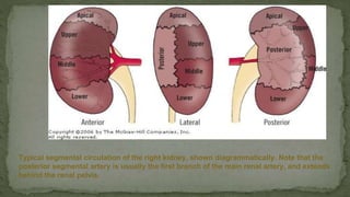
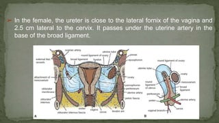
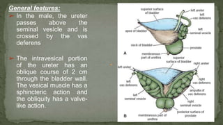
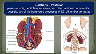
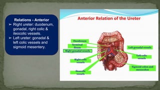
The document provides an overview of the radiological anatomy of the kidneys and adrenal glands. It describes the location, structure and blood supply of the kidneys. The document outlines the pelvicalyceal system and nephron anatomy. It also discusses congenital anomalies, imaging modalities including ultrasound, CT, MRI, and angiography. For the ureters, the document notes their course from the kidneys to the bladder and relationships in the male and female pelvis.
6. ANATOMY OF THE KIDNEY, URETER & POSTERIOR.pdfmarkmuiruri581
Anatomy of Urinary System
Urinary System Organs
Kidneys (2)
Ureters (2)
Urinary bladder
Urethra
Kidney Functions
Control blood volume and composition.
Filter blood plasma, eliminate wastes.
Regulate blood volume, pressure, and fluid osmolarity.
Secrete renin and erythropoietin (EPO).
Regulate PCO2, acid-base balance.
Synthesize calcitriol (Vitamin D).
Detoxify free radicals and drugs.
Perform gluconeogenesis.
Kidney Anatomy
Renal Fascia: Attaches to the abdominal wall.
Adipose Capsule: Provides fat cushioning for the kidney.
Renal Capsule: Fibrous sac that protects from trauma and infection.
Renal Sinus: Contains blood vessels, lymphatics, nerves, and urine-collecting structures.
Renal Parenchyma:
Outer Cortex
Inner Medulla
Renal Pyramids: Extensions of cortex dividing medulla.
Renal Columns: Connect cortex and medulla.
Renal Pelvis: Collects urine from pyramids.
Ureter: Carries urine to the bladder.
Remember, the kidneys play a crucial role in maintaining homeostasis by regulating fluid balance, electrolytes, and waste elimination. Ureter Anatomy
Overview
The ureters are bilateral, muscular, tubular structures responsible for transporting urine from the kidneys to the urinary bladder for storage and eventual excretion.
After blood filtration in the kidneys, the filtrate undergoes reabsorption and exudation along the convoluted tubules.
The urine then passes through the collecting tubules and enters the collecting ducts.
From the collecting ducts, it flows through the calyces into the renal pelvis, marking the beginning of the ureters.
Histology of Ureter
The lumen of each ureter is lined by a mucosal layer of urothelium (transitional epithelium).
The ureteral wall contains two muscular layers:
Longitudinal layer
Circular layer
In the lower segment of the ureters, an additional longitudinal layer is found proximal to the bladder.
Urine is propelled along the ureters by peristaltic motions initiated by pacemaker cells in the proximal renal pelvis.
Relations
Both ureters pass inferiorly over the abdominal surface of the psoas major muscle.
The right ureter travels posterior to the duodenum and is crossed by branches of the superior mesenteric vessels.
The left ureter is also posterior to the psoas major and is crossed by branches of the inferior mesenteric vessels.
Posterior Abdominal Wall
Construction
Bony: Extends from the 12th rib above to the pelvic brim below.
Muscular part: Composed of muscles and fasciae.
Fasciae: Provides stability and support for retroperitoneal organs, vessels, and nerves.
Remember, understanding the anatomy of the ureter and posterior abdominal wall is essential for clinical pracPosterior Abdominal Wall
Construction
Bony: Extends from the 12th rib above to the pelvic brim below.
Muscular part: Composed of muscles and fasciae.
Fasciae: Provides stability and support for retroperitoneal organs, vessels, and nerves.
Muscles of Posterior Abdominal Wall
Psoas Major:
Origin: Continuously attached from T12 (lower border) to L5
The kidneys are paired retroperitoneal organs located on the posterior abdominal wall. Each kidney has an outer renal cortex and inner renal medulla divided into renal pyramids. The kidneys receive blood supply from the renal arteries which branch into segmental and lobar arteries before branching further. Blood exits via interlobar, arcuate and interlobular veins into the renal veins which drain into the inferior vena cava. The kidneys are surrounded by fibrous capsules and perirenal fat and have anterior relations to other abdominal organs and posterior relations to the vertebral column and muscles.
The kidney is bean shaped and measures approximately 10-12cm vertically and 5-7cm transversely. It has an outer renal cortex and inner renal medulla composed of renal pyramids. The renal artery branches within the kidney to form segmental arteries which further branch into interlobar arteries that supply blood to glomeruli. Renal veins mirror the arterial supply and drainage exits via the renal vein. Lymphatic drainage is into lymph nodes near the renal hilum and varies between left and right kidneys.
The kidney is bean shaped and measures approximately 10-12 cm vertically and 5-7 cm transversely. It has an outer renal cortex and inner renal medulla composed of renal pyramids. Blood vessels and collecting ducts enter the kidney through the renal sinus and branch throughout the organ before draining into the renal pelvis. The kidney is surrounded by Gerota fascia and has complex vascular and lymphatic supply.
Radiological anatomy of kidney, ureter & bladderrajss007
The document describes the anatomy and locations of the kidneys, ureters, and urinary bladder. It discusses that the kidneys are retroperitoneal bean-shaped organs located in the posterior abdominal cavity between vertebrae T12 and L3. The ureters are tubular structures that carry urine from the kidneys to the bladder. The urinary bladder is a muscular organ in the pelvis that temporarily stores urine until urination. Imaging modalities like CT, MRI, ultrasound can be used to visualize these structures and evaluate for any abnormalities.
The kidneys are paired retroperitoneal organs located in the posterior abdominal cavity. Each kidney is reddish-brown and ovoid in shape, measuring approximately 10-12 cm long. The kidneys receive blood supply from the renal arteries and drain into the renal veins. The internal anatomy of the kidneys includes the renal cortex, medulla, pyramids, calyces and pelvis which collect urine drained from nephrons. Variations can occur in the number and branching pattern of renal vessels.
The kidneys are paired retroperitoneal organs located on the posterior abdominal wall. Each kidney is bean-shaped and measures around 12cm x 6cm x 3cm. Internally, each kidney contains an outer renal cortex and inner renal medulla divided into renal pyramids. The kidneys receive blood supply from the renal arteries and drain into the renal veins, which join the inferior vena cava. They have both anterior and posterior relations separated by perirenal fat and fascia. The kidneys filter blood and remove waste through complex vascular and tubular systems within the renal cortex and medulla.
The kidneys are retroperitoneal paired organs located on the posterior abdominal wall. Each kidney has an outer renal cortex and inner renal medulla divided into renal pyramids. The kidneys receive blood supply from the renal arteries which branch into segmental arteries then further branch into interlobar arteries and arcuate arteries. Venous drainage occurs through interlobular veins, arcuate veins, and interlobar veins which drain into the renal veins. The kidneys are surrounded by perirenal fat and renal fascia and have anterior relations to other abdominal organs and posterior relations to the diaphragm and vertebral column.
The kidneys are paired retroperitoneal organs located on the posterior abdominal wall. The left kidney is slightly higher than the right kidney. Kidney size varies with gender and stature. Each kidney has an oblique orientation with the hilum angled anteriorly. The kidneys are surrounded by renal fascia and covered by a fibrous capsule. The kidneys contain an outer cortex and inner medulla divided into renal pyramids drained by minor calyces that join to form major calyces and eventually the renal pelvis. The kidneys receive nerve supply from both the sympathetic and parasympathetic nervous systems.
This document provides details on the anatomy and histology of the kidneys. It discusses the location, structure, blood supply and drainage of the kidneys. Some key points include:
- The kidneys are located retroperitoneally on either side of the vertebral column.
- Internally, they contain an outer cortex and inner medulla, separated by renal columns and arches.
- They are supplied by renal arteries which branch numerous times to form the renal microvasculature. Renal veins drain into the vena cava.
- Nephrons are the functional units of the kidney, each containing a renal corpuscle for filtration and tubules for reabsorption and secretion to form urine
The kidneys are paired organs located retroperitoneally on either side of the vertebral column. Each kidney is bean-shaped and surrounded by three layers - a fibrous capsule, perirenal fat, and renal fascia. The kidneys receive their blood supply from the renal arteries and drain into the renal veins. They have an extensive arterial supply that branches into segments and interlobar arteries within renal columns. The kidneys also have a complex calyceal system that drains urine into the renal pelvis and ureters. Percutaneous access for procedures must consider the location of calyces and vascular anatomy to avoid complications.
The kidneys are bean-shaped organs located retroperitoneally between vertebrae T12 and L3. Each kidney has an outer renal cortex and inner renal medulla divided into renal pyramids. The kidneys are supplied by renal arteries and drained by renal veins and lymphatics. Urine drains from the kidneys through the ureters into the bladder. The ureters have several points of narrowing as they course retroperitoneally from the kidneys to the bladder.
The urinary system includes the kidneys, ureters, urinary bladder, and urethra. The kidneys filter the blood to remove wastes and produce urine. The ureters are tubes that carry urine from the kidneys to the bladder. The bladder stores urine until urination. The urethra then carries urine from the bladder to the outside of the body. Key structures of the urinary system were described in detail including locations, blood supply, and clinical relevance.
The document discusses renal vascular anatomy and its implications for surgery. It notes that the renal arteries typically enter the kidney through the hilum and branch into segmental arteries that do not anastomose. It also describes variations such as accessory renal arteries. The segmental arteries further branch within the kidney and an avascular plane exists between anterior and posterior branches. Venous drainage parallels the arterial supply. Imaging like CTA can accurately map the vasculature preoperatively to aid surgery. Understanding variations and collateral circulation is important for procedures like donor nephrectomy.
The document provides an overview of the urinary system. It describes the main components which include the kidneys, ureters, urinary bladder, and urethra. The kidneys filter waste from the blood to form urine, which is then transported via the ureters to the bladder. The bladder stores urine until urination, at which point urine exits the body through the urethra. The document also discusses the structures and functions of the kidneys and nephrons in more detail.
he kidneys are a vital organ critical to the human body. From filtering waste from blood to produce red blood cells, it serves a crucial role. With cells and tissue that work together in synchronized form for common function
Kidneys and adrenal glands,it's functionSaiSai875502
The kidneys and adrenal glands are located in the retroperitoneal space near the spine. The kidneys filter blood to remove waste and regulate electrolytes and fluid balance. Each kidney receives blood supply from the renal arteries and drains into the renal veins. The adrenal glands sit above the kidneys and consist of an outer cortex and inner medulla. The cortex secretes corticosteroids and androgens while the medulla secretes catecholamines. Kidney stones may cause pain and block urine flow.
The kidneys are a pair of excretory organs located retroperitoneally on either side of the vertebral column. They remove waste and regulate water and electrolyte balance. Each kidney contains an inner medulla and outer cortex. The kidneys receive blood supply from the renal arteries and drain into the renal veins. They are important for regulating blood pressure and red blood cell production. Kidney diseases can cause hypertension, renal failure and require dialysis in severe cases.
The kidneys filter waste from the blood to produce urine, which then travels down the ureters into the urinary bladder. The bladder stores urine until urination, when it is expelled through the urethra. The kidneys and urinary system work together to remove waste from the body and regulate fluid balance and blood pressure.
The kidneys are paired retroperitoneal organs that vary in size between individuals. The left kidney typically sits higher than the right. Each kidney contains an outer cortical region and inner medullary pyramids drained by minor calyces that join to form major calyces, eventually draining into the renal pelvis. Kidney anatomy is important for surgical and interventional procedures. Radiological imaging such as ultrasound, CT and MRI can evaluate kidney size, structure and enhancement following contrast administration.
Radiological anatomy of hepatobiliary systemPankaj Kaira
The document provides an overview of the radiological anatomy of the hepatobiliary system. In 3 sentences:
It describes the anatomy of the liver including its lobes, ligaments, blood supply from the hepatic artery and portal vein, and segmentation. The pancreas and biliary apparatus are also discussed, including the gallbladder, cystic duct, common hepatic duct, and common bile duct. Diagrams and images are included to illustrate the structures and their relationships.
Rectum means straight as if ruled. This is a misnorma,for it is curved in conformity with the hollow of the sacrum.
Rectum is continuous with the sigmoid colon and there is no change of structure at the junction. The distinction is a matter of peritoneal attachment; where there is a mesocolon, the gut is called sigmoid colon and where there is no mesentery, it is called rectum . Where the muscle coats are replaced by sphincters it becomes the anal canal.
The rectum begins in the hollow of the sacrum at the level of its 3rd. Piece and it curves forwards over coccyx and ano-coccygeal raphe.
It is 15 cm long.
The 3 tinea of the sigmoid colon come together over the rectum invest it in a complete outer layer of the longitudinal muscle.
The upper and lower ends of the rectum lie in the midline but the ampulla is convex to the left.
Rectal valves of Houston,2 on the left and one on the Right are produced by circular muscles of the gut.
The document provides an overview of kidney anatomy, physiology, and pathology. It describes the macro and micro structure of the kidneys, including nephrons, blood supply, drainage, and innervation. Key functions discussed include regulation of water and electrolytes through filtration, reabsorption, and concentration processes. Hormonal control of acid-base balance and water resorption is also summarized.
The document provides information on the surgical anatomy of the kidney and ureter. It discusses the embryology, gross anatomy including orientation and position of the kidneys. It describes the microscopic anatomy including the nephron. It details the coverings of the kidney including the fibrous capsule, perinephric fat, Gerota's fascia and paranephric fat. It outlines the relations of the kidney to surrounding structures like ribs, diaphragm and pleura. It also discusses the blood supply, lymphatic drainage and nerve supply of the kidneys.
Muktapishti is a traditional Ayurvedic preparation made from Shoditha Mukta (Purified Pearl), is believed to help regulate thyroid function and reduce symptoms of hyperthyroidism due to its cooling and balancing properties. Clinical evidence on its efficacy remains limited, necessitating further research to validate its therapeutic benefits.
Osteoporosis - Definition , Evaluation and Management .pdfJim Jacob Roy
Osteoporosis is an increasing cause of morbidity among the elderly.
In this document , a brief outline of osteoporosis is given , including the risk factors of osteoporosis fractures , the indications for testing bone mineral density and the management of osteoporosis
Does Over-Masturbation Contribute to Chronic Prostatitis.pptxwalterHu5
In some case, your chronic prostatitis may be related to over-masturbation. Generally, natural medicine Diuretic and Anti-inflammatory Pill can help mee get a cure.
share - Lions, tigers, AI and health misinformation, oh my!.pptxTina Purnat
• Pitfalls and pivots needed to use AI effectively in public health
• Evidence-based strategies to address health misinformation effectively
• Building trust with communities online and offline
• Equipping health professionals to address questions, concerns and health misinformation
• Assessing risk and mitigating harm from adverse health narratives in communities, health workforce and health system
8 Surprising Reasons To Meditate 40 Minutes A Day That Can Change Your Life.pptxHolistified Wellness
We’re talking about Vedic Meditation, a form of meditation that has been around for at least 5,000 years. Back then, the people who lived in the Indus Valley, now known as India and Pakistan, practised meditation as a fundamental part of daily life. This knowledge that has given us yoga and Ayurveda, was known as Veda, hence the name Vedic. And though there are some written records, the practice has been passed down verbally from generation to generation.
Recomendações da OMS sobre cuidados maternos e neonatais para uma experiência pós-natal positiva.
Em consonância com os ODS – Objetivos do Desenvolvimento Sustentável e a Estratégia Global para a Saúde das Mulheres, Crianças e Adolescentes, e aplicando uma abordagem baseada nos direitos humanos, os esforços de cuidados pós-natais devem expandir-se para além da cobertura e da simples sobrevivência, de modo a incluir cuidados de qualidade.
Estas diretrizes visam melhorar a qualidade dos cuidados pós-natais essenciais e de rotina prestados às mulheres e aos recém-nascidos, com o objetivo final de melhorar a saúde e o bem-estar materno e neonatal.
Uma “experiência pós-natal positiva” é um resultado importante para todas as mulheres que dão à luz e para os seus recém-nascidos, estabelecendo as bases para a melhoria da saúde e do bem-estar a curto e longo prazo. Uma experiência pós-natal positiva é definida como aquela em que as mulheres, pessoas que gestam, os recém-nascidos, os casais, os pais, os cuidadores e as famílias recebem informação consistente, garantia e apoio de profissionais de saúde motivados; e onde um sistema de saúde flexível e com recursos reconheça as necessidades das mulheres e dos bebês e respeite o seu contexto cultural.
Estas diretrizes consolidadas apresentam algumas recomendações novas e já bem fundamentadas sobre cuidados pós-natais de rotina para mulheres e neonatos que recebem cuidados no pós-parto em unidades de saúde ou na comunidade, independentemente dos recursos disponíveis.
É fornecido um conjunto abrangente de recomendações para cuidados durante o período puerperal, com ênfase nos cuidados essenciais que todas as mulheres e recém-nascidos devem receber, e com a devida atenção à qualidade dos cuidados; isto é, a entrega e a experiência do cuidado recebido. Estas diretrizes atualizam e ampliam as recomendações da OMS de 2014 sobre cuidados pós-natais da mãe e do recém-nascido e complementam as atuais diretrizes da OMS sobre a gestão de complicações pós-natais.
O estabelecimento da amamentação e o manejo das principais intercorrências é contemplada.
Recomendamos muito.
Vamos discutir essas recomendações no nosso curso de pós-graduação em Aleitamento no Instituto Ciclos.
Esta publicação só está disponível em inglês até o momento.
Prof. Marcus Renato de Carvalho
www.agostodourado.com
Adhd Medication Shortage Uk - trinexpharmacy.comreignlana06
The UK is currently facing a Adhd Medication Shortage Uk, which has left many patients and their families grappling with uncertainty and frustration. ADHD, or Attention Deficit Hyperactivity Disorder, is a chronic condition that requires consistent medication to manage effectively. This shortage has highlighted the critical role these medications play in the daily lives of those affected by ADHD. Contact : +1 (747) 209 – 3649 E-mail : sales@trinexpharmacy.com
Basavarajeeyam is an important text for ayurvedic physician belonging to andhra pradehs. It is a popular compendium in various parts of our country as well as in andhra pradesh. The content of the text was presented in sanskrit and telugu language (Bilingual). One of the most famous book in ayurvedic pharmaceutics and therapeutics. This book contains 25 chapters called as prakaranas. Many rasaoushadis were explained, pioneer of dhatu druti, nadi pareeksha, mutra pareeksha etc. Belongs to the period of 15-16 century. New diseases like upadamsha, phiranga rogas are explained.
4. General features:
➢ The kidneys are paired retroperitoneal organs that lie at the
level of the T12 to L3 vertebral bodies.
➢ They lie obliquely with their upper poles more medial and
more posterior than their lower.
➢ The kidneys measure 10-15 cm in length, the left being
commonly 1.5 cm longer than the right
➢ Their size is approximately that of three-and-a-half lumbar
vertebrae and their associated discs on a radiograph.
5. General features:.
➢ On coronal cross-section
each kidney is seen to
have an outer cortex and
an inner medulla.
➢ Extensions of the cortex
centrally as columns of
Bertin separate the
medulla into pyramids
whose apices, jutting into
the calyces, are called the
papillae.
6. Pelvicalyceal arrangement:
➢ There are usually seven pairs of minor calyces, each
pair having an anterior and a posterior calyx, although
there is wide variation
➢ Minor calyx pairs combine to form two or three major
calyces, which in turn drain via their infundibula to the
pelvis.
➢ This arrangement is quite variable, but when there are
two infundibula these usually drain four pairs of calyces
from the upper pole and three pairs from the lower.
➢ When there are three infundibula there are usually
three pairs of upper pole calyces, and two sets of two
pairs of calyces draining the midpolar region and lower
pole.
➢ A simple calyx has one papilla indenting it; a
compound calyx has more than one.
7. Extrarenal Pelvis:
➢ Refers to the presence of the
renal pelvis outside the confines
of the renal hilum; it is a normal
anatomic variant
➢ It is found in about 10% of the
population.
➢ Ultrasound an extra-renal
pelvis usually appears dilated,
suggesting obstructive
pathology. Subsequent
investigation with CT usually
clarifies a false interpretation on
ultrasound.
8. Renal hilum:
➢ Right renal hilum L1/L2
vertebral level.
➢ Left renal hilum L1 vertebral
level.
➢ Structures from anterior to
posterior:
• Renal vein.
• Renal artery.
• Renal pelvis.
➢ The artery may branch early and
a posterior arterial branch may
enter the hilum posterior to the
pelvis.
➢ Lymph vessels and nerves also
9. Nephron:
• The functional subunit of the
kidney is called the nephron
and consists of
• Renal corpuscle: Bowman’s
capsule & Glomerulus.
• Proximal convoluted tubule.
• Loop of Henle.
• Distal convoluted tubule.
• Collecting duct.
➢ The collecting duct, which
empties into the calyx at the
tip of the medulla.
➢ The kidney has
approximately 1 million
nephrons.
10. Relations of the Kidneys:
➢ Superiorly: the adrenal gland –
more medial on the right
kidney.
➢ Anteriorly:
▪ Right kidney liver, 2nd
part of the duodenum,
ascending colon, small
intestinal loops.
▪ Left kidney stomach,
pancreas and its vessels,
spleen, splenic flexure of the
colon, jejunal loops.
11. ➢ Posteriorly:
▪ Upper third diaphragm,12th rib and the costodiaphragmatic
recess of the pleura.
▪ Lower third medial to lateral: psoas, quadratus lumborum
and transversus abdominis muscles.
12. Blood supply of the kidneys:
Main renal artery:
➢ The renal arteries normally arise from
the abdominal aorta at L1/2 interspace.
➢ Each renal artery lies anterior to renal
pelvis & posterior to renal vein.
➢ Right renal artery:
▪ Longer than left.
▪ Downward course and passes posterior
to IVC.
➢ Left renal artery:
▪ Arises higher than the right renal artery
▪ Has a more horizontal orientation.
▪ Accessory arteries occur in 20–25% of
people. A lower pole artery is the
commonest and bilateral in 15%.
13. Intra-renal arterial anatomy:
➢ The main renal artery divides into segmental arteries
near the hilum.
➢ The first division is classically a posterior segmental
branch, supplying the posterior and apical kidney.
➢ The main renal artery then divides into 4 further
segmental branches at the hilum (apical, upper,
middle and lower anterior), supplying the anterior
surface, lower pole and variable portion of the apex.
➢ Segmental arteries branch into lobar arteries.
➢ Lobar arteries divide into interlobar branches (lie
between pyramids/lobes), these branch into arcuate
arteries that run along the base of the pyramids.
➢ Arcuate arteries branch laterally into the terminal
intralobular arteries.
14. Typical segmental circulation of the right kidney, shown diagrammatically. Note that the
posterior segmental artery is usually the first branch of the main renal artery, and extends
behind the renal pelvis.
15. Avascular plane of Brodel:
➢ It is the section of renal parenchyma
between anterior 2/3 and posterior 1/3
of the kidney on the cross-section.
➢ It is relatively avascular because it
represents the plane where the
anterior and posterior segmental renal
artery branches meet.
➢ It is located just posterior to the lateral
convex border of the kidney and
permits a relatively safe access route
to the pelvicalyceal system for
nephrostomy insertion.
16. Venous drainage:
➢ Renal venules and branch renal veins freely
communicate throughout the parenchyma, but
finally drain only into the main renal vein.
➢ The left renal vein:
▪ 6–10 cm in length. Three times longer than the
right renal vein.
▪ Courses anteriorly, between the superior
mesenteric artery and aorta.
▪ Enters the medial aspect of inferior vena cava.
▪ Three tributaries:
❖ left adrenal vein
❖ left gonadal vein
❖ lumbar veins (variable number).
➢ The right renal vein is 2–4 cm in length and has no
tributaries.
17. Renal vein variants & anomalies:
Circum-aortic left renal vein:
commonest left renal vein
anomaly, seen in 5–7% of
individuals, where the vein
bifurcates into anterior and
posterior limbs that encircle the
aorta.
18. Renal vein variants & anomalies:
Retro-aortic left renal vein: seen in 3% of individuals, the vein may have an
abnormal caudal course, entering the iliac vein.
19. ➢.
Nutcracker syndrome
➢vascular compression disorder
➢compression of the left renal vein
most commonly between the
superior mesenteric artery (SMA)
and aorta
➢can lead to renal venous
hypertension, resulting in rupture
of thin-walled veins into the
collecting system with resultant
hematuria.
➢slightly greater female predilection
21. Retroperitoneum & fascial spaces around the kidneys:
➢ The retroperitoneum is divided into three spaces:
▪ Perinephric space.
▪ Anterior pararenal space.
▪ Posterior pararenal space.
22. the largest of the three divisions of the
retroperitoneum and is the most easily
identified. It contains the kidneys, renal
vessels, proximal collecting systems,
adrenal glands and an adequate amount of
fat to allow identification on CT scanning
The space is surrounded by the perirenal
fascia and is in continuity with the opposite
perirenal space across the midline.
Perirenal fascia consists of two layers:
Gerota’s fascia anteriorly &
Zuckerkandl’s fascia posteriorly. These
fascial layers are fused laterally as the
lateral conal fascia, which is continuous
with the fascia transversalis.
Retroperitoneum & fascial spaces around the kidneys
23.
24. ➢ Anterior pararenal space:
▪ Lies anterior to the anterior renal
fascia and behind the posterior
peritoneum. It is continuous across
the midline.
▪ Contains the pancreas, duodenum
and ascending and descending
colon.
▪ Superiorly, the space is limited
where the anterior renal fascia
blends with the posterior
peritoneum, but inferiorly the space
is open to the pelvic extraperitoneal
spaces.
Retroperitoneum & fascial spaces around the kidneys
25. ➢ Posterior pararenal space:
▪ Lies posterior to the posterior
renal fascia and anterior to the
muscles of the posterior
abdominal wall.
▪ This is limited medially by the
attachment of the renal fascia to
the psoas muscle, but is
continuous laterally with the
extraperitoneal fatty tissue
(properitoneal fat plane) deep to
the transversalis fascia.
▪ It contains only fat
Retroperitoneum & fascial spaces around the kidneys
26. Congenital anomalies of the kidney:
Pelvic Kidney:
• IVU demonstrating right pelvic
kidney.
• Pelvic kidney occurs secondary
to failure of migration, with an
incidence of 1:900 to 1:1200
with no sex predilection.
27. Congenital anomalies of the kidney:
Crossed fused ectopia:
• IVU demonstrating left crossed fused
ectopia due to fusion of the lower pole
of the left kidney with the upper pole
of the ectopic right kidney, but note
that the ureters are normally sited.
• These kidneys invariably have an
aberrant vascular supply.
28. Horseshoe kidney:
• IVU and coronal MIP CT urogram
image of a horseshoe kidney
• Note the lower poles of the kidney
cross the midline and are fused,
the hallmark of a horseshoe
kidney; the fused tissue may be
non-functional fibrous tissue.
• Horseshoe kidneys are prone to
traumatic damage and are the
commonest fusion anomaly,
associated with Turner’s syndrome
and trisomy 18.
Congenital anomalies of the
kidney:
29. Bifid renal pelvis:
• IVU illustrating a bifid renal pelvis of
the left kidney .
Congenital anomalies of the kidney:
30. Partial duplex:
• IVU illustrating a
partial duplex
collecting system of
the left kidney with
fusion of the
ureters in the distal
third.
Congenital anomalies of the kidney:
31. Complete duplex:
• IVU demonstrating a duplex left
kidney with complete ureteric
duplication. The upper moiety ureter
is seen entering the bladder as a
ureterocele with a typical ‘cobra’s
head’ appearance (arrow).
• Complete duplex systems are more
common with the ureter of the lower
pole moiety inserting normally in the
bladder and the upper pole moiety
having an ectopic insertion in the
bladder, urethra or elsewhere
(Weigert-Meyer law).
Congenital anomalies of the kidney:
32.
33. Plain films of the abdomen:
➢ Perirenal fat often makes part
or all of the renal outlines
visible.
➢ Renal size is variable, with a
normal range of 10–15 cm on a
radiograph or approximately
three-and-a-half vertebral
bodies in height(renal size is
magnified by 15% on
radiograph )
➢ The left kidney is usually
larger, but a difference in size
of more than 2 cm is abnormal.
.
34. Intravenous urography:
➢ After opacification by intravenous contrast, the renal
parenchyma and outline can be assessed in the early
or nephrographic phase, and the collecting system and
ureteric anatomy in the urographic phase
➢ In the urographic phase the calyceal system can be
seen. Minor and major calyces are seen. These are
connected to the pelvis of the kidney by infundibula ,
which may be long or short.
35.
36. Ultrasound examination of the kidneys:
Normal kidney appearance in adult:
➢ The renal size is normally 9–12 cm,Cortex is less echogenic than the liver.
➢ Medullary pyramids are slightly less echogenic than the cortex.
➢ Cortex thickness equals/is more than 6 mm, If the pyramids are difficult to
differentiate, the parenchymal thickness can be measured instead and should
be 15-20 mm.
➢ Central renal sinus, consisting of the calyces, renal pelvis and fat, is more
echogenic than the cortex,Renal pelvis may appear as a central slit of
anechoic fluid at the hilum
37. Ultrasound examination of the
kidneys:
➢ Ultrasonographic differences of
neonatal kidneys from older children
and adults:
▪ Increased cortical echogenicity
(maybe similar to liver or spleen).
▪ Larger and more hypoechoic
pyramids.
▪ Little or no sinus fat.
▪ Fetal lobulation maybe seen.
➢ Note adult pattern is attained at 6
months of age.
38. CT & MRI
➢ The kidneys are seen on slices from T12 to L3 vertebral levels
➢ Posterior relations and anterior relations can be seen on axial
CT images, but are very well appreciated on sagittal and
coronal MR images.
➢ The renal substance is homogeneous on unenhanced CT.
39. ➢ On MR, the intrinsic contrast between cortex and medulla is seen on T1W and
T2W images. On T1W images the renal cortex has a slightly higher signal than
the medulla. On T2W images the renal cortex is slightly lower in signal than the
medulla and intrinsic renal contrast is superior.
40. ➢ On both CT and MRI three phases of enhancement can
be appreciated:
▪ Arterial corticomedullary phase, where the cortex
enhances strongly and contrast between cortex and
medulla is greatest,
▪ Venous nephrographic phase, where the contrast is
homogeneous throughout the kidney.
▪ Delayed excretory phase, where contrast is seen in the
collecting system.
41.
42. ➢ The renal vessels can be identified
on unenhanced images, but are best
seen after contrast
➢ The arteries are best seen early in a
contrast bolus (first 25 seconds).
➢ The veins are best seen after
approximately 60 seconds.
➢ With MR, the renal arteries and
veins can also be imaged without
intravenous contrast using flow-
sensitive imaging sequences.
43. Arteriography of the
kidneys:
➢ Direct arteriography
allows assessment of
vascular and other lesions
of the kidneys, but is
primarily used to facilitate
interventional procedures
such as renal artery
angioplasty or stent
placement.
44. Renal venography
This is performed via the inferior
vena cava Although it is rarely used,
it may be required to identify the
location of a renin-producing tumour
.
The left adrenal and left gonad are
also imaged via left renal venography
because of the common drainage of
veins from these organs on this side
The renal veins are seen to have
valves These are more common on
the left side
The right renal vein is multiple in
10% of venograms and receives the
right gonadal vein in 6% of cases.
45.
46. ➢ lateral
General features:
➢ Retroperitoneal and extraperitoneal structure.
➢ 25–30 cm long.
➢ Diameter of approximately 3 mm but has three ‘functionally’ narrow regions:
▪ Pelviureteric junction.
▪ As the ureter crosses bony pelvic brim.
▪ Vesicouretric junction.
➢ The ureter enters the pelvis at the bifurcation of the common iliac artery anterior to
the sacroiliac joint.
➢ It then lies on the lateral wall of the pelvis in front of the internal iliac artery to a
point just anterior to the ischial spine, where it turns forwards and medially to enter
the bladder
47. ➢ In the female, the ureter is close to the lateral fornix of the vagina and
2.5 cm lateral to the cervix. It passes under the uterine artery in the
base of the broad ligament.
48. General features:
➢ In the male, the ureter
passes above the
seminal vesicle and is
crossed by the vas
deferens
➢ The intravesical portion
of the ureter has an
oblique course of 2 cm
through the bladder wall.
The vesical muscle has a
sphincteric action and
the obliquity has a valve-
like action.
49. Relations – Posterior
psoas muscle, genitofemoral nerve, sacroiliac joint and common iliac
vessels, tips of the transverse processes of L2–L5 lumbar vertebrae
50. Relations - Anterior
➢ Right ureter: duodenum,
gonadal, right colic &
ileocolic vessels.
➢ Left ureter: gonadal &
left colic vessels and
sigmoid mesentery.
51. Blood supply & lymphatics:
➢ Arterial supply is highly variable:
▪ Upper ureter: branch from renal
artery.
▪ Mid ureter: small medial branches
from the aorta.
▪ Lower ureter: small branches from
the superior and inferior vesical,
middle rectal and uterine arteries.
➢ Venous drainage is highly variable
and not defined.
➢ Lymphatic drainage:
▪ Abdominal ureter drains to aorto-
caval and common iliac nodes.
▪ Pelvic ureter drains to internal and
external iliac nodes.
52. Duplication :
occurs in about 4% of subjects
It is the commonest significant congenital anomaly of the urinary tract
Duplication
two to three times commoner in females
When complete duplication occurs, the ureter serving the upper renal
moiety drains fewer calyces and is inserted lower into the bladder than that
draining the lower moiety
The low insertion may extend to the bladder neck or the urethra or, in
females, the vestibule or vagina
53.
54. Ureterocele :
is a dilation of the
intramural portion of
the ureter due to
narrowing of its orifice
This is most common in
a duplicated system,
when it occurs in the
ureter draining the
upper renal moiety that
is usually ectopic
56. Plain films of the abdomen:
➢ The ureter is not visible, but a knowledge of its course in relation to
the skeleton is necessary when looking for radio-opaque calculi.
57. Intravenous urography:
➢ The ureters are either
completely or partly
visible when filled with
contrast.
➢ Prone views aid
ureteric filling.
➢ Distension of the
upper part of the
collecting system can
also be aided by
applying a
compression band
across the abdomen.
58. Ultrasound:
➢ The proximal and distal ureters may be visible on ultrasound when well
distended.
➢ Intestinal gas generally obscures the mid-portion unless it is abnormally dilated.
59. CT:
➢ Ureteric calculi not visible
on radiographs are readily
visible on CT scans, and
non-contrast CT has largely
replaced the IVU for
diagnosis of ureteric calculi
➢ The normal ureter can be
identified on non-contrast
scans, although it is easier
to identify if it contains
contrast medium.
.
60. Coronal reformatted MIP image from a CT
urogram demonstrates retrocaval ureter
Axial image from "stone protocol" CT
showing left ureteral stone.
61. MR urography:
➢ Static fluid MR urography: by using a heavily T2 weighted sequence (similar to MRCP). However,
because the ureters are intermittently collapsed due to peristalsis, parts of the ureter may not be
distended with urine and thus not imaged using these techniques. But, this technique is at its best in
obstructed, fluid-filled systems
➢ MR contrast urography: can be performed where the ureters are imaged during the excretory phase
after intravenous gadolinium and is aided by concurrent administration of a diuretic.
Static fluid MR Technique
63. General features:
➢ Paired retroperitoneal glands,
supero-medial to the kidneys
within the perinephric space, but
outside the renal capsule.
➢ Each gland is composed of a
body and medial and lateral limbs.
➢ The adrenals do not develop with
the kidneys. They develop in the
retroperitoneum and descend,
whereas the kidneys develop in
the pelvis and ascend.
➢ In cases where the kidneys fail to
ascend normally, the adrenal
glands are still found in the
expected position, although their
shape may be more discoid owing
to lack of moulding by the kidneys
during development.
64. Right adrenal gland.
➢ More consistent in location.
➢ It lies posterior to IVC, medial to the right lobe of the liver and lateral to the right diaphragmatic crus.
➢ It is lower and more medial in relation to the spine than the left.
➢ On cross-section, it is linear or V-shaped, with a larger medial limb and a smaller lateral limb
Left adrenal gland:
➢ Less constant in location.
➢ Usually lies posterior to the splenic vein and lateral to the diaphragmatic crus.
➢ More semilunar than the right and it extends down the superomedial border of the kidney towards the
hilum.
➢ On cross-section it is triangular or Y-shaped.
65. Blood supply:
➢ Three arteries on each side:
▪ Superior adrenal artery from the
inferior phrenic artery (which is a
branch of the abdominal aorta).
▪ Middle adrenal artery arises from
the abdominal aorta.
▪ Inferior adrenal artery from the
renal artery
➢ A single vein drains each gland.
▪ Right adrenal vein: shorter,
drains directly into the IVC.
▪ Left adrenal vein: longer, drains
into the left renal vein and may
be joined by the inferior phrenic
vein.
67. Plain films of the abdomen:
➢ The adrenal glands are visible
only if calcified, and they are
then seen to be lateral to the
spine at the level of the upper
pole of the kidneys.
Bilateral triangular foci of
calcifications near the adrenal
lodge, compatible with bilateral
adrenal calcifications.
This patient had a past medical
history of adrenal hemorrhage.
68. Ultrasound:
➢ In thin individuals the adrenal glands can sometimes be seen between the
kidney and liver on the right and between the kidney and pancreatic tail on the
left using high-resolution scanning.
➢ They are readily seen in neonates and usually seen in children.
69. CT:
➢ The shape of the adrenal gland on CT cuts is variable, with a linear, inverted V shape
being commonest on the right and a triangular or Y shape commonest on the left.
➢ Craniocaudal extent is less than 4 cm.
➢ Limb thickness is usually less than 1 cm.
70.
71. MRI:
➢ The adrenals are very well
seen on MRI because of
surrounding fat (more
easily than with CT)
➢ They are iso- or slightly
hypointense compared to
liver on both T1W and
T2W images.
➢ They lose signal on fat
suppression or fat
subtraction techniques,
depending on the
cholesterol content of the
adrenal cortex.
72.
73. Name the study
IVU
A . Left ureter
B. Left renal pelvis
C . Urinary bladder
D . Right lower pole major
calyx
E . Right sacroiliac joint
74. (a) The right kidney is usually larger by about 1.5 cm than the
left.
(F)
(b) The columns of Bertin extend medially within the substance of
the kidney separating the medulla into pyramids.
(T)
(c) The anterior division of the renal artery supplies both
upper and lower portions of the kidney.
(T)
(d) The segmental branches divide into interlobar arteries
between the pyramids.
(T)