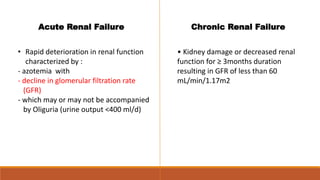
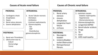
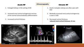
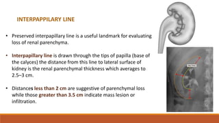
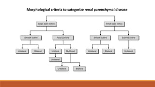
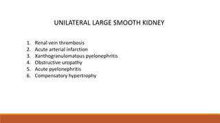
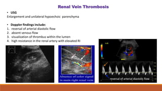
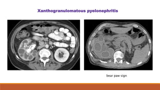
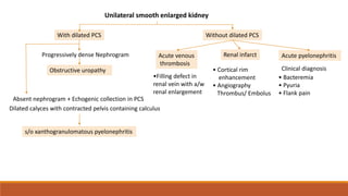
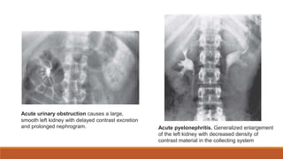
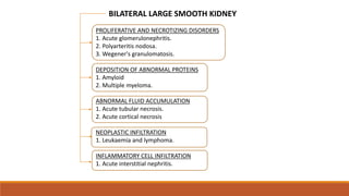
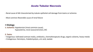
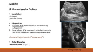
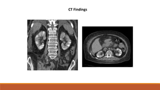
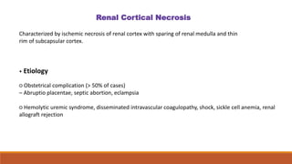
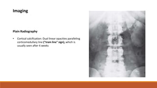
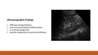
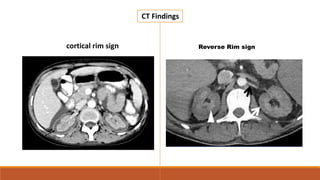
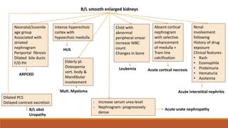
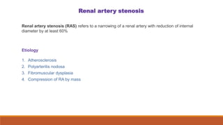
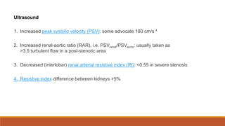
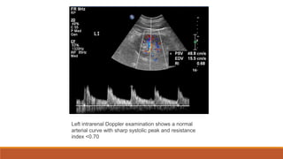
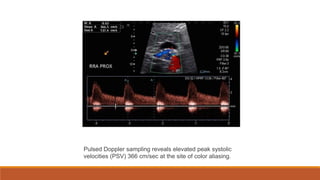
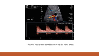
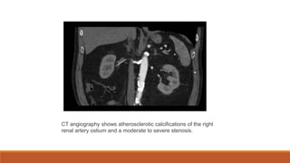
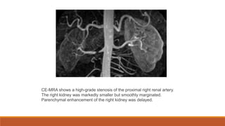
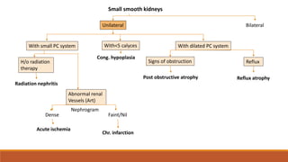
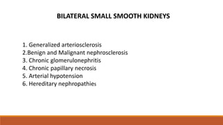
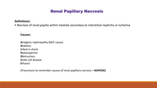
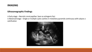
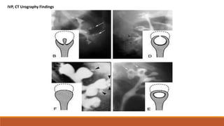
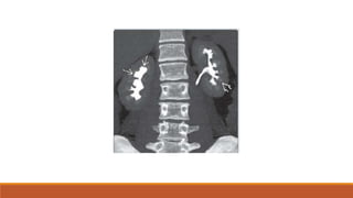
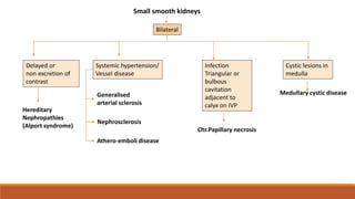
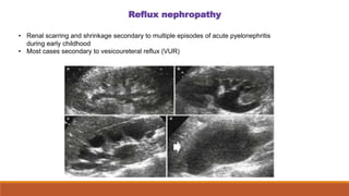
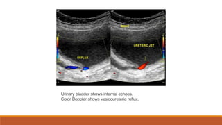
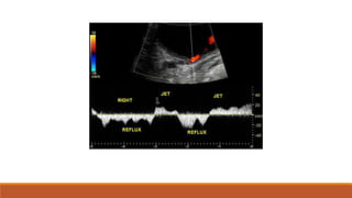
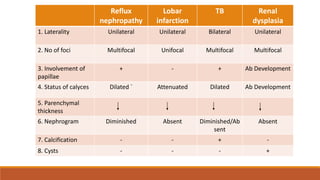
Renal Failure
&
Renal Parenchymal Disease
Dr. Niranjan Patil
DEPARTMENT OF RADIODIAGNOSIS
DR D Y PATIL HOSPITAL KOLHAPUR
Acute Renal Failure
• Rapid deterioration in renal function
characterized by :
- azotemia with
- decline in glomerular filtration rate
(GFR)
- which may or may not be accompanied
by Oliguria (urine output <400 ml/d)
Chronic Renal Failure
• Kidney damage or decreased renal
function for ≥ 3months duration
resulting in GFR of less than 60
mL/min/1.17m2
Causes of Acute renal failure
PRERENAL
1. Cardiogenic shock
2. Anaphylaxis
3. Sepsis
4. Renal artery stenosis
5. Severe dehydration
INTRARENAL
1. Acute Tubular necrosis
-Hemolysis
-Antibiotics
-Contrast nephrotoxicty
2. Glomerulopathy
-SLE
-HSP
-Postinfective
glomerulonephritis
POSTRENAL
1. Renal vein Thrombosis
2. Obstruction due to;
-Calculi
-Hemorrahge
-Tumor
Causes of Chronic renal failure
PRERENAL
1. Renal artery
stenosis
2. chronic
dehydration
3. congestive heart
failure
4. cirrhosis
INTRARENAL
1. common causes
-Diabetic nephropathy
-Hypertensive
-Glomerulosclerosis
2. Glomerulonephritis
3. congenital ADPKD
4. Renal tubular acidosis
5. Drugs
6. TB
7. Gout
8. AIDS nephropathy
POSTRENAL
1. Neurogenic
bladder
2. Ureteric/bladder
outlet Obstruction
3. Retroperitoneal
fibrosis
1. Enlarged kidneys in the setting of AKI
2. increased renal cortical echogenicity and loss
of the normal corticomedullary differentiation
3. Increased cortical thickness
1. Small or atrophic kidneys are often seen with
CKD
2. Markedly increased renal cortical echogenicity
is seen in CKD
3. Decreased cortical thickness
reduced renal cortical thickness <6 mm
Ultrasonography
Acute RF Chronic RF
• Preserved interpapillary line is a useful landmark for evaluating
loss of renal parenchyma.
• Interpapillary line is drawn through the tips of papilla (base of
the calyces) the distance from this line to lateral surface of
kidney is the renal parenchymal thickness which averages to
2.5–3 cm.
• Distances less than 2 cm are suggestive of parenchymal loss
while those greater than 3.5 cm indicate mass lesion or
infiltration.
INTERPAPPILARY LINE
Morphological criteria to categorize renal parenchymal disease
UNILATERAL LARGE SMOOTH KIDNEY
1. Renal vein thrombosis
2. Acute arterial infarction
3. Xanthogranulomatous pyelonephritis
4. Obstructive uropathy
5. Acute pyelonephritis
6. Compensatory hypertrophy
Renal Vein Thrombosis
• USG
Enlargement and unilateral hypoechoic parenchyma
• Doppler findings include:
1. reversal of arterial diastolic flow
2. absent venous flow
3. visualization of thrombus within the lumen
4. high resistance in the renal artery with elevated RI
reversal of arterial diastolic flow
the thrombosis is observed as a
filling defect during venous
phase imaging following
intravenous contrast
Long linear filling defect in the left renal vein.
CT findings
Renal infarction
Renal infarction results from interruption of the normal blood supply to part of,
or to the whole kidney.
• Causes of renal infarction include
1. Thromboembolism
2. Aortic dissection
3. Renal artery stenosis
Power doppler Colour doppler
Ultrasound
• Hyperechoic
• Absence of flow in the renal artery
• Infarcts will appear as wedge-shaped regions of nonperfusion
contrast-enhanced CT demonstrate hypoattenuation of the posteroinferior part of the
kidney with surrounding stranding.
There is preservation of the very outer layer of parenchyma (cortical rim sign).
cortical rim sign
Xanthogranulomatous pyelonephritis
bear paw sign
Without dilated PCS
Unilateral smooth enlarged kidney
Progressively dense Nephrogram
Obstructive uropathy
With dilated PCS
Absent nephrogram + Echogenic collection in PCS
Dilated calyces with contracted pelvis containing calculus
s/o xanthogranulomatous pyelonephritis
Acute venous
thrombosis
Renal infarct
• Cortical rim
enhancement
• Angiography
Thrombus/ Embolus
•Filling defect in
renal vein with a/w
renal enlargement
Acute pyelonephritis
Clinical diagnosis
• Bacteremia
• Pyuria
• Flank pain
Acute urinary obstruction causes a large,
smooth left kidney with delayed contrast excretion
and prolonged nephrogram. Acute pyelonephritis. Generalized enlargement
of the left kidney with decreased density of
contrast material in the collecting system
PROLIFERATIVE AND NECROTIZING DISORDERS
1. Acute glomerulonephritis.
2. Polyarteritis nodosa.
3. Wegener's granulomatosis.
DEPOSITION OF ABNORMAL PROTEINS
1. Amyloid
2. Multiple myeloma.
ABNORMAL FLUID ACCUMULATION
1. Acute tubular necrosis.
2. Acute cortical necrosis
NEOPLASTIC INFILTRATION
1. Leukaemia and lymphoma.
INFLAMMATORY CELL INFILTRATION
1. Acute interstitial nephritis.
BILATERAL LARGE SMOOTH KIDNEY
- Renal cause of AKI characterized by tubular epithelial cell damage from toxins or ischemia
- Most common Reversible cause of renal failure
• Etiology
1. Ischemia: Hypotension (most common cause),
hypovolemia, renal vasoconstriction, DIC
2. Toxins
– Exogenous: Iodinated contrast media, antibiotics, chemotherapeutic drugs, organic solvents, heavy metals
– Endogenous: Hemolysis, rhabdomyolysis, uric acid, oxalate
Acute Tubular Necrosis
 Ultrasonographic Findings
 Morphology
- Enlarged
- Smooth outline
 Echogenicity
1. Ischemic ATN- Normal cortical and medullary
echogenicity
2. Drug related ATN- increased cortical echogenicity
and maintained corticomedullary differentiation
○ Perirenal hypoechoic rim ("kidney sweat")
 Color Doppler
- Resistive index: ↑ (> 0.7)
IMAGING
"kidney sweat“ sign
CT Findings
Characterized by ischemic necrosis of renal cortex with sparing of renal medulla and thin
rim of subcapsular cortex.
• Etiology
○ Obstetrical complication (> 50% of cases)
– Abruptio placentae, septic abortion, eclampsia
○ Hemolytic uremic syndrome, disseminated intravascular coagulopathy, shock, sickle cell anemia, renal
allograft rejection
Renal Cortical Necrosis
Plain Radiography
• Cortical calcification: Dual linear opacities paralleling
corticomedullary line ("tram line" sign), which is
usually seen after 4 weeks
Imaging
Ultrasonographic Findings
• Diffusely enlarged kidneys
• Loss of corticomedullary differentiation
• ↓ cortical echogenicity
• acoustic shadow due to cortical calcification
CT Findings
cortical rim sign Reverse Rim sign
B/L smooth enlarged kidneys
Neonatal/Juvenile
age group
Associated with
striated
nephrogram
Periportal fibrosis
Dilated bile ducts
F/O PH
ARPCKD
Dilated PCS
Delayed contrast excretion
B/L obst
Uropathy
- Increase serum urea level
- Nephrogram- progressively
dense
Intense hyperechoic
cortex with
hypoechoic medulla
HUS
Elderly pt.
Osteopenia
vert. body &
Mandibular
involvement
Mult. Myeloma
Child with
abnormal
peripheral smear
increase WBC
count
Changes in bone
Leukemia
Absent cortical
nephrogram
with selective
enhancement
of medulla +
Tram line
calcification
Acute cortical necrosis
Renal
involvement
following
History of drug
exposure
Clinical features-
• Rash
• Eosinophila
• Proteinuria
• Hematuria
• Azotemia
Acute interstitial nephritis
Acute urate nephropathy
UNILATERAL SMALL SMOOTH KIDNEYS
1. Ischemia due to renal artery stenosis
2. Post-obstructive atrophy
3. End result of renal infarction
4. Congenital hypoplasia
5.Reflux atrophy
Renal artery stenosis
Renal artery stenosis (RAS) refers to a narrowing of a renal artery with reduction of internal
diameter by at least 60%
Etiology
1. Atherosclerosis
2. Polyarteritis nodosa
3. Fibromuscular dysplasia
4. Compression of RA by mass
Ultrasound
1. Increased peak systolic velocity (PSV): some advocate 180 cm/s 4
2. Increased renal-aortic ratio (RAR), i.e. PSVrenal/PSVaorta: usually taken as
>3.5 turbulent flow in a post-stenotic area
3. Decreased (interlobar) renal arterial resistive index (RI): <0.55 in severe stenosis
4. Resistive index difference between kidneys >5%
Left intrarenal Doppler examination shows a normal
arterial curve with sharp systolic peak and resistance
index <0.70
Pulsed Doppler sampling reveals elevated peak systolic
velocities (PSV) 366 cm/sec at the site of color aliasing.
Turbulent flow is seen downstream in the mid renal artery.
CT angiography shows atherosclerotic calcifications of the right
renal artery ostium and a moderate to severe stenosis.
CE-MRA shows a high-grade stenosis of the proximal right renal artery.
The right kidney was markedly smaller but smoothly marginated.
Parenchymal enhancement of the right kidney was delayed.
Small smooth kidneys
Unilateral Bilateral
With small PC system With<5 calyces With dilated PC system
Cong. hypoplasia
H/o radiation
therapy
Radiation nephritis
Abnormal renal
Vessels (Art)
Dense Faint/Nil
Acute ischemia
Signs of obstruction
Post obstructive atrophy
Reflux
Reflux atrophy
Chr. infarction
Nephrogram
1. Generalized arteriosclerosis
2.Benign and Malignant nephrosclerosis
3. Chronic glomerulonephritis
4. Chronic papillary necrosis
5. Arterial hypotension
6. Hereditary nephropathies
BILATERAL SMALL SMOOTH KIDNEYS
Definitions:
• Necrosis of renal papilla within medulla secondary to interstitial nephritis or ischemia
Renal Papillary Necrosis
Causes:
Analgesic nephropathy (M/C cause)
Diabetes
Infant in shock
Pyelonephritis
Obstruction,
Sickle cell disease
Ethanol
(Pneumonic to remember causes of renal papillary necrosis—ADIPOSE)
IMAGING
Ultrasonographic Findings
○ Early stage – Necrotic renal papillae: Seen as echogenic foci
○ Advanced stage – Single or multiple cystic cavities in medullary pyramids continuous with calyces ±
calcification
IVP, CT Urography Findings
Small smooth kidneys
Bilateral
Delayed or
non excretion of
contrast
Hereditary
Nephropathies
(Alport syndrome)
Systemic hypertension/
Vessel disease
Generalised
arterial sclerosis
Nephrosclerosis
Athero-emboli disease
Infection
Triangular or
bulbous
cavitation
adjacent to
calyx on IVP
Chr.Papillary necrosis
Cystic lesions in
medulla
Medullary cystic disease
1.Reflux nephropathy
2.Lobar infarction
3.Tuberculosis
4.Renal dysplasia
SMALL SCARRED KIDNEYS
Reflux nephropathy
• Renal scarring and shrinkage secondary to multiple episodes of acute pyelonephritis
during early childhood
• Most cases secondary to vesicoureteral reflux (VUR)
Urinary bladder shows internal echoes.
Color Doppler shows vesicoureteric reflux.
Reflux
nephropathy
Lobar
infarction
TB Renal
dysplasia
1. Laterality Unilateral Unilateral Bilateral Unilateral
2. No of foci Multifocal Unifocal Multifocal Multifocal
3. Involvement of
papillae
+ - + Ab Development
4. Status of calyces Dilated ` Attenuated Dilated Ab Development
5. Parenchymal
thickness
6. Nephrogram Diminished Absent Diminished/Ab
sent
Absent
7. Calcification - - + -
8. Cysts - - - +
THANK YOU
Renal Parenchymal Dieases.pptx

Renal Parenchymal Dieases.pptx

  • 1.
    Renal Failure & Renal ParenchymalDisease Dr. Niranjan Patil DEPARTMENT OF RADIODIAGNOSIS DR D Y PATIL HOSPITAL KOLHAPUR
  • 2.
    Acute Renal Failure •Rapid deterioration in renal function characterized by : - azotemia with - decline in glomerular filtration rate (GFR) - which may or may not be accompanied by Oliguria (urine output <400 ml/d) Chronic Renal Failure • Kidney damage or decreased renal function for ≥ 3months duration resulting in GFR of less than 60 mL/min/1.17m2
  • 3.
    Causes of Acuterenal failure PRERENAL 1. Cardiogenic shock 2. Anaphylaxis 3. Sepsis 4. Renal artery stenosis 5. Severe dehydration INTRARENAL 1. Acute Tubular necrosis -Hemolysis -Antibiotics -Contrast nephrotoxicty 2. Glomerulopathy -SLE -HSP -Postinfective glomerulonephritis POSTRENAL 1. Renal vein Thrombosis 2. Obstruction due to; -Calculi -Hemorrahge -Tumor Causes of Chronic renal failure PRERENAL 1. Renal artery stenosis 2. chronic dehydration 3. congestive heart failure 4. cirrhosis INTRARENAL 1. common causes -Diabetic nephropathy -Hypertensive -Glomerulosclerosis 2. Glomerulonephritis 3. congenital ADPKD 4. Renal tubular acidosis 5. Drugs 6. TB 7. Gout 8. AIDS nephropathy POSTRENAL 1. Neurogenic bladder 2. Ureteric/bladder outlet Obstruction 3. Retroperitoneal fibrosis
  • 4.
    1. Enlarged kidneysin the setting of AKI 2. increased renal cortical echogenicity and loss of the normal corticomedullary differentiation 3. Increased cortical thickness 1. Small or atrophic kidneys are often seen with CKD 2. Markedly increased renal cortical echogenicity is seen in CKD 3. Decreased cortical thickness reduced renal cortical thickness <6 mm Ultrasonography Acute RF Chronic RF
  • 5.
    • Preserved interpapillaryline is a useful landmark for evaluating loss of renal parenchyma. • Interpapillary line is drawn through the tips of papilla (base of the calyces) the distance from this line to lateral surface of kidney is the renal parenchymal thickness which averages to 2.5–3 cm. • Distances less than 2 cm are suggestive of parenchymal loss while those greater than 3.5 cm indicate mass lesion or infiltration. INTERPAPPILARY LINE
  • 6.
    Morphological criteria tocategorize renal parenchymal disease
  • 7.
    UNILATERAL LARGE SMOOTHKIDNEY 1. Renal vein thrombosis 2. Acute arterial infarction 3. Xanthogranulomatous pyelonephritis 4. Obstructive uropathy 5. Acute pyelonephritis 6. Compensatory hypertrophy
  • 8.
    Renal Vein Thrombosis •USG Enlargement and unilateral hypoechoic parenchyma • Doppler findings include: 1. reversal of arterial diastolic flow 2. absent venous flow 3. visualization of thrombus within the lumen 4. high resistance in the renal artery with elevated RI reversal of arterial diastolic flow
  • 9.
    the thrombosis isobserved as a filling defect during venous phase imaging following intravenous contrast Long linear filling defect in the left renal vein. CT findings
  • 10.
    Renal infarction Renal infarctionresults from interruption of the normal blood supply to part of, or to the whole kidney. • Causes of renal infarction include 1. Thromboembolism 2. Aortic dissection 3. Renal artery stenosis
  • 11.
    Power doppler Colourdoppler Ultrasound • Hyperechoic • Absence of flow in the renal artery • Infarcts will appear as wedge-shaped regions of nonperfusion
  • 12.
    contrast-enhanced CT demonstratehypoattenuation of the posteroinferior part of the kidney with surrounding stranding. There is preservation of the very outer layer of parenchyma (cortical rim sign). cortical rim sign
  • 13.
  • 14.
    Without dilated PCS Unilateralsmooth enlarged kidney Progressively dense Nephrogram Obstructive uropathy With dilated PCS Absent nephrogram + Echogenic collection in PCS Dilated calyces with contracted pelvis containing calculus s/o xanthogranulomatous pyelonephritis Acute venous thrombosis Renal infarct • Cortical rim enhancement • Angiography Thrombus/ Embolus •Filling defect in renal vein with a/w renal enlargement Acute pyelonephritis Clinical diagnosis • Bacteremia • Pyuria • Flank pain
  • 15.
    Acute urinary obstructioncauses a large, smooth left kidney with delayed contrast excretion and prolonged nephrogram. Acute pyelonephritis. Generalized enlargement of the left kidney with decreased density of contrast material in the collecting system
  • 16.
    PROLIFERATIVE AND NECROTIZINGDISORDERS 1. Acute glomerulonephritis. 2. Polyarteritis nodosa. 3. Wegener's granulomatosis. DEPOSITION OF ABNORMAL PROTEINS 1. Amyloid 2. Multiple myeloma. ABNORMAL FLUID ACCUMULATION 1. Acute tubular necrosis. 2. Acute cortical necrosis NEOPLASTIC INFILTRATION 1. Leukaemia and lymphoma. INFLAMMATORY CELL INFILTRATION 1. Acute interstitial nephritis. BILATERAL LARGE SMOOTH KIDNEY
  • 17.
    - Renal causeof AKI characterized by tubular epithelial cell damage from toxins or ischemia - Most common Reversible cause of renal failure • Etiology 1. Ischemia: Hypotension (most common cause), hypovolemia, renal vasoconstriction, DIC 2. Toxins – Exogenous: Iodinated contrast media, antibiotics, chemotherapeutic drugs, organic solvents, heavy metals – Endogenous: Hemolysis, rhabdomyolysis, uric acid, oxalate Acute Tubular Necrosis
  • 18.
     Ultrasonographic Findings Morphology - Enlarged - Smooth outline  Echogenicity 1. Ischemic ATN- Normal cortical and medullary echogenicity 2. Drug related ATN- increased cortical echogenicity and maintained corticomedullary differentiation ○ Perirenal hypoechoic rim ("kidney sweat")  Color Doppler - Resistive index: ↑ (> 0.7) IMAGING "kidney sweat“ sign
  • 19.
  • 20.
    Characterized by ischemicnecrosis of renal cortex with sparing of renal medulla and thin rim of subcapsular cortex. • Etiology ○ Obstetrical complication (> 50% of cases) – Abruptio placentae, septic abortion, eclampsia ○ Hemolytic uremic syndrome, disseminated intravascular coagulopathy, shock, sickle cell anemia, renal allograft rejection Renal Cortical Necrosis
  • 21.
    Plain Radiography • Corticalcalcification: Dual linear opacities paralleling corticomedullary line ("tram line" sign), which is usually seen after 4 weeks Imaging
  • 22.
    Ultrasonographic Findings • Diffuselyenlarged kidneys • Loss of corticomedullary differentiation • ↓ cortical echogenicity • acoustic shadow due to cortical calcification
  • 23.
    CT Findings cortical rimsign Reverse Rim sign
  • 24.
    B/L smooth enlargedkidneys Neonatal/Juvenile age group Associated with striated nephrogram Periportal fibrosis Dilated bile ducts F/O PH ARPCKD Dilated PCS Delayed contrast excretion B/L obst Uropathy - Increase serum urea level - Nephrogram- progressively dense Intense hyperechoic cortex with hypoechoic medulla HUS Elderly pt. Osteopenia vert. body & Mandibular involvement Mult. Myeloma Child with abnormal peripheral smear increase WBC count Changes in bone Leukemia Absent cortical nephrogram with selective enhancement of medulla + Tram line calcification Acute cortical necrosis Renal involvement following History of drug exposure Clinical features- • Rash • Eosinophila • Proteinuria • Hematuria • Azotemia Acute interstitial nephritis Acute urate nephropathy
  • 25.
    UNILATERAL SMALL SMOOTHKIDNEYS 1. Ischemia due to renal artery stenosis 2. Post-obstructive atrophy 3. End result of renal infarction 4. Congenital hypoplasia 5.Reflux atrophy
  • 26.
    Renal artery stenosis Renalartery stenosis (RAS) refers to a narrowing of a renal artery with reduction of internal diameter by at least 60% Etiology 1. Atherosclerosis 2. Polyarteritis nodosa 3. Fibromuscular dysplasia 4. Compression of RA by mass
  • 27.
    Ultrasound 1. Increased peaksystolic velocity (PSV): some advocate 180 cm/s 4 2. Increased renal-aortic ratio (RAR), i.e. PSVrenal/PSVaorta: usually taken as >3.5 turbulent flow in a post-stenotic area 3. Decreased (interlobar) renal arterial resistive index (RI): <0.55 in severe stenosis 4. Resistive index difference between kidneys >5%
  • 28.
    Left intrarenal Dopplerexamination shows a normal arterial curve with sharp systolic peak and resistance index <0.70
  • 29.
    Pulsed Doppler samplingreveals elevated peak systolic velocities (PSV) 366 cm/sec at the site of color aliasing.
  • 30.
    Turbulent flow isseen downstream in the mid renal artery.
  • 31.
    CT angiography showsatherosclerotic calcifications of the right renal artery ostium and a moderate to severe stenosis.
  • 32.
    CE-MRA shows ahigh-grade stenosis of the proximal right renal artery. The right kidney was markedly smaller but smoothly marginated. Parenchymal enhancement of the right kidney was delayed.
  • 33.
    Small smooth kidneys UnilateralBilateral With small PC system With<5 calyces With dilated PC system Cong. hypoplasia H/o radiation therapy Radiation nephritis Abnormal renal Vessels (Art) Dense Faint/Nil Acute ischemia Signs of obstruction Post obstructive atrophy Reflux Reflux atrophy Chr. infarction Nephrogram
  • 34.
    1. Generalized arteriosclerosis 2.Benignand Malignant nephrosclerosis 3. Chronic glomerulonephritis 4. Chronic papillary necrosis 5. Arterial hypotension 6. Hereditary nephropathies BILATERAL SMALL SMOOTH KIDNEYS
  • 35.
    Definitions: • Necrosis ofrenal papilla within medulla secondary to interstitial nephritis or ischemia Renal Papillary Necrosis Causes: Analgesic nephropathy (M/C cause) Diabetes Infant in shock Pyelonephritis Obstruction, Sickle cell disease Ethanol (Pneumonic to remember causes of renal papillary necrosis—ADIPOSE)
  • 36.
    IMAGING Ultrasonographic Findings ○ Earlystage – Necrotic renal papillae: Seen as echogenic foci ○ Advanced stage – Single or multiple cystic cavities in medullary pyramids continuous with calyces ± calcification
  • 37.
  • 39.
    Small smooth kidneys Bilateral Delayedor non excretion of contrast Hereditary Nephropathies (Alport syndrome) Systemic hypertension/ Vessel disease Generalised arterial sclerosis Nephrosclerosis Athero-emboli disease Infection Triangular or bulbous cavitation adjacent to calyx on IVP Chr.Papillary necrosis Cystic lesions in medulla Medullary cystic disease
  • 40.
  • 41.
    Reflux nephropathy • Renalscarring and shrinkage secondary to multiple episodes of acute pyelonephritis during early childhood • Most cases secondary to vesicoureteral reflux (VUR)
  • 42.
    Urinary bladder showsinternal echoes. Color Doppler shows vesicoureteric reflux.
  • 44.
    Reflux nephropathy Lobar infarction TB Renal dysplasia 1. LateralityUnilateral Unilateral Bilateral Unilateral 2. No of foci Multifocal Unifocal Multifocal Multifocal 3. Involvement of papillae + - + Ab Development 4. Status of calyces Dilated ` Attenuated Dilated Ab Development 5. Parenchymal thickness 6. Nephrogram Diminished Absent Diminished/Ab sent Absent 7. Calcification - - + - 8. Cysts - - - +
  • 45.

Editor's Notes

  • #5 first-choice imaging modality for the kidneys in the context of AKI is US Doppler findings higher RI is associated with intrarenal causes of AKI RI greater than 0.70 (segmental and interlobular arteries)
  • #6 Schematic diagram of normal interpapillary line: This line is drawn by joining the tips of renal papillae; smooth uninterrupted interpapillary line is seen in normal kidney
  • #7 Morphological criteria are useful to categorize the wide spectrum of renal parenchymal diseases. Evaluation is done based on renal size, contour and laterality of involvement
  • #9 Renal vein thrombosis The kidney is greatly enlarged with a hazy, irregular hypoechoic structure. • Nephrotic syndrome and underlying malignancy (usually RCC) are the most common etiologies in adults • Dehydration and sepsis are the most common etiologies in children
  • #10 Axial C+ portal venous phase CT Long linear filling defect in the left renal vein
  • #12 Power Doppler: avascular area of the upper pole of the right kidney depicting the infarction. Color Doppler: the wedge-shaped avascular region confirms the infarct. On Color Doppler : Complete absence of perfusion when the entire kidney is affected, or patchy if segmental arteries are involved  Absence of flow in the renal artery Over time the regions of infarctions shrink, becoming hyperechoic scars  Infarcts will appear as wedge-shaped regions of nonperfusion 
  • #13 absence of contrast enhancement in the cortex and relatively hyperdense medulla and subcapsular cortex, the characteristic ‘cortical rim sign’ Preserved enhancement of thin rim of subcapsular cortical tissue due to separate capsular blood supply
  • #14 CT scan demonstrates low density rounded parenchymal collections throughout the right kidney, without hydronephrosis, with a central large (presumably staghorn) calculus.  Also note the enlarged periaortic and pericaval lymph nodes. The bear paw sign refers to the cross-sectional appearance of the kidney affected by xanthogranulomatous pyelonephritis. There is a radial arrangement of multiple, low attenuation rounded spaces representing dilated calyces, surrounded by thin renal parenchyma that has higher attenuation or contrast enhancement, mimicking the appearance of the dark toe pads on a polar bear's paw.
  • #15 *Nephrogram=IVP Obstructive uropathy- ivu progressively becomes dense Xanthogranulomatous pyelonephritis- ivu fails to fill in presence of good thickness of renal substance (parenchymal inflammation due to foamy histiocytes) Acute venous thrombosis- ivu varies from absent to normal Renal infarct- ivu absent to diminished density
  • #19 USG- The kidney sweat sign refers to the presence of thin, hypoechoic, extracapsular fluid collections around kidneys in renal failure patients.  This fluid is thought to represent perirenal edema. It can be appreciated on ultrasound, CT and MRI.  T2W MRI- demonstrating kidney sweat sign
  • #20 Striated nephrogram is a descriptive term indicating an appearance of alternating linear bands of high and low attenuation in a radial pattern extending through the corticomedullary layers of the kidney on iodine-based intravenous contrast enhanced imaging.
  • #22 Fig: Right kidney- There is punctate calcification in right kidney Left kidney- And a peripheral rim of calcification surrounding left kidney
  • #23 USG- Right kidney ultrasonography showed a hypoechoic renal cortex.
  • #24 Axial post-contrast CT scan (corticomedullary phase) absence of contrast enhancement in the cortex and relatively hyperdense medulla and subcapsular cortex, the characteristic ‘cortical rim sign’ Preserved enhancement of thin rim of subcapsular cortical tissue due to separate capsular blood supply a bilateral hypoattenuating renal cortex compared with intact medullary enhancement, which is called “reverse rim sign” (white arrowhead), and In contrast enhanced ct In arterial phase the ct scan shows senhancement of inter lobular and arcuate arteries adjacent to the non enchancing cortex. In the portovenous phase the CT shows enchnacement of the renal medulla with a hypoantttenuating non enchanging cortex ,this is called the reverse rim sign
  • #25 Drugs AIN– methicillin, penicillin, amphotericin, nsaids, sulfonamides, phenytoin, thiazides
  • #38 B. Ball-on-tee/egg in cup appearance: Contrast material filling central excavations in the papilla of the interpolar region gives ball-on-tee appearance. D. Lobster claw sign: Excavation extending from the caliceal fornices produces the lobster claw deformity. F. Club shaped saccular calyx: Due to sloughed papilla E. Signet ring sign: The necrotic papillary tip may remain within the excavated calyx, producing the signet ring sign when the calyx is filled with contrast material.
  • #39 Coronal CECT urography in a 33-yearold man with history of IV drug use - shows loss of the normal renal pyramids in the upper pole of the right kidney with blunted, rounded calyces ſt. Compare these with the cupped, normal calyces on the left and the pyramids, with their rays of contrast-opacified tubules.
  • #40 Generalized arteriosclerosis involving most of the interlobar and arcuate arteries causes uniform shrinkage of both kidneys. Benign nephrosclerosis :Thickening and subendothelial hyalinization of afferent arterioles associated with hypertension. Atheroembolic renal disease:Caused by the dislodgment from the aorta of multiple atheromatous emboli that occlude intrarenal arteries.
  • #42 USG img- (a) First grade of hydronephrosis with mild dilatation of the intrarenal urinary tract (arrow).  (b) Second grade of hydronephrosis with pyelocaliectasis and normal morphology of the renal calyx. (c) Third grade of hydronephrosis with pyelocaliectasis and renal calyces with a balloon shape. (d) Fourth grade of hydronephrosis with a progressive thinning of the renal parenchyma (arrow)
  • #43 .