
More Related Content
What's hot
What's hot (20)
Similar to 1-Sternum-Osteology- 1 & 2.pdf
Similar to 1-Sternum-Osteology- 1 & 2.pdf (20)
Recently uploaded
Recently uploaded (20)
1-Sternum-Osteology- 1 & 2.pdf
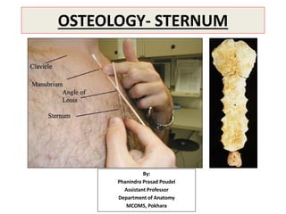
- 1. OSTEOLOGY- STERNUM By: Phanindra Prasad Poudel Assistant Professor Department of Anatomy MCOMS, Pokhara
- 2. STERNUM • Sternum (breast bone) is an elongated flat bone. • It lies in the anterior median part of the chest wall. • It is about 17 cm long.
- 3. • Sternum consists of 3 parts: 1. Manubrium (episternum) 2. Body (mesosternum) 3. Xiphoid process (metasternum)
- 4. • Sternum resembles a dagger or a small sword in shape. • Its 3 parts- manubrium, body and xiphoid process represent the handle, blade and point of the sword respectively.
- 5. • Upper part of sternum is broad and thick. • Lower part is thin and pointed. • Anterior surface is slightly rough and convex. • Posterior surface is smooth and slightly concave.
- 6. • Manubrium and body of sternum lie at an angle of 163° to each other. • Which increases slightly during inspiration and decreases during expiration. • Angle between long axis of manubrium and long axis of body of sternum is about 17°.
- 7. Anatomical Position of Sternum: • It is directed downwards and inclined slightly forward with its rough convex surface facing anteriorly. • Its broad end is directed upwards and lower pointed end is directed downwards.
- 8. MANUBRIUM (EPISTERNUM): • It is roughly quadrilateral in shape. • It lies opposite to the T4 & T5 vertebrae. • It is the thickest and strongest part of the sternum and presents the following features: • Two surfaces: – anterior and – posterior. • Four borders: – superior – Inferior and – lateral (right and left).
- 9. Anterior surface: • Provides attachment to: – sternal head of Sternocleidomastoid and – Pectoralis major.
- 10. Posterior surface: It is smooth and forms the anterior boundary of superior mediastinum. Attachments and relations: • Sternohyoid at the level of clavicular notch. • Sternothyroid at the level of facet for 1st costal cartilage. • Lower half is related to the arch of aorta. • Upper half is related to 3 branches of arch of aorta, viz. brachiocephalic artery, left common carotid artery, left subclavian artery, and left brachiocephalic vein.
- 11. Upper border: • It is thick, rounded and concave. • It presents a notch called suprasternal notch or jugular notch. • It provides attachment to the interclavicular ligament. • Clavicular notch on either side of the suprasternal notch articulates with the clavicle to form sternoclavicular joint.
- 12. Lateral border: • It presents two articular facets: • Upper facet articulates with the 1st costal cartilage to form primary cartilaginous joint. • Lower demifacet along with other demifacet in the body of sternum articulates with the 2nd costal cartilage.
- 13. Lower border: • It articulates with the upper end of the body of sternum to form secondary cartilaginous joint called manubriosternal joint. • Manubrium makes a slight angle with the body at this junction called sternal angle or angle of Louis. • Lies at the level of lower border of T4. • It is recognized by the presence of a transverse ridge on the anterior aspect of the sternum.
- 14. II CLASS
- 15. BODY (MESOSTERNUM): • It is longer, narrower and thinner than the manubrium. • It is broadest at its lower end. • Upper end articulates with the manubrium at the sternal angle to form manubriosternal joint.
- 16. • Lower end articulates with the xiphoid process to form primary cartilaginous xiphisternal joint. • Anterior surface presents 3 faint transverse ridges indicating the lines of fusion of 4 small segments called sternebrae. • Anterior surface on each side gives origin to the pectoralis major muscle. • Posterior surface is smooth and slightly concave. • Lower part of posterior surface gives origin to sternocostalis muscle.
- 17. • On the right side of median plane, posterior surface is related to pleura, which separates it from the lung. • On the left side of median plane, upper half of the body is related to the pleura and lower half to the pericardium.
- 18. • Lateral border articulates with the 2nd–7th costal cartilages to form synovial joints. • 2nd costal cartilage articulates at the side of manubriosternal junction and 7th costal cartilage articulates at the xiphisternal junction.
- 19. XIPHOID PROCESS (METASTERNUM): • It is the lowest and smallest part of the sternum. • It varies greatly in size and shape. • It may be bifid or perforated. • Anterior surface provides insertion to the medial fibres of the rectus abdominis. • Posterior surface gives origin to the sternal fibres of the diaphragm. • Tip provides attachment to the upper end of linea alba.
- 20. Muscles attached on the anterior & posterior surfaces of sternum: • Muscles attached on the anterior surface: – Sternal head of sternocleidomastoid – Pectoralis major – Rectus abdominis • Muscles attached on the posterior surface: – Sternohyoid – Sternothyroid – Sternocostalis – Diaphragm (sternal fibres)
- 21. N.B. Features of interest at the sternal angle: • It can be felt as a transverse ridge on the sternum about 5 cm below the suprasternal notch. • Sternal angle is an important landmark for many anatomical events at this level, these are: • Articulation of second costal cartilage with sternum, hence this level is used for counting the ribs. • It lies at the level of intervertebral disc between T4 and T5 vertebrae.
- 22. • Horizontal plane passing through this level separates superior mediastinum from inferior mediastinum. • Ascending aorta ends at this level. • Arch of aorta begins and ends at this level. • Descending aorta begins at this level.
- 23. • Trachea bifurcates into right and left principal bronchi at this level. • Pulmonary trunk divides into right and left pulmonary arteries at this level. • Upper border of heart lies at this level. • Azygos vein arches over the root of right lung to end in the superior vena cava.
- 24. Sternal puncture: • Manubrium sterni is the preferred site for bone marrow aspiration because it is subcutaneous and readily accessible. • The bone marrow sample is required for hematological examination. • A thick needle is inserted into the upper part of manubrium to avoid injury to arch of aorta which lies behind the lower part. • Sternal puncture is not advisable in children because in them the plates of compact bone of sternum are very thin and if needle passes through the manubrium it will damage the arch of aorta and its branches, leading to fatal hemorrhage.
- 25. Mid-sternotomy: To gain access to the mediastinum for: • Surgical operations on heart and great blood vessels. • Sternum is often divided in the median plane called midsternotomy.
- 26. Funnel chest (pectus excavatum): • Chest is compressed anteroposteriorly • Sternum is pushed backward by the overgrowth of the ribs. • May compress the heart. Pigeon chest (pectus carinatum): • Chest is compressed from side-to- side. • Sternum projects forward and downward like a keel of a boat.
- 27. Sternal fracture: • It is common in automobile accidents. • When the driver’s chest is hit against the steering wheel, the sternum is often fractured at the sternal angle. • The backward displacement of fractured fragments may damage aorta, heart, or liver and cause severe bleeding which may prove fatal.
- 28. OSSIFICATION: • Sternum develops from two vertical cartilaginous plates (sternal plates), which fuse in the midline. • It ossifies from six double centers. – One for manubrium. – Four for body. – One for xiphoid process.
- 29. Appearance of ossification centers: • Ossification centers appear in descending order for different parts of sternum as follows: • Manubrium: 5th month • Body: – First sternebra: 6th month – Second sternebra: 7th month – Third sternebra: 8th month – Fourth sternebra: 9th month • Xiphoid process: 3rd year
- 30. Fusion: Fusion occurs as follows: • Fusion between sternal plates takes place from below upwards. • It begins at puberty and completed by 25 years. • Xiphoid process fuses with the body at the age of 40 years. • Manubrium does not fuse with the body throughout life.
- 31. Sternal foramen and cleft sternum: • Two sternal plates fuse in caudocranial direction. • Sometimes sternebrae fail to fuse in the midline, as a result defect occurs in the body of sternum in the form of sternal foramen or cleft sternum. • The cleft sternum is often associated with ectopia cordis.
- 32. ASSIGNMENT WITH IN 24 HOURS- MCQs 1. Sternal angle lies at which of the following vertebral level? a. Upper border of T4 b. Lower border of T4 c. Upper border of T5 d. Lower border of T5 2. Pick up a muscle which is attached on the posterior surface of the manubrium sterni. a. Sternothyroid b. Sternocleidomastoid c. Pectoralis major d. Pectoralis minor
- 33. 3. What is the time of completion of fusion between two sternal plates of the body of sternum? a. Puberty b. 23 years c. 24 years d. 25 years 4. Which part of the sternum is commonly used for the bone marrow aspiration? a. Upper part of manubrium b. Lower part of manubrium c. Upper part of body of sternum d. Lower part of body of sternum
- 34. HOME ASSIGNMENT: 1. Describe the features of different parts of sternum with the help of well labeled diagrams? 2. Write about the ossification and clinical anatomy related to the sternum. THE END