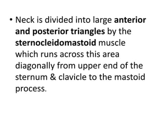
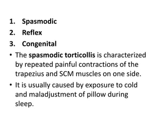
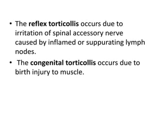
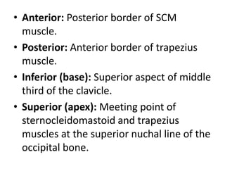
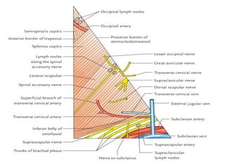
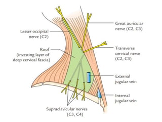
1. The posterior triangle is the triangular space behind the sternocleidomastoid muscle in the neck. It contains nerves, arteries, veins and lymph nodes.
2. The spinal accessory nerve and occipital artery pass through the upper part, while the subclavian artery and brachial plexus pass through the lower part.
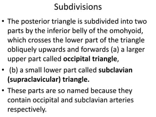
3. The posterior triangle is divided into an upper occipital triangle and lower subclavian triangle by the omohyoid muscle. Structures in each triangle and potential spaces are described.