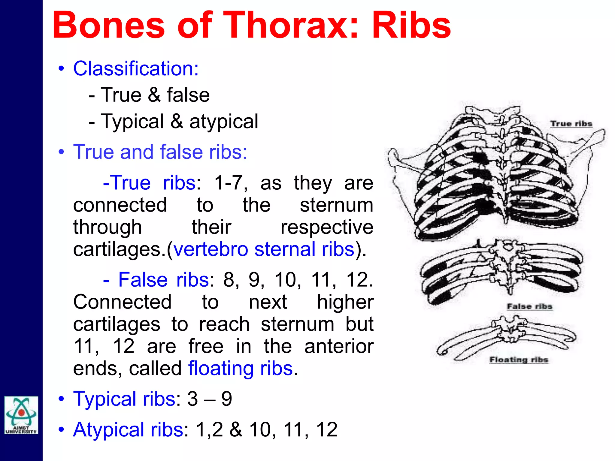
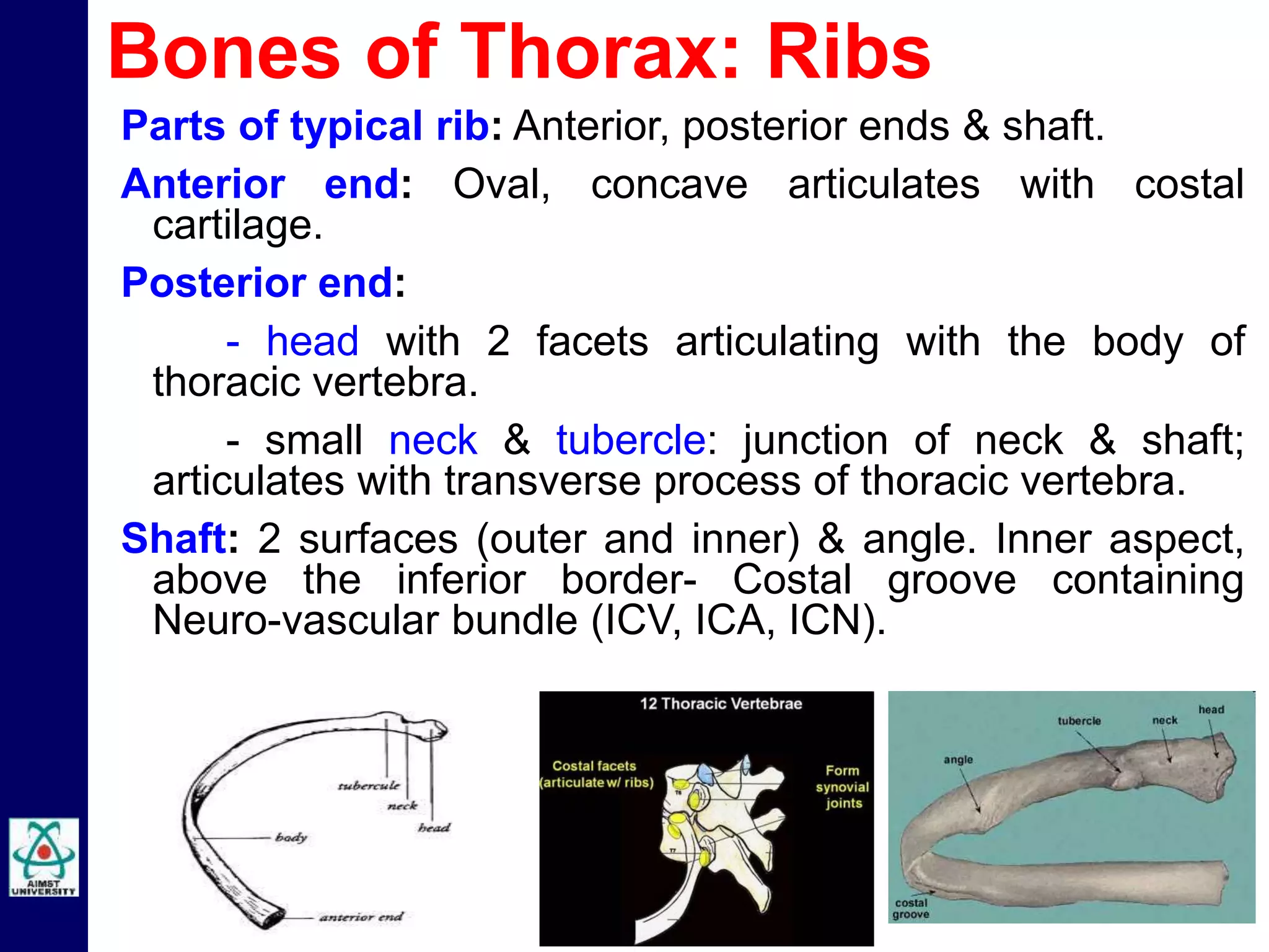
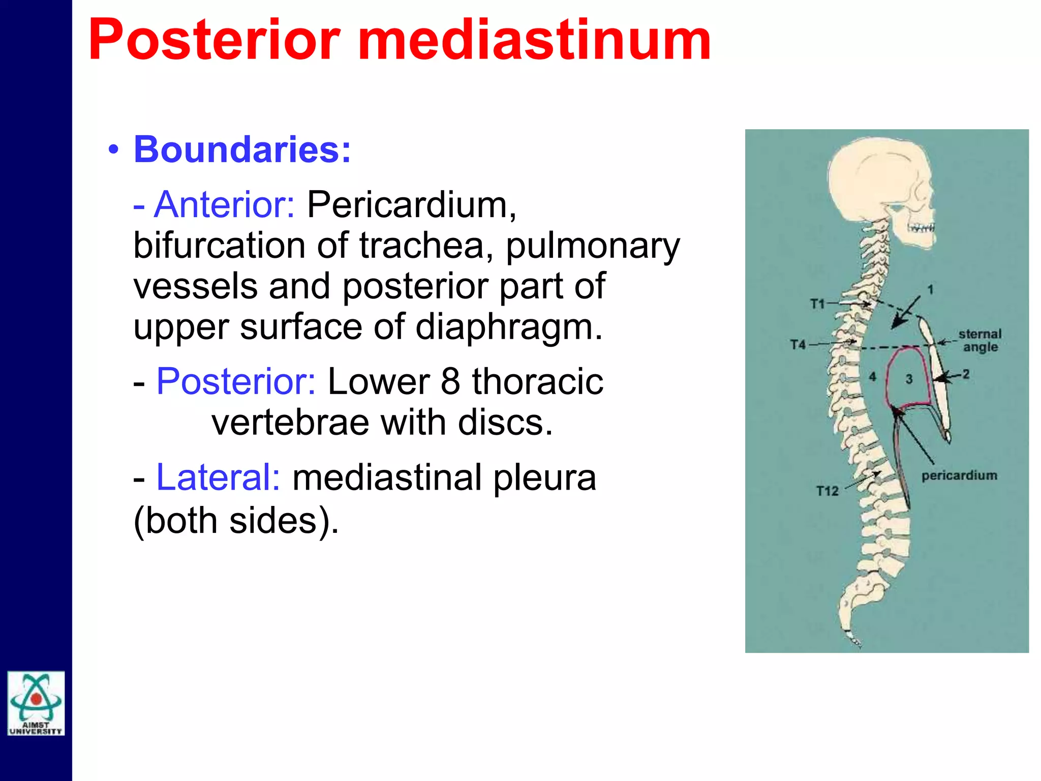
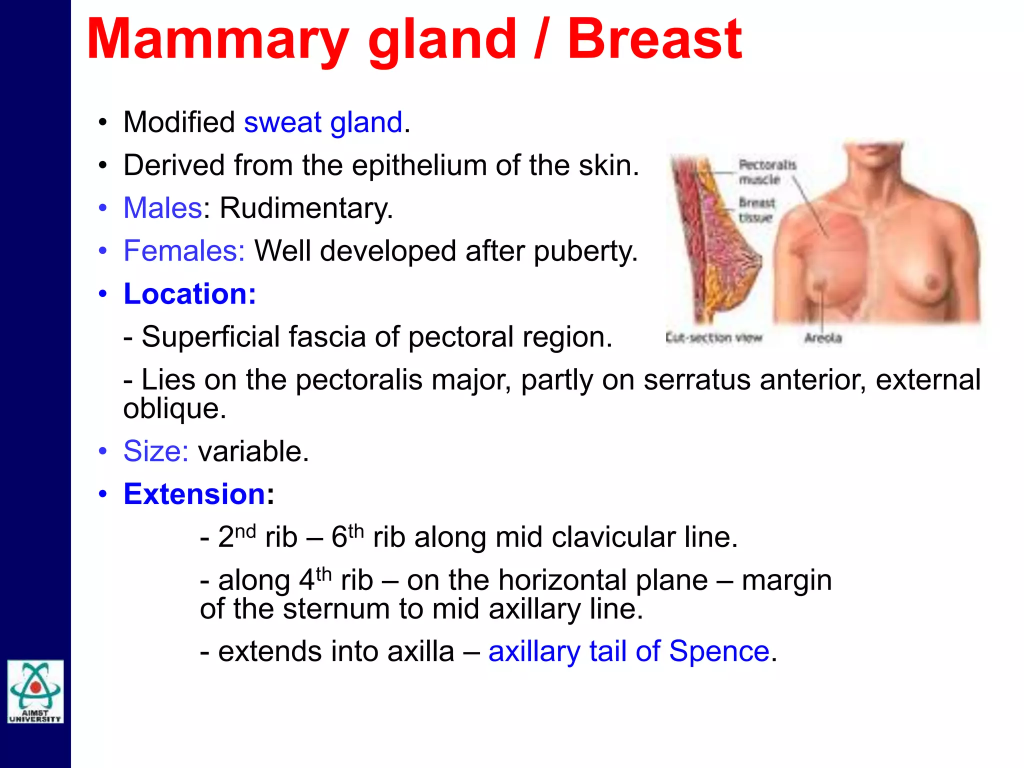
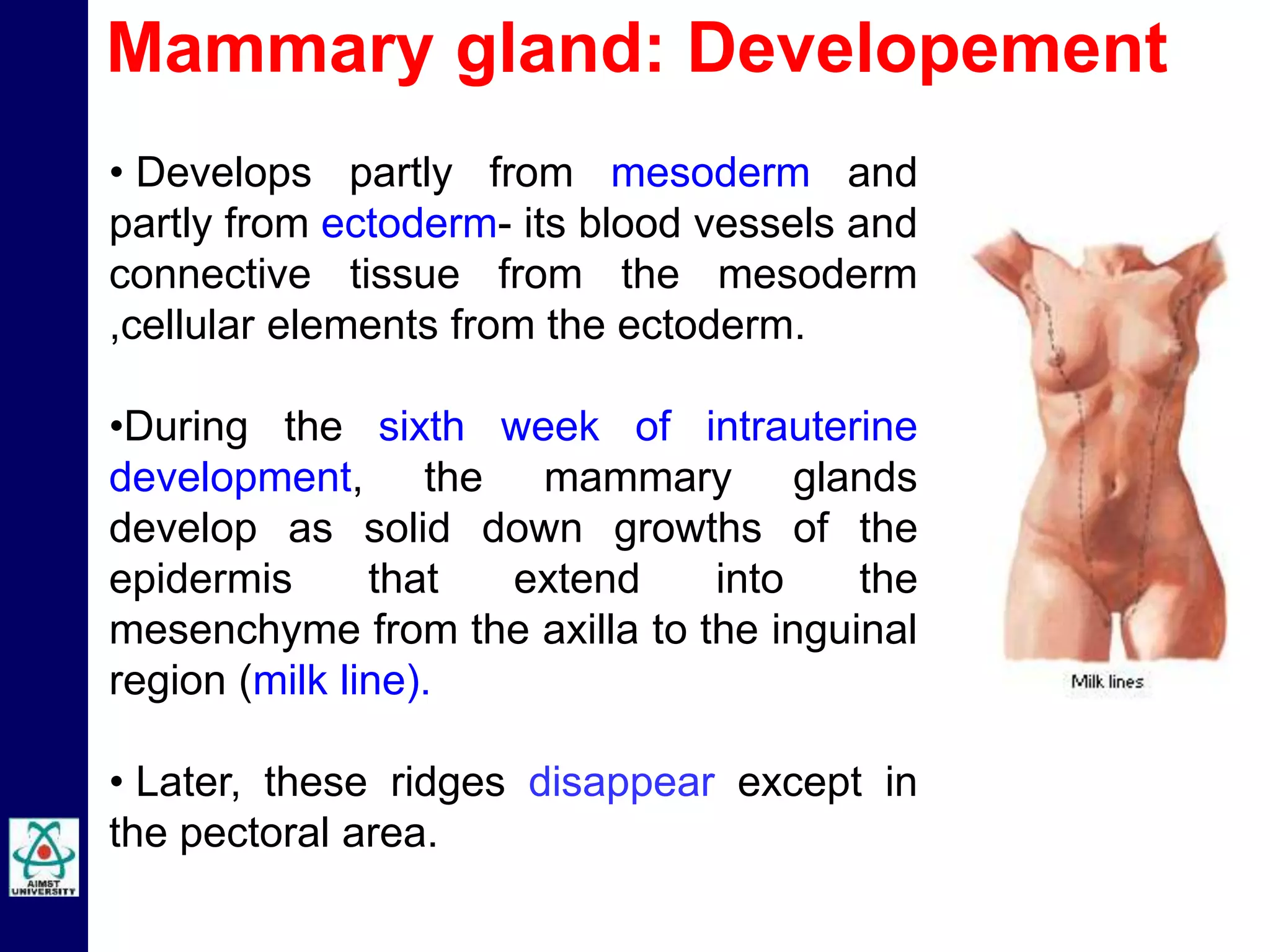
Dr S M Niazur Rahman
Anatomy Unit, Faculty of Medicine
L 18: THORAX
ANATOMY L 18: MBBS B 21: Element 1: 07.12.2015
Learning outcomes
• List and describe the bones and muscles of
thoracic cage and wall.
• Discuss the contents of intercostal space.
• Describe the boundaries,subdivisions and
contents of mediastinum.
• Explain the structure,blood supply and
lymphatic drainage of breast.
• Describe the origin, insertion, nerve supply,
major openings and action of diaphragm.
 Thoracic cage
 Thoracic wall
 Mediastinum
 Breast
 Diaphragm
Thorax
Thoracic cage
Made up of bones
• Ribs – 12 pairs.
• Sternum – manubrium,
body & xiphoid.
• Vertebral column – 12
thoracic vertebrae.
• Classification:
- True & false
- Typical & atypical
• True and false ribs:
-True ribs: 1-7, as they are
connected to the sternum
through their respective
cartilages.(vertebro sternal ribs).
- False ribs: 8, 9, 10, 11, 12.
Connected to next higher
cartilages to reach sternum but
11, 12 are free in the anterior
ends, called floating ribs.
• Typical ribs: 3 – 9
• Atypical ribs: 1,2 & 10, 11, 12
Bones of Thorax: Ribs
Parts of typical rib: Anterior, posterior ends & shaft.
Anterior end: Oval, concave articulates with costal
cartilage.
Posterior end:
- head with 2 facets articulating with the body of
thoracic vertebra.
- small neck & tubercle: junction of neck & shaft;
articulates with transverse process of thoracic vertebra.
Shaft: 2 surfaces (outer and inner) & angle. Inner aspect,
above the inferior border- Costal groove containing
Neuro-vascular bundle (ICV, ICA, ICN).
Bones of Thorax: Ribs
Parts of vertebra:
• Body: heart-shaped
• Vertebral or Neural arch:
- Pedicle
- Transverse process
- Lamina
- Spinous process and
- Articulating facets
-Vertebral canal for spinal cord
 12 in number: Typical- 2 to 8 &
Atypical- 1,9,10,11 & 12.
 Has articulating surfaces/
costal facet on the body and
transverse processes for the
ribs.
Bones of Thorax: Thoracic vertebra
• Sternum is a flat bone, present in front of
the thoracic cage.
• Parts: Manubrium, Body & Xiphoid
process.
• Manubrium:
- Upper part of the sternum.
- Joins with medial end of clavicle and first
costal cartilage.
-Has jugular or interclavicular or
suprasternal notch.
Body : Larger part of the sternum. On each
side, it receives ribs through their costal
cartilages (3rd to 6th rib).
Xiphoid process: Most variable part of the
sternum. Cartilaginous structure in adults.
Bones of Thorax: Sternum
Thoracic Cage: Applied Anatomy
Funnel chest: Lower end of
sternum is depressed and xiphoid
process projects forwards.
Rickets: Pigeon chest associated
with lack of calcification in the costal
cartilages at the costo-chondral
junction.
Barrel shaped chest: In chronic
obstructive pulmonary disease
(COPD)- asthma & emphysema.
Funnel chest
Barrel chest
Pigeon chest
• Thoracic cage is formed by bony frame work.
• Bones include sternum anteriorly,12 pairs of ribs laterally
& posteriorly and 12 thoracic vertebrae posteriorly.
• Sternum has manubrium,body & xiphoid process.
• Ribs have head, neck & shaft.
• Vertebra has body, pedicle, transverse process and
spine.
Thoracic Cage: Summary
Thoracic Wall
• Skeletal frame work.
• Intercostal spaces filled with
intercostal muscles, intercostal
nerves, arteries and veins.
• Intercostal muscles:
1.External intercostal
2.Internal intercostal
3.Transversus thoracis : 3 parts
- Subcostalis,
- Intercostalis intimi
- Sternocostalis.
1. External intercostal muscles:
• Extends from the tubercle of rib
posteriorly to costo chondral
junction anteriorly.
• Between costochondral junction
and the sternum the muscle is
replaced by external intercostal
membrane.
• Origin:From the lower border of
rib.
• Insertion:Into the upper border of
the rib.
• Direction of fibres- downwards,
forwards and medially.
Thoracic Wall: Intercostal muscles
2. Internal intercostal muscle:
• Extends from the lateral
border of sternum to angle of
rib. Beyond the angle the
muscle is replaced by
posterior or internal
intercostal membrane.
• Origin: From the lower border
of rib.
• Insertion: Into the upper
border of the rib.
• Direction of fibres-
downwards, backwards and
laterally at right angles to the
external intercostalis.
Thoracic Wall: Intercostal muscles
3. Transversus thoracis: 3 parts-
- Sternocostalis – present in the
anterior parts of upper intercostal
spaces.
- Subcostalis - confined to the
posterior part of lower intercostal
spaces.
- Intercostalis intimi or innermost
intercostal: present in the middle
third of the intercostal spaces.
Direction of fibres is similar to
internal intercostal.
Thoracic Wall: Intercostal muscles
• Nerve supply:
Intercostal nerves.
• Action:
- Prevent retraction of intercostal spaces during
expiration, bulging outwards during inspiration.
- Portion of external & internal intercostal
muscles elevate the ribs during inspiration.
- Internal intercostal and transverse thoracis
depress the ribs during expiration.
Thoracic Wall: Intercostal muscles
• Neurovascular bundle consists of intercostal vein,
intercostal artery and intercostal nerve (VAN).
• Neurovascular plane lying between the internal intercostal
and innermost intercostal muscles.
Thoracic Wall: Intercostal spaces
• 11 pairs of intercostal and 1
pair of subcostal nerves.
• Origin:Ventral rami of
thoracic spinal nerves.
• Course:Passes from
posterior to anterior in the
costal groove in the
neurovascular plane.
• Branches:
1. Lateral cutaneous
2. Anterior cutaneous
3. Muscular branches.
Thoracic Wall: Intercostal nerve
• Each intercostal space
contains 1 posterior & 2
anterior intercostal arteries.
• Posterior intercostal arteries
arise from costocervical
artery and thoracic aorta.
• Anterior intercostal arteries
arise from internal thoracic
artery.
Thoracic Wall: Intercostal artery
• Each intercostal space
contains 1 posterior and 2
anterior intercostal veins.
• Posterior intercostal veins
drain into:
- Right side -right
brachiocephalic vein
and azygos vein.
- Left side - left
brachiocephalic vein,
hemiazygos and accessory
hemiazygos vein.
• Anterior intercostal veins
drain into internal thoracic
vein.
Thoracic Wall: Intercostal vein
• Paracentesis thoracis-removal
of fluid from the pleural cavity.
• Needle is inserted into the
middle of the intercostal space.
• Intercostal nerve block at the
intercostal groove.
• Pus from the thoracic bodies
tracks along the neurovascular
bundle and point of termination
of the intercostal nerve.
• Herpes Zoster – viral disease,
affects thoracic ganglia leading
painful vesicles all along the
intercostal nerve distribution.
Thoracic Wall: Applied Anatomy
Mediastinum
• Septum or space between
the two lungs in the thoracic
cavity.
• Boundaries:
Anterior: sternum.
Posterior: Thoracic vertebral
column.
Superior: Thoracic inlet.
Inferior: Diaphragm.
Lateral: Mediastinal pleura
(both sides).
Mediastinum
Mediastinum
• Divisions: imaginary line
passing at sternal angle -T4
divides it into superior & inferior
mediastinum
• Inferior mediastinum is further
divided into:
- Anterior: Area in front of
pericardium.
- Middle: Area with pericardium
& its contents.
- Posterior: Area behind
pericardium.
Boundaries –
Anterior: Manubrium sterni.
Posterior: T4 upper portion.
Superior: Thoracic inlet.
Inferior: Imaginary line at sternal
angle – T4 behind.
Superior mediastinum
Contents:
• Trachea & oesophagus.
• Muscles: sternohyoid, sternothyroid.
• Arteries: Arch of aorta, brachiocephalic artery, left
common carotid artery, left subclavian artery.
• Veins: Right & left brachiocephalic veins, SVC (upper
part), left superior intercostal vein.
• Nerves: Vagus, phrenic, cardiac N (both sides),left
recurrent laryngeal nerve.
• Thymus.
• Thoracic duct .
• Lymph nodes: Paratracheal, brachiocephalic and
tracheobronchial.
Superior mediastinum
• Narrow space in front of pericardium,
overlapped by thin anterior borders of lung.
• Continuous with pretracheal space of the
neck.
• Boundaries:
Anterior: Body of sternum.
Posterior: Pericardium.
Superior: Imaginary plane separating the
superior from inferior mediastinum.
Inferior: Superior surface of diaphragm.
Lateral : Mediastinal pleura (both sides)
• Contents: sternopericardial ligaments,
lymph nodes, branches of internal thoracic
artery, lower part of thymus.
Anterios mediastinum
• Pericardium and contents along with
phrenic nerve, percardiophrenic
vessels.
• Contents:
- Heart enclosed with pericardium.
- Arteries: ascending aorta, pulmonary
trunk and two pulmonary arteries.
- Veins: lower part SVC, terminal part of
azygous vein, right & left
pulmonary veins.
- Nerves: phrenic and deep cardiac
plexus.
- Tubes: bifurcation of trachea, right &
left principal bronchi.
Middle mediastinum
• Boundaries:
- Anterior: Pericardium,
bifurcation of trachea, pulmonary
vessels and posterior part of
upper surface of diaphragm.
- Posterior: Lower 8 thoracic
vertebrae with discs.
- Lateral: mediastinal pleura
(both sides).
Posterior mediastinum
• Contents:
- Oesophagus.
- Arteries: Descending thoracic aorta &
branches.
- Veins: Azygos vein, hemiazygos vein,
Accessory hemiazygos vein.
- Nerves: Vagus, splanchnic, greater & lesser
and least arising from lower 8 thoracic ganglia
of the sympathetic chain.
- Lymph nodes and lymphatics: Posterior
mediastinal nodes along aorta, thoracic duct.
Posterior mediastinum
• Modified sweat gland.
• Derived from the epithelium of the skin.
• Males: Rudimentary.
• Females: Well developed after puberty.
• Location:
- Superficial fascia of pectoral region.
- Lies on the pectoralis major, partly on serratus anterior, external
oblique.
• Size: variable.
• Extension:
- 2nd rib – 6th rib along mid clavicular line.
- along 4th rib – on the horizontal plane – margin
of the sternum to mid axillary line.
- extends into axilla – axillary tail of Spence.
Mammary gland / Breast
• Nipple is the conical elevation.
• Areola is pigmented area
around the nipple – has
modified sebaceous glands,
which enlarge during
pregnancy – tubercles of
Montgomery that lubricate the
breast during breast feeding.
• Suspensory ligament of
Cooper - fibrous tissue
extends from the skin to the
underlying fascia covering the
muscle –becomes weak in
elderly female and makes it
pendulous (sagging).
Mammary gland / Breast
Internal structure:
• 15-20 lobes separated by a
fibrous connective tissue.
• Each lobe drained by –
lactiferous duct.
- ducts from each lobe converge
to open at the summit of
the nipple.
- lactiferous ducts show a
dilatation called – lactiferous
sinus.
Mammary gland / Breast
• Arterial supply:
1.Superior thoracic artery- branch of axillary artery –
supplies upper part of the breast.
2.Lateral thoracic artery – branch of axillary artery –
supplies lateral part of the breast.
3. Internal thoracic artery – branch of subclavian
artery – with their medial mammary branch supplies
medial part of the breast .
4. Posterior intercostal artery – arise from thoracic
aorta – lateral part of the gland.
• Venous drainage: Veins drain into internal
thoacic,axillary and posterior intercostal veins.
Mammary gland / Breast
• Skin:
- Outer side – axillary group of nodes.
- Upper quadrant – supra clavicular nodes.
- Inner quadrant – internal mammary nodes.
• Parenchyma, nipple and areola:
- Accompany lateral thoracic artery –anterior set of
axillary nodes.
- Posterior sets (few) via axillary tail spence – efferents
to central and apical nodes.
- From medial side and lateral side - internal thoracic
artery – parasternal nodes.
- ateral and posterior side – posterior inter costal nodes.
Mammary gland: Lymphatic drainage
• Anterior and lateral cutaneous branches of the
fourth, fifth, and sixth thoracic nerves(T4,5,6).
• Sensation to the nipple derives largely from
the lateral cutaneous branch of T4.
Mammary gland: Nerve supply
• Develops partly from mesoderm and
partly from ectoderm- its blood vessels and
connective tissue from the mesoderm
,cellular elements from the ectoderm.
•During the sixth week of intrauterine
development, the mammary glands
develop as solid down growths of the
epidermis that extend into the
mesenchyme from the axilla to the inguinal
region (milk line).
• Later, these ridges disappear except in
the pectoral area.
Mammary gland: Developement
Mammary gland: Anomalies
Amastia (absence of breast) athelia (absence of nipple) and
may occur bilaterally or unilaterally.
Polymastia: Extra breast most commonly occur along the milk
line
Polythelia: Extra nipple
Gynecomastia is a benign and usually self-limited condition that
occurs in 50-60% of boys during early adolescence.
Gynecomastia occurs in Klinefelter syndrome, testicular
feminization, hormone-secreting tumors, hyperthyroidism,
hypothyroidism.
• Musculo-tendinous partition between
the thorax and abdomen.
Parts: vertebral, sternal and costal.
Origin:
Verterbral origin:
Right crus: L1,2,3
Left crus: L1,2
Joined by median arcuate ligament.
Costal origin: Inner aspects of lower
six costal cartilages.
Sternal origin: Posterior surface of
xiphoid process.
Insertion: All the 3 parts converge on
central tendon.
Action: Increases the vertical diameter
of the thoracic cavity in inspiration.
Diaphragm
• Nerve supply:
- Phrenic nerve (C3, 4, 5) –
motor and sensory to central
part.
- Lower six intercostal nerves –
sensory to peripheral part.
• Major openings in the
diaphragm and structures
passing through diaphragm:
- T12 - aorta, azygos vein, and
thoracic duct.
- T10 - oesophagus, branches of
left gastric artery,vein and vagul
trunks.
- T8 – inferior vena cava, right
phrenic nerve.
Diaphragm
Suggested/Further Reading:
1. Essentials of Anatomy-A K Datta (part 1 & 3)
2. B D Chaurasia’s Human Anatomy- volume 1
Anatomy of the Thorax by Dr Niazur Rahman

Anatomy of the Thorax by Dr Niazur Rahman

  • 1.
    Dr S MNiazur Rahman Anatomy Unit, Faculty of Medicine L 18: THORAX ANATOMY L 18: MBBS B 21: Element 1: 07.12.2015
  • 2.
    Learning outcomes • Listand describe the bones and muscles of thoracic cage and wall. • Discuss the contents of intercostal space. • Describe the boundaries,subdivisions and contents of mediastinum. • Explain the structure,blood supply and lymphatic drainage of breast. • Describe the origin, insertion, nerve supply, major openings and action of diaphragm.
  • 3.
     Thoracic cage Thoracic wall  Mediastinum  Breast  Diaphragm Thorax
  • 4.
    Thoracic cage Made upof bones • Ribs – 12 pairs. • Sternum – manubrium, body & xiphoid. • Vertebral column – 12 thoracic vertebrae.
  • 5.
    • Classification: - True& false - Typical & atypical • True and false ribs: -True ribs: 1-7, as they are connected to the sternum through their respective cartilages.(vertebro sternal ribs). - False ribs: 8, 9, 10, 11, 12. Connected to next higher cartilages to reach sternum but 11, 12 are free in the anterior ends, called floating ribs. • Typical ribs: 3 – 9 • Atypical ribs: 1,2 & 10, 11, 12 Bones of Thorax: Ribs
  • 6.
    Parts of typicalrib: Anterior, posterior ends & shaft. Anterior end: Oval, concave articulates with costal cartilage. Posterior end: - head with 2 facets articulating with the body of thoracic vertebra. - small neck & tubercle: junction of neck & shaft; articulates with transverse process of thoracic vertebra. Shaft: 2 surfaces (outer and inner) & angle. Inner aspect, above the inferior border- Costal groove containing Neuro-vascular bundle (ICV, ICA, ICN). Bones of Thorax: Ribs
  • 7.
    Parts of vertebra: •Body: heart-shaped • Vertebral or Neural arch: - Pedicle - Transverse process - Lamina - Spinous process and - Articulating facets -Vertebral canal for spinal cord  12 in number: Typical- 2 to 8 & Atypical- 1,9,10,11 & 12.  Has articulating surfaces/ costal facet on the body and transverse processes for the ribs. Bones of Thorax: Thoracic vertebra
  • 8.
    • Sternum isa flat bone, present in front of the thoracic cage. • Parts: Manubrium, Body & Xiphoid process. • Manubrium: - Upper part of the sternum. - Joins with medial end of clavicle and first costal cartilage. -Has jugular or interclavicular or suprasternal notch. Body : Larger part of the sternum. On each side, it receives ribs through their costal cartilages (3rd to 6th rib). Xiphoid process: Most variable part of the sternum. Cartilaginous structure in adults. Bones of Thorax: Sternum
  • 9.
    Thoracic Cage: AppliedAnatomy Funnel chest: Lower end of sternum is depressed and xiphoid process projects forwards. Rickets: Pigeon chest associated with lack of calcification in the costal cartilages at the costo-chondral junction. Barrel shaped chest: In chronic obstructive pulmonary disease (COPD)- asthma & emphysema. Funnel chest Barrel chest Pigeon chest
  • 10.
    • Thoracic cageis formed by bony frame work. • Bones include sternum anteriorly,12 pairs of ribs laterally & posteriorly and 12 thoracic vertebrae posteriorly. • Sternum has manubrium,body & xiphoid process. • Ribs have head, neck & shaft. • Vertebra has body, pedicle, transverse process and spine. Thoracic Cage: Summary
  • 11.
    Thoracic Wall • Skeletalframe work. • Intercostal spaces filled with intercostal muscles, intercostal nerves, arteries and veins. • Intercostal muscles: 1.External intercostal 2.Internal intercostal 3.Transversus thoracis : 3 parts - Subcostalis, - Intercostalis intimi - Sternocostalis.
  • 12.
    1. External intercostalmuscles: • Extends from the tubercle of rib posteriorly to costo chondral junction anteriorly. • Between costochondral junction and the sternum the muscle is replaced by external intercostal membrane. • Origin:From the lower border of rib. • Insertion:Into the upper border of the rib. • Direction of fibres- downwards, forwards and medially. Thoracic Wall: Intercostal muscles
  • 13.
    2. Internal intercostalmuscle: • Extends from the lateral border of sternum to angle of rib. Beyond the angle the muscle is replaced by posterior or internal intercostal membrane. • Origin: From the lower border of rib. • Insertion: Into the upper border of the rib. • Direction of fibres- downwards, backwards and laterally at right angles to the external intercostalis. Thoracic Wall: Intercostal muscles
  • 14.
    3. Transversus thoracis:3 parts- - Sternocostalis – present in the anterior parts of upper intercostal spaces. - Subcostalis - confined to the posterior part of lower intercostal spaces. - Intercostalis intimi or innermost intercostal: present in the middle third of the intercostal spaces. Direction of fibres is similar to internal intercostal. Thoracic Wall: Intercostal muscles
  • 15.
    • Nerve supply: Intercostalnerves. • Action: - Prevent retraction of intercostal spaces during expiration, bulging outwards during inspiration. - Portion of external & internal intercostal muscles elevate the ribs during inspiration. - Internal intercostal and transverse thoracis depress the ribs during expiration. Thoracic Wall: Intercostal muscles
  • 16.
    • Neurovascular bundleconsists of intercostal vein, intercostal artery and intercostal nerve (VAN). • Neurovascular plane lying between the internal intercostal and innermost intercostal muscles. Thoracic Wall: Intercostal spaces
  • 17.
    • 11 pairsof intercostal and 1 pair of subcostal nerves. • Origin:Ventral rami of thoracic spinal nerves. • Course:Passes from posterior to anterior in the costal groove in the neurovascular plane. • Branches: 1. Lateral cutaneous 2. Anterior cutaneous 3. Muscular branches. Thoracic Wall: Intercostal nerve
  • 18.
    • Each intercostalspace contains 1 posterior & 2 anterior intercostal arteries. • Posterior intercostal arteries arise from costocervical artery and thoracic aorta. • Anterior intercostal arteries arise from internal thoracic artery. Thoracic Wall: Intercostal artery
  • 19.
    • Each intercostalspace contains 1 posterior and 2 anterior intercostal veins. • Posterior intercostal veins drain into: - Right side -right brachiocephalic vein and azygos vein. - Left side - left brachiocephalic vein, hemiazygos and accessory hemiazygos vein. • Anterior intercostal veins drain into internal thoracic vein. Thoracic Wall: Intercostal vein
  • 20.
    • Paracentesis thoracis-removal offluid from the pleural cavity. • Needle is inserted into the middle of the intercostal space. • Intercostal nerve block at the intercostal groove. • Pus from the thoracic bodies tracks along the neurovascular bundle and point of termination of the intercostal nerve. • Herpes Zoster – viral disease, affects thoracic ganglia leading painful vesicles all along the intercostal nerve distribution. Thoracic Wall: Applied Anatomy
  • 21.
  • 22.
    • Septum orspace between the two lungs in the thoracic cavity. • Boundaries: Anterior: sternum. Posterior: Thoracic vertebral column. Superior: Thoracic inlet. Inferior: Diaphragm. Lateral: Mediastinal pleura (both sides). Mediastinum
  • 23.
    Mediastinum • Divisions: imaginaryline passing at sternal angle -T4 divides it into superior & inferior mediastinum • Inferior mediastinum is further divided into: - Anterior: Area in front of pericardium. - Middle: Area with pericardium & its contents. - Posterior: Area behind pericardium.
  • 24.
    Boundaries – Anterior: Manubriumsterni. Posterior: T4 upper portion. Superior: Thoracic inlet. Inferior: Imaginary line at sternal angle – T4 behind. Superior mediastinum
  • 25.
    Contents: • Trachea &oesophagus. • Muscles: sternohyoid, sternothyroid. • Arteries: Arch of aorta, brachiocephalic artery, left common carotid artery, left subclavian artery. • Veins: Right & left brachiocephalic veins, SVC (upper part), left superior intercostal vein. • Nerves: Vagus, phrenic, cardiac N (both sides),left recurrent laryngeal nerve. • Thymus. • Thoracic duct . • Lymph nodes: Paratracheal, brachiocephalic and tracheobronchial. Superior mediastinum
  • 26.
    • Narrow spacein front of pericardium, overlapped by thin anterior borders of lung. • Continuous with pretracheal space of the neck. • Boundaries: Anterior: Body of sternum. Posterior: Pericardium. Superior: Imaginary plane separating the superior from inferior mediastinum. Inferior: Superior surface of diaphragm. Lateral : Mediastinal pleura (both sides) • Contents: sternopericardial ligaments, lymph nodes, branches of internal thoracic artery, lower part of thymus. Anterios mediastinum
  • 27.
    • Pericardium andcontents along with phrenic nerve, percardiophrenic vessels. • Contents: - Heart enclosed with pericardium. - Arteries: ascending aorta, pulmonary trunk and two pulmonary arteries. - Veins: lower part SVC, terminal part of azygous vein, right & left pulmonary veins. - Nerves: phrenic and deep cardiac plexus. - Tubes: bifurcation of trachea, right & left principal bronchi. Middle mediastinum
  • 28.
    • Boundaries: - Anterior:Pericardium, bifurcation of trachea, pulmonary vessels and posterior part of upper surface of diaphragm. - Posterior: Lower 8 thoracic vertebrae with discs. - Lateral: mediastinal pleura (both sides). Posterior mediastinum
  • 29.
    • Contents: - Oesophagus. -Arteries: Descending thoracic aorta & branches. - Veins: Azygos vein, hemiazygos vein, Accessory hemiazygos vein. - Nerves: Vagus, splanchnic, greater & lesser and least arising from lower 8 thoracic ganglia of the sympathetic chain. - Lymph nodes and lymphatics: Posterior mediastinal nodes along aorta, thoracic duct. Posterior mediastinum
  • 30.
    • Modified sweatgland. • Derived from the epithelium of the skin. • Males: Rudimentary. • Females: Well developed after puberty. • Location: - Superficial fascia of pectoral region. - Lies on the pectoralis major, partly on serratus anterior, external oblique. • Size: variable. • Extension: - 2nd rib – 6th rib along mid clavicular line. - along 4th rib – on the horizontal plane – margin of the sternum to mid axillary line. - extends into axilla – axillary tail of Spence. Mammary gland / Breast
  • 31.
    • Nipple isthe conical elevation. • Areola is pigmented area around the nipple – has modified sebaceous glands, which enlarge during pregnancy – tubercles of Montgomery that lubricate the breast during breast feeding. • Suspensory ligament of Cooper - fibrous tissue extends from the skin to the underlying fascia covering the muscle –becomes weak in elderly female and makes it pendulous (sagging). Mammary gland / Breast
  • 32.
    Internal structure: • 15-20lobes separated by a fibrous connective tissue. • Each lobe drained by – lactiferous duct. - ducts from each lobe converge to open at the summit of the nipple. - lactiferous ducts show a dilatation called – lactiferous sinus. Mammary gland / Breast
  • 33.
    • Arterial supply: 1.Superiorthoracic artery- branch of axillary artery – supplies upper part of the breast. 2.Lateral thoracic artery – branch of axillary artery – supplies lateral part of the breast. 3. Internal thoracic artery – branch of subclavian artery – with their medial mammary branch supplies medial part of the breast . 4. Posterior intercostal artery – arise from thoracic aorta – lateral part of the gland. • Venous drainage: Veins drain into internal thoacic,axillary and posterior intercostal veins. Mammary gland / Breast
  • 34.
    • Skin: - Outerside – axillary group of nodes. - Upper quadrant – supra clavicular nodes. - Inner quadrant – internal mammary nodes. • Parenchyma, nipple and areola: - Accompany lateral thoracic artery –anterior set of axillary nodes. - Posterior sets (few) via axillary tail spence – efferents to central and apical nodes. - From medial side and lateral side - internal thoracic artery – parasternal nodes. - ateral and posterior side – posterior inter costal nodes. Mammary gland: Lymphatic drainage
  • 35.
  • 36.
    • Anterior andlateral cutaneous branches of the fourth, fifth, and sixth thoracic nerves(T4,5,6). • Sensation to the nipple derives largely from the lateral cutaneous branch of T4. Mammary gland: Nerve supply
  • 37.
    • Develops partlyfrom mesoderm and partly from ectoderm- its blood vessels and connective tissue from the mesoderm ,cellular elements from the ectoderm. •During the sixth week of intrauterine development, the mammary glands develop as solid down growths of the epidermis that extend into the mesenchyme from the axilla to the inguinal region (milk line). • Later, these ridges disappear except in the pectoral area. Mammary gland: Developement
  • 38.
    Mammary gland: Anomalies Amastia(absence of breast) athelia (absence of nipple) and may occur bilaterally or unilaterally. Polymastia: Extra breast most commonly occur along the milk line Polythelia: Extra nipple Gynecomastia is a benign and usually self-limited condition that occurs in 50-60% of boys during early adolescence. Gynecomastia occurs in Klinefelter syndrome, testicular feminization, hormone-secreting tumors, hyperthyroidism, hypothyroidism.
  • 39.
    • Musculo-tendinous partitionbetween the thorax and abdomen. Parts: vertebral, sternal and costal. Origin: Verterbral origin: Right crus: L1,2,3 Left crus: L1,2 Joined by median arcuate ligament. Costal origin: Inner aspects of lower six costal cartilages. Sternal origin: Posterior surface of xiphoid process. Insertion: All the 3 parts converge on central tendon. Action: Increases the vertical diameter of the thoracic cavity in inspiration. Diaphragm
  • 40.
    • Nerve supply: -Phrenic nerve (C3, 4, 5) – motor and sensory to central part. - Lower six intercostal nerves – sensory to peripheral part. • Major openings in the diaphragm and structures passing through diaphragm: - T12 - aorta, azygos vein, and thoracic duct. - T10 - oesophagus, branches of left gastric artery,vein and vagul trunks. - T8 – inferior vena cava, right phrenic nerve. Diaphragm
  • 41.
    Suggested/Further Reading: 1. Essentialsof Anatomy-A K Datta (part 1 & 3) 2. B D Chaurasia’s Human Anatomy- volume 1