SlideShare a Scribd company logo
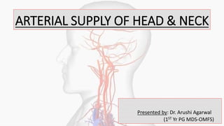
ARTERIAL SUPPLY OF HEAD & NECK
Presented by: Dr. Arushi Agarwal
(1ST Yr PG MDS-OMFS)
CONTENTS
• Artery- structure & function
• Clinical correlation
• Aorta
• Aortic Arch
• Subclavian artery
• Common carotid artery
• Carotid sinus & body
• Carotid sheath
• External carotid artery
• Internal carotid artery
• Ligation
ARTERY-STRUCTURE & FUNCTION
• Arteries are the blood vessels that deliver oxygen-rich blood from the heart to the tissues of the body
• Artery means ‘air tube’
• When rigor mortis passes over, liquid blood collects in dilated veins & arteries become empty*
Types:
• Conducting vessels : Large arteries (elastic)
• Distributing vessels : small arteries (muscular)
• Resistance vessels : arterioles
Terminal arterioles Meta arterioles
MICROSCOPIC STRUCTURE OF ARTERY
Made of 3 layers:
1. Tunica intima : made of endothelium, consisting of flattened cells & basal lamina, supported by internal
elastic lamina
2. Tunica media : thickest layer & made of alternate layers of circularly arranged smooth muscle fibers &
elastic fibers, supported by external elastic lamina
3. Tunica adventitia/externa : thin & strongest, made of longitudinally arranged connective tissue fibers &
cells
CLINICAL CORRELATION
• Arteriosclerosis: age related progressive degeneration of walls seen in middle age causing loss of elasticity &
thickening of tunica intima due to increase in collagen and lipid
• Atherosclerosis: plaques of lipid, fibrous tissue, macrophages called atheroma accumulate in tunica intima,
initiating clot formation (thrombus)
• Embolus is a thrombus which is dislodged from the wall of vessel and moves in the bloodstream
• Blood supply to arteries is by vasa vasorum & luminal blood thru diffusion
• Syphilitic aneurysm: vasa vasorum is involved in tertiary syphilis, leading to poor blood supply of wall of artery
leading to ischemic degeneration. Therefore, weakness leads to aneurysm.
• Arterial pulse: pulse is a palpable impulse of pressure wave of blood flow
initiated by ventricular systole.
AORTA
• Aorta-main & largest artery originating from left ventricle that carries blood away from heart to body.
• Blood leaves heart through aortic valve & travels through aorta, making a cane-shaped curve, allows
other major arteries to deliver oxygen-rich blood to brain, muscles & other cells.
• Segments of aorta:
1. Ascending aorta (supply the heart)
2. Aortic arch (supply arms, head & neck)
3. Descending aorta (supply chest)
4. Abdominal aorta (supply abdomen)
AORTIC ARCH
• 3 major branches:
1. Brachiocephalic trunk – supplies right side of head & neck, right arm & chest wall
a. Right subclavian b. Right common carotid
2. Left common carotid
3. Left subclavian artery
Supply left side of head & neck, arm & chest wall
SUBCLAVIAN ARTERY
• Found below the clavicle therefore named so
• Right subclavian: branch of brachiocephalic artery
• Left subclavian: direct branch of aorta
Course:
Arises posterior to sternoclavicular joint, travels lateral to trachea into root of neck.
Scalenus anterior muscles crosses the artery and divides it into 3 parts:
PARTS COMMENTS
1st Part • Extends from beginning of subclavian to medial border of anterior
scalene.
• Anteriorly related to CCA, vagus nerve, IJV, SCM, sternohyoid &
sternothyroid muscles.
• All branches arise from 1st part except left costocervical trunk.
2nd Part • Located posterior to anterior scalene & SCM
• Posterioinferiorly related to suprapleural memb. , cervical pleura & apex
of lung.
3rd Part • Located on lateral margin of anterior scalene m. to lateral border of 1st
rib where it gives of axillary a.
• BRANCHES OF FIRST PART OF SUBCLAVIAN ARTERY
BRANCH OF PART 1 COMMENTS
Vertebral • 1st and largest branch
• Ascends to enter foramen transversarium of C6
• Passes around atlas and foramen magnum to enter skull, unites with opposite vertebral
a. to form basilar a. supplying brain
Thyrocervical • Short & wide vessel
• Divides into 3 branches:
1. Inferior thyroid a. – travels along medial aspect of anterior scalene m. & posterior to
carotid sheath. Gives rise to inferior laryngeal a. to larynx & ascending cervical
supplying muscles in the area.
2. Suprascapular a. – travels inferior & laterally across anterior scalene m. & deep to
SCM m. , crosses posterior triangle to reach scapula.
3. Transverse cervical a. – travels across posterior triangle to reach anterior border of
trapezius m.
Right costocervical • Divides into 2 branches:
1. Deep cervical a. – travels superiorly along posterior of neck to supply muscles
2. Supreme intercostal a. – travles to supply 1st & 2nd intercostal spaces
Internal thoracic • Arises from inferior aspect of the artery
• Runs downwards & medially in front of cervical pleura & enters thorax by passing
behind 1st intercostal space
• BRANCHES OF SECOND & THIRD PART OF SUBCLAVIAN ARTERY
BRANCH COMMENTS
Left costocervical Branch of 2nd part of left subclavian a.
Dorsal scapular Branch of 2nd or 3rd part
Arises occasionally from subclavian a.
COMMON CAROTID ARTERY
COURSE:
• Rt common carotid – branch of brachiocephalic a. , begins in neck behind rt sternoclavicular joint.
• Lt common carotid – branch of arch of aorta, begins in the thorax in front of trachea. It ascends back to left
sternoclavicular joint & enters neck.
• Each artery runs upwards within carotid sheath under anterior border of SCM m.
• Bifurcates at superior border of thyroid cartilage of C3 into:
1. External carotid a.
2. Internal carotid a.
RELATIONS:
ANTERIOR:
Superior belly of omohyoid at level of cricoid cartilage
Below omohyoid it is covered by:
SCM, ant. Jugular vein, sternohyoid, sternothyroid, middle
thyroid v.
POSTERIOR:
Transverse process of vertebrae C4-8 & muscles (longus colli,
longus capitis, scalenus anterior)
Inferior thyroid a.
Vertebral a.
Thoracic duct
MEDIAL:
Thyroid gland
Larynx & pharynx, trachea, oesophagus, recurrent laryngeal n.
LATERAL:
IJV
• CAROTID SINUS
Termination of CCA shows a slight dilatation k/a carotid sinus. This region receives rich innervation from
glossopharyngeal (Hering’s n.) & sympathetic nerves.
Carotid sinus acts as a baroreceptor or pressure receptor & regulates blood pressure.
Carotid sinus syndrome (syncopal episodes due to inadvertant triggering of the carotid sinus) is a
pathology of the carotid sinus, in addition, carotid massage triggers the carotid sinus pathway
(increased pressure on carotid sinus due to massage → sends signal to decrease systemic BP)
• CAROTID BODY
Small, oval reddish brown structure situated behind the bifurcation of CCA. It receives rich nerve supply from
glossopharyngeal, vagus & sympathetic nerves
Carotid body acts as a chemoreceptor & responds to changes in Oxygen, Carbon dioxide & Ph of blood
CAROTID SHEATH
• Condensation of fibroaerolar tissue around main vessels of neck
• Formed on anterior aspect by pretracheal fascia & on posterior aspect by prevertebral fascia
• Contents: IJV, ICA, vagus n.
• Upper part of sheath has IX, XI, XII nerves. These nerves pierce along ECA
Relations:
1. Ansa cervicalis in anterior wall
2. Cervical sympathetic chain behind sheath
3. Sheath overlapped by anterior border of SCM & fused to layers of deep cervical fascia
EXTERNAL CAROTID ARTERY
• Gives rise to majority of branches in neck
• Arises from superior border of thyroid cartilage at C3
• Located anterior to carotid sheath & travels anteriorly &
superiorly in neck posterior to mandible & deep to posterior belly of
digastric & stylohyoid m. to enter parotid gland.
• In the carotid triangle, it is superficial, lies under anterior border
of SCM m., crossed by facial and hypoglossal nerves & facial,
lingual, superior thyroid veins. Deep to ECA lies wall of pharynx,
superior laryngeal nerve & ascending pharyngeal artery.
• Above the carotid triangle, it lies deep in substance of parotid gland.
Within gland it is related to retromandibular vein, facial nerve.
Deep to ECA lies, internal carotid a., styloglossus, stylopharyngeus,
9th nerve, styloid process. Others being, sup. laryngeal nerve &
sup. cervical sympathetic ganglia.
BRANCHES OF ECA:
ANTERIOR:
• Superior thyroid
• Lingual
• Facial
POSTERIOR:
• Occipital
• Posterior auricular
MEDIAL:
• Ascending pharyngeal
TERMINAL:
• Maxillary
• Superficial temporal
BRANCHES COMMENTS
ANTERIOR SUPERIOR THYROID • First branch
• Arises in carotid triangle below level of greater
cornua of hyoid
• Passes deep to infrahyoid m. to reach upper pole of
lateral lobe of thyroid
• Of importance in thyroid gland surgery as it runs
parallel to external laryngeal n. therefore to be
ligated.
• BRANCHES: superior laryngeal a.
sternocleidomastoid a.
cricothyroid a.
LINGUAL • Passes superiorly & medially towards greater
cornua of hyoid
• Course divided into 3 parts by hyoglossus m.
1. First: in carotid triangle, forms loop, helps in
free movement of hyoid bone
2. Second: deep to hyoglossus
3. Third: arteria profunda linguae, runs
upwards along anterior border of hyoglossus
& horizontally on undersurface of tongue.
Lies b/w geniglossus and inferior
longitudinal m.
• During surgical removal of tongue, it is
ligated
FACIAL • Arises above tip of greater cornua of hyoid
• Runs upwards in neck (cervical part) on superior
constrictor of pharynx deep to posterior belly of
digastric with stylohyoid to ramus
• Grooves along the posterior border of submandibular
salivary gland , makes S-bend
winding down over gland & then up over
base of mandible
• Facial part begins as it enters at anteroinferior angle
of masseter, runs upwards close to angle of mouth,
side of nose till medial angle of eye.
• BRANCHES: ascending palatine
superior & inferior labial
lateral nasal
angular
tonsillar
submental (supplies submental
triangle & sublingual gland)
branch to submandibular gland & lymph node
• Tortuous course – allows free movements of pharynx
during deglutition
on face allows free movement of mandible,
lips, cheeks for expressions
OCCIPITAL • Arises opposite to origin of facial a.
• Crossed by hypoglossal n.
• Runs backward & upward deep to lower border of
posterior belly of digastric , crossing carotid sheath
• Runs deep to mastoid process and muscles attached
to it
• BRANCHES: upper sternocleidomastoid
lower sternocleidomastoid
mastoid
meningeal
POSTERIOR AURICULAR • Arises from posterior aspect
• Runs upwards & backwards deep to parotid gland
but superficial to styloid process.
• Crosses base of mastoid process, ascends behind
auricle.
• Supplies back of auricle, skin over mastoid
process, back of scalp.
• Cut in incisions for mastoid operations.
• BRANCH: stylomastoid (enters stylomastoid
foramen & supplies middle ear & mastoid antrum,
air cells, semicircular canals and facial n.)
ASCENDING PHARYNGEAL
SUPERFICIAL TEMPORAL
• Arises from medial aspect of ECA
• Runs vertically upwards b/w side wall
of pharynx, tonsil, medial wall of
middle ear & auditory tube
• BRANCH: meningeal
• Begins behind neck of mandible
under cover of parotid gland
• Runs vertically upwards crossing root
of zygoma or preauricular point
where pulsation is felt
• About 5cm above zygoma it divides
into branches ton supply scalp and
temple
• BRANCHES: transverse facial
middle temporal artery
MAXILLARY ARTERY • Arises behind of neck of mandible
• Supplies: external & middle ear, auditory tube
dura mater
upper & lower jaws & teeth
muscles of temporal & infratemporal region
nose & paranasal air sinuses
palate
root of pharynx
COURSE & RELATIONS:
1st part: Mandibular, runs horizontally forward b/w neck
of mandible & sphenomandibular ligament below
auriculotemporal n. and then along lower border of
lateral pterygoid
2nd part: Pterygoid, runs upward and forward superficial
to lower head of lateral pterygoid
3rd part: Pterygopalatine, passes b/w two heads of
lateral pterygoid and thru pterygomaxillary fissure to
enter pterygopalatine fossa.
INTERNAL CAROTID ARTERY
• No branches of ICA in neck
• Passes superiorly in neck within carotid sheath along with IJV, & vagus n. anterior to
transverse processes of upper cervical vertebrae
• Principal artery of brain and eye, also supplies relates bones & meninges
• Carotid sinus located at the beginning of ICA
• Course divided into 4 parts:
1. Cervical part in neck
2. Petrous part in petrous temporal bone
3. Cavernous part in cavernous sinus
4. Cerebral part irt. base of skull
RELATIONS:
ANTERIOR:
• In carotid triangle, present at anterior border of SCM ,ECA lies anteromedially.
• Above carotid triangle, relates to posterior belly of digastric, stylohyoid, stylopharyngeus,
styloid process, parotid gland
POSTERIOR:
• Superior cervical ganglia
• Carotid sheath
• Glossopharyngeal, vagus, accessory & hypoglossal n.
MEDIAL:
• Pharynx
• ECA below parotid
LATERAL:
• IJV
• TMJ
SEGMENTS OF ICA:
• C1 – Cervical Segment
• C2 – Petrous Segment
• C3 – Lacerum Segment
• C4 – Cavernous Segment
• C5 – Clinoid Segment
• C6 – Ophthalmic (Supraclinoid) Segment
• C7 – Communicating (Terminal) Segment
BRANCHES OF ICA:
PETROUS PART:
• Caroticotympanic a.
• Pterygoid a.
CAVERNOUS PART:
• Hypophyseal a.
• Meningeal a.
• Ganglionic a.
CEREBRAL PART:
• Ophthalmic a.
• Anterior choroidal a.
• Posterior communicating a.
• Anterior cerebral a.
• Middle cerebral a.
• Anterior choroidal a.
• ICA enters cranial cavity after
traversing carotid canal &
superior aspect of foramen
lacerum
• It then courses thru cavernous
sinus, pierces dural roof of sinus
& ends immediately lateral to
optic chiasma & divides into
middle & anterior cerebral
arteries.
BRANCH COURSE SUB-BRANCHES
CAROTICOTYMPANIC Enters middle ear & anastomoses with ant. & post.
Tympanic a.
PTERYGOID Enters pterygoid canal with nerve of canal &
anastomoses with greater palatine n.
OPHTHALMIC Emerges from cavernous sinus on medial side of
anterior clinoid process close to optic canal.
Central a. of retina, post.
ciliary a., lacrimal, small
muscular branches etc.
ANTERIOR CHOROIDAL Passes posterolaterally, supplies crus cerebri. Turns to
medial aspect of temporal lobe to supply choroid
plexus of lateral ventricle
POSTERIOR COMMUNICATING Passes posterior to crus cerebri to join post. cerebral
a. & completes arterial circle
Crus cerebri, optic tract,
hypophysis, hypothalamus
ANTERIOR CEREBRAL Terminal branch. Runs above optic n., joined by
anterior communicating a.
Orbital, frontal, parietal
MIDDLE CEREBRAL Terminal branch. Lies in line with ICA. Frontal, parietal. Temporal,
deep branches
OPHTHALMIC ARTERY
• Emerges from cavernous sinus on medial side of anterior clinoid process close to optic canal.
• Artery enters orbit thru optic canal, lying inferolateral to otic n.
both artery & nerve lie in same dural sheath
• In orbit artery pierces dura mater, ascends over lateral side
of optic n. & crosses above nerve lateral to medial side along nasociliary n.
• It runs forward along medial wall of orbit b/w superior
oblique & medial rectus muscles.
• Terminates near medial angle of eye by dividing.
BRANCHES COMMENTS
Central artery of retina • First branch
• End artery
• Lies below optic n.
• Pierces dural sheath of nerve & runs forward for a short distance & enters substance of
n. to reach optic disc and divides into branches
• Obstruction can cause blindness
Lacrimal artery • Arises near optic foramen
• Follow lacrimal n. along superior border of lateral rectus m. of eye to reach & supply
lacrimal gland
• BRANCHES: zygomaticotemporal & zygomaticofacial (supplies respective region)
lateral palpebral (supplies eyelids)
recurrent meningeal
muscular branches
Supratrochlear • Exits orbit at medial angle accompanied by spratrochlear n.
• Supplies skin of forehead
Supraorbital • Passes medially to levator palpebrae superioris & superior rectus m.
• Passes thru supraorbital foramen and ascends towards scalp
• Supplies skin of forehead
BRANCHES COMMENTS
Posterior long & short • Supplies choroid & iris
Anterior ciliary • Supplies eyeball
Anterior ethmoidal • Travels thru ant. Ethmoidal canal
• Supplies anterior & middle ethmoidal air sinuses
• BRANCHES: meningeal
external nasal (supplies lateral nasal cartilage &
septum of nose)
Posterior ethmoidal • Travels thru post. Ethmoidal canal
• Supplies posterior ethmoidal sinuses
Medial palpebral • Arises near trochlea & exits orbit to pass along upper & lower eyelids
• Supplies face in the region
Dorsal nasal (infratrochlear) • Supplies upper part of nose
CIRCLE OF WILLIS
• Arterial circle situated at base of brain in the
interpeduncular fossa
• Comprises of :
Internal carotid artery
Anterior cerebral artery
Anterior communicating artery
Posterior communicating artery
Posterior cerebral artery
Vertebral artery
Basilar artery
The circle of Willis acts to provide collateral blood flow
between the anterior and posterior circulations of the
brain, protecting against ischemia in the event of vessel
disease or damage in one or more areas.
LIGATION OF ARTERIES
LIGATION OF FACIAL ARTERY
• exposed at point where it crosses lower border of mandible to pass from submandibular region into face
• Situated anterior to attachment of masseter, pulse can be felt.
• Runs irt. facial vein and marginal mandibular n., covered by platysma, subcutaneous tissue & skin.
LIGATION OF LINGUAL ARTERY
• Exposed in submandibular triangle, bounded by lower border of mandible & 2 bellies of
digastric m.
• Posterior corner of triangle is behind angle of mandible in communication with
retromandibular fossa.
LIGATION OF ECA
2 points where artery can be exposed and tied
1. Exposure of artery at its origin from common carotid a., ligature being placed
above origin of superior thyroid a
2. Ligation of ECA in carotid triangle , exposed behind angle of mandible
PROCEDURE
• With a blood vessel the surgeon will clamp the vessel perpendicular to the axis of the artery or vein
with a hemostat, then secure it by ligating it; i.e. using a piece of suture around it before dividing the
structure and releasing the hemostat.
REFERENCES
• Netter’s head and neck anatomy
• Oral anatomy by Sicher
• BD Chaurasia’s Human Anatomy
• Vishram singh – General Anatomy
ARTERIAL SUPPLY.pptx

More Related Content

What's hot

The pericardium
The pericardiumThe pericardium
The pericardium
Idris Siddiqui
 
Axillary artery and axillary vein
Axillary artery and axillary veinAxillary artery and axillary vein
Axillary artery and axillary vein
kamil khan
 
Vascular system in the head and neck
Vascular system in the head and neckVascular system in the head and neck
Vascular system in the head and neck
Hamzeh AlBattikhi
 
Scalp Anatomy
Scalp AnatomyScalp Anatomy
Head and neck anatomy
Head and neck anatomyHead and neck anatomy
Head and neck anatomy
Buttsa
 
Submandibular Region (Anatomy of the Neck)
Submandibular Region (Anatomy of the Neck)Submandibular Region (Anatomy of the Neck)
Submandibular Region (Anatomy of the Neck)
Dr. Sherif Fahmy
 
Vena cava anatomy and variants
Vena cava anatomy and variantsVena cava anatomy and variants
Vena cava anatomy and variants
Milan Silwal
 
Anatomy of temporal region
Anatomy of temporal regionAnatomy of temporal region
Anatomy of temporal region
Dr. Mohammad Mahmoud
 
Venous drainage of upper limb
Venous drainage of upper limbVenous drainage of upper limb
Venous drainage of upper limb
Dr. sana yaseen
 
Carotid Sheath
Carotid SheathCarotid Sheath
Carotid Sheath
Festo D Ngadaya
 
Arteries of Head and Neck
Arteries of Head and NeckArteries of Head and Neck
Arteries of Head and Neck
Himanshu Soni
 
Blood vessels and lymphatic of the thoracic wall
Blood vessels and lymphatic of the thoracic wallBlood vessels and lymphatic of the thoracic wall
Blood vessels and lymphatic of the thoracic wall
Dr. Mohammad Mahmoud
 
Head and neck blood vessels 2
Head and neck blood vessels 2Head and neck blood vessels 2
Head and neck blood vessels 2
mgmcri1234
 
posterior triangle of neck
posterior triangle of neck posterior triangle of neck
posterior triangle of neck
drhimanshuupadhyay
 
Tm joint, mandibular nerve
Tm joint, mandibular nerveTm joint, mandibular nerve
Tm joint, mandibular nerve
DrPratik Mistry
 
Mediastinum
MediastinumMediastinum
Mediastinum
Idris Siddiqui
 
Arteries of the head and neck
Arteries of the head and neckArteries of the head and neck
Arteries of the head and neck
Dr. Mohammad Mahmoud
 
Pericardium and heart
Pericardium and heartPericardium and heart
Pericardium and heart
Egoist el
 
Posterior abdominal wall
Posterior abdominal wallPosterior abdominal wall
Posterior abdominal wall
Suraj Dhara
 
Cranial fossa-SCALP
Cranial fossa-SCALPCranial fossa-SCALP
Cranial fossa-SCALP
Mosese HULKSTAH Tuapati JNR
 

What's hot (20)

The pericardium
The pericardiumThe pericardium
The pericardium
 
Axillary artery and axillary vein
Axillary artery and axillary veinAxillary artery and axillary vein
Axillary artery and axillary vein
 
Vascular system in the head and neck
Vascular system in the head and neckVascular system in the head and neck
Vascular system in the head and neck
 
Scalp Anatomy
Scalp AnatomyScalp Anatomy
Scalp Anatomy
 
Head and neck anatomy
Head and neck anatomyHead and neck anatomy
Head and neck anatomy
 
Submandibular Region (Anatomy of the Neck)
Submandibular Region (Anatomy of the Neck)Submandibular Region (Anatomy of the Neck)
Submandibular Region (Anatomy of the Neck)
 
Vena cava anatomy and variants
Vena cava anatomy and variantsVena cava anatomy and variants
Vena cava anatomy and variants
 
Anatomy of temporal region
Anatomy of temporal regionAnatomy of temporal region
Anatomy of temporal region
 
Venous drainage of upper limb
Venous drainage of upper limbVenous drainage of upper limb
Venous drainage of upper limb
 
Carotid Sheath
Carotid SheathCarotid Sheath
Carotid Sheath
 
Arteries of Head and Neck
Arteries of Head and NeckArteries of Head and Neck
Arteries of Head and Neck
 
Blood vessels and lymphatic of the thoracic wall
Blood vessels and lymphatic of the thoracic wallBlood vessels and lymphatic of the thoracic wall
Blood vessels and lymphatic of the thoracic wall
 
Head and neck blood vessels 2
Head and neck blood vessels 2Head and neck blood vessels 2
Head and neck blood vessels 2
 
posterior triangle of neck
posterior triangle of neck posterior triangle of neck
posterior triangle of neck
 
Tm joint, mandibular nerve
Tm joint, mandibular nerveTm joint, mandibular nerve
Tm joint, mandibular nerve
 
Mediastinum
MediastinumMediastinum
Mediastinum
 
Arteries of the head and neck
Arteries of the head and neckArteries of the head and neck
Arteries of the head and neck
 
Pericardium and heart
Pericardium and heartPericardium and heart
Pericardium and heart
 
Posterior abdominal wall
Posterior abdominal wallPosterior abdominal wall
Posterior abdominal wall
 
Cranial fossa-SCALP
Cranial fossa-SCALPCranial fossa-SCALP
Cranial fossa-SCALP
 

Similar to ARTERIAL SUPPLY.pptx

Subclavian vessels.pptx
Subclavian vessels.pptxSubclavian vessels.pptx
Subclavian vessels.pptx
Sundip Charmode
 
arterial supply of head and neck
arterial supply of head and neckarterial supply of head and neck
arterial supply of head and neck
shalini sampreethi
 
External carotid artery, branches and ligation
External carotid artery, branches and ligationExternal carotid artery, branches and ligation
External carotid artery, branches and ligation
benjamin Emmanuel
 
Blood supply of upper limb
Blood supply of upper limbBlood supply of upper limb
Blood supply of upper limb
Idris Siddiqui
 
Posterior triangle
Posterior trianglePosterior triangle
Posterior triangle
Chitransha03
 
External carotid artery
External carotid arteryExternal carotid artery
External carotid artery
Dr Rayan Malick
 
axilla.pptx
axilla.pptxaxilla.pptx
axilla.pptx
Atul Saini
 
Anterior traingle of neck -1.pptx
Anterior traingle of neck -1.pptxAnterior traingle of neck -1.pptx
Anterior traingle of neck -1.pptx
Sundip Charmode
 
Seminar Presentation On PMMC By Dr Abdul Qahar Qureshi
Seminar Presentation On PMMC By Dr Abdul Qahar QureshiSeminar Presentation On PMMC By Dr Abdul Qahar Qureshi
Seminar Presentation On PMMC By Dr Abdul Qahar Qureshi
Dr. Abdul Qahar Qureshi
 
Mediastinum.pptx
Mediastinum.pptxMediastinum.pptx
Mediastinum.pptx
Abhishek840813
 
Anatomy of the Thorax by Dr Niazur Rahman
Anatomy of the Thorax by Dr Niazur RahmanAnatomy of the Thorax by Dr Niazur Rahman
Anatomy of the Thorax by Dr Niazur Rahman
S M Niazur Rahman
 
Arterial supply of the head and neck
Arterial supply of the head and neckArterial supply of the head and neck
Arterial supply of the head and neck
Mahima Shanker
 
THORAX SCS.pptx1111111111111111111111111111
THORAX SCS.pptx1111111111111111111111111111THORAX SCS.pptx1111111111111111111111111111
THORAX SCS.pptx1111111111111111111111111111
marrahmohamed33
 
Axilla - 1.pptx
Axilla - 1.pptxAxilla - 1.pptx
Axilla - 1.pptx
Sundip Charmode
 
externalcarotidartery-presented-dr meher moin khan.pptx
externalcarotidartery-presented-dr meher moin khan.pptxexternalcarotidartery-presented-dr meher moin khan.pptx
externalcarotidartery-presented-dr meher moin khan.pptx
mehermoinkhan
 
Arteria venous and lymphatic drinage of head and neck basics
Arteria venous and lymphatic drinage of head and neck basicsArteria venous and lymphatic drinage of head and neck basics
Arteria venous and lymphatic drinage of head and neck basics
Manoj Kumar
 
Posterior triangle (1) Dental student / general anatomy .pdf
Posterior triangle (1) Dental student / general anatomy .pdfPosterior triangle (1) Dental student / general anatomy .pdf
Posterior triangle (1) Dental student / general anatomy .pdf
sanjeevani34
 
The Upper ggUpper limbs stuffAXILLA.pptx
The Upper ggUpper limbs stuffAXILLA.pptxThe Upper ggUpper limbs stuffAXILLA.pptx
The Upper ggUpper limbs stuffAXILLA.pptx
AlabiDavid4
 
Arteries of carotid triangle
Arteries of carotid triangleArteries of carotid triangle
Arteries of carotid triangle
GeethaHari3
 
Axillary fossa_12.7.23.pptx
Axillary fossa_12.7.23.pptxAxillary fossa_12.7.23.pptx
Axillary fossa_12.7.23.pptx
Nikolay Uzunov
 

Similar to ARTERIAL SUPPLY.pptx (20)

Subclavian vessels.pptx
Subclavian vessels.pptxSubclavian vessels.pptx
Subclavian vessels.pptx
 
arterial supply of head and neck
arterial supply of head and neckarterial supply of head and neck
arterial supply of head and neck
 
External carotid artery, branches and ligation
External carotid artery, branches and ligationExternal carotid artery, branches and ligation
External carotid artery, branches and ligation
 
Blood supply of upper limb
Blood supply of upper limbBlood supply of upper limb
Blood supply of upper limb
 
Posterior triangle
Posterior trianglePosterior triangle
Posterior triangle
 
External carotid artery
External carotid arteryExternal carotid artery
External carotid artery
 
axilla.pptx
axilla.pptxaxilla.pptx
axilla.pptx
 
Anterior traingle of neck -1.pptx
Anterior traingle of neck -1.pptxAnterior traingle of neck -1.pptx
Anterior traingle of neck -1.pptx
 
Seminar Presentation On PMMC By Dr Abdul Qahar Qureshi
Seminar Presentation On PMMC By Dr Abdul Qahar QureshiSeminar Presentation On PMMC By Dr Abdul Qahar Qureshi
Seminar Presentation On PMMC By Dr Abdul Qahar Qureshi
 
Mediastinum.pptx
Mediastinum.pptxMediastinum.pptx
Mediastinum.pptx
 
Anatomy of the Thorax by Dr Niazur Rahman
Anatomy of the Thorax by Dr Niazur RahmanAnatomy of the Thorax by Dr Niazur Rahman
Anatomy of the Thorax by Dr Niazur Rahman
 
Arterial supply of the head and neck
Arterial supply of the head and neckArterial supply of the head and neck
Arterial supply of the head and neck
 
THORAX SCS.pptx1111111111111111111111111111
THORAX SCS.pptx1111111111111111111111111111THORAX SCS.pptx1111111111111111111111111111
THORAX SCS.pptx1111111111111111111111111111
 
Axilla - 1.pptx
Axilla - 1.pptxAxilla - 1.pptx
Axilla - 1.pptx
 
externalcarotidartery-presented-dr meher moin khan.pptx
externalcarotidartery-presented-dr meher moin khan.pptxexternalcarotidartery-presented-dr meher moin khan.pptx
externalcarotidartery-presented-dr meher moin khan.pptx
 
Arteria venous and lymphatic drinage of head and neck basics
Arteria venous and lymphatic drinage of head and neck basicsArteria venous and lymphatic drinage of head and neck basics
Arteria venous and lymphatic drinage of head and neck basics
 
Posterior triangle (1) Dental student / general anatomy .pdf
Posterior triangle (1) Dental student / general anatomy .pdfPosterior triangle (1) Dental student / general anatomy .pdf
Posterior triangle (1) Dental student / general anatomy .pdf
 
The Upper ggUpper limbs stuffAXILLA.pptx
The Upper ggUpper limbs stuffAXILLA.pptxThe Upper ggUpper limbs stuffAXILLA.pptx
The Upper ggUpper limbs stuffAXILLA.pptx
 
Arteries of carotid triangle
Arteries of carotid triangleArteries of carotid triangle
Arteries of carotid triangle
 
Axillary fossa_12.7.23.pptx
Axillary fossa_12.7.23.pptxAxillary fossa_12.7.23.pptx
Axillary fossa_12.7.23.pptx
 

Recently uploaded

The Electrocardiogram - Physiologic Principles
The Electrocardiogram - Physiologic PrinciplesThe Electrocardiogram - Physiologic Principles
The Electrocardiogram - Physiologic Principles
MedicoseAcademics
 
Osteoporosis - Definition , Evaluation and Management .pdf
Osteoporosis - Definition , Evaluation and Management .pdfOsteoporosis - Definition , Evaluation and Management .pdf
Osteoporosis - Definition , Evaluation and Management .pdf
Jim Jacob Roy
 
CHEMOTHERAPY_RDP_CHAPTER 2 _LEPROSY.pdf1
CHEMOTHERAPY_RDP_CHAPTER 2 _LEPROSY.pdf1CHEMOTHERAPY_RDP_CHAPTER 2 _LEPROSY.pdf1
CHEMOTHERAPY_RDP_CHAPTER 2 _LEPROSY.pdf1
rishi2789
 
Dehradun #ℂall #gIRLS Oyo Hotel 8107221448 #ℂall #gIRL in Dehradun
Dehradun #ℂall #gIRLS Oyo Hotel 8107221448 #ℂall #gIRL in DehradunDehradun #ℂall #gIRLS Oyo Hotel 8107221448 #ℂall #gIRL in Dehradun
Dehradun #ℂall #gIRLS Oyo Hotel 8107221448 #ℂall #gIRL in Dehradun
chandankumarsmartiso
 
The Best Ayurvedic Antacid Tablets in India
The Best Ayurvedic Antacid Tablets in IndiaThe Best Ayurvedic Antacid Tablets in India
The Best Ayurvedic Antacid Tablets in India
Swastik Ayurveda
 
Journal Article Review on Rasamanikya
Journal Article Review on RasamanikyaJournal Article Review on Rasamanikya
Journal Article Review on Rasamanikya
Dr. Jyothirmai Paindla
 
NVBDCP.pptx Nation vector borne disease control program
NVBDCP.pptx Nation vector borne disease control programNVBDCP.pptx Nation vector borne disease control program
NVBDCP.pptx Nation vector borne disease control program
Sapna Thakur
 
Netter's Atlas of Human Anatomy 7.ed.pdf
Netter's Atlas of Human Anatomy 7.ed.pdfNetter's Atlas of Human Anatomy 7.ed.pdf
Netter's Atlas of Human Anatomy 7.ed.pdf
BrissaOrtiz3
 
Integrating Ayurveda into Parkinson’s Management: A Holistic Approach
Integrating Ayurveda into Parkinson’s Management: A Holistic ApproachIntegrating Ayurveda into Parkinson’s Management: A Holistic Approach
Integrating Ayurveda into Parkinson’s Management: A Holistic Approach
Ayurveda ForAll
 
Top-Vitamin-Supplement-Brands-in-India List
Top-Vitamin-Supplement-Brands-in-India ListTop-Vitamin-Supplement-Brands-in-India List
Top-Vitamin-Supplement-Brands-in-India List
SwisschemDerma
 
Histololgy of Female Reproductive System.pptx
Histololgy of Female Reproductive System.pptxHistololgy of Female Reproductive System.pptx
Histololgy of Female Reproductive System.pptx
AyeshaZaid1
 
Efficacy of Avartana Sneha in Ayurveda
Efficacy of Avartana Sneha in AyurvedaEfficacy of Avartana Sneha in Ayurveda
Efficacy of Avartana Sneha in Ayurveda
Dr. Jyothirmai Paindla
 
8 Surprising Reasons To Meditate 40 Minutes A Day That Can Change Your Life.pptx
8 Surprising Reasons To Meditate 40 Minutes A Day That Can Change Your Life.pptx8 Surprising Reasons To Meditate 40 Minutes A Day That Can Change Your Life.pptx
8 Surprising Reasons To Meditate 40 Minutes A Day That Can Change Your Life.pptx
Holistified Wellness
 
Part II - Body Grief: Losing parts of ourselves and our identity before, duri...
Part II - Body Grief: Losing parts of ourselves and our identity before, duri...Part II - Body Grief: Losing parts of ourselves and our identity before, duri...
Part II - Body Grief: Losing parts of ourselves and our identity before, duri...
bkling
 
Vestibulocochlear Nerve by Dr. Rabia Inam Gandapore.pptx
Vestibulocochlear Nerve by Dr. Rabia Inam Gandapore.pptxVestibulocochlear Nerve by Dr. Rabia Inam Gandapore.pptx
Vestibulocochlear Nerve by Dr. Rabia Inam Gandapore.pptx
Dr. Rabia Inam Gandapore
 
#cALL# #gIRLS# In Dehradun ꧁❤8107221448❤꧂#cALL# #gIRLS# Service In Dehradun W...
#cALL# #gIRLS# In Dehradun ꧁❤8107221448❤꧂#cALL# #gIRLS# Service In Dehradun W...#cALL# #gIRLS# In Dehradun ꧁❤8107221448❤꧂#cALL# #gIRLS# Service In Dehradun W...
#cALL# #gIRLS# In Dehradun ꧁❤8107221448❤꧂#cALL# #gIRLS# Service In Dehradun W...
chandankumarsmartiso
 
OCT Training Course for clinical practice Part 1
OCT Training Course for clinical practice Part 1OCT Training Course for clinical practice Part 1
OCT Training Course for clinical practice Part 1
KafrELShiekh University
 
share - Lions, tigers, AI and health misinformation, oh my!.pptx
share - Lions, tigers, AI and health misinformation, oh my!.pptxshare - Lions, tigers, AI and health misinformation, oh my!.pptx
share - Lions, tigers, AI and health misinformation, oh my!.pptx
Tina Purnat
 
CHEMOTHERAPY_RDP_CHAPTER 4_ANTI VIRAL DRUGS.pdf
CHEMOTHERAPY_RDP_CHAPTER 4_ANTI VIRAL DRUGS.pdfCHEMOTHERAPY_RDP_CHAPTER 4_ANTI VIRAL DRUGS.pdf
CHEMOTHERAPY_RDP_CHAPTER 4_ANTI VIRAL DRUGS.pdf
rishi2789
 
Aortic Association CBL Pilot April 19 – 20 Bern
Aortic Association CBL Pilot April 19 – 20 BernAortic Association CBL Pilot April 19 – 20 Bern
Aortic Association CBL Pilot April 19 – 20 Bern
suvadeepdas911
 

Recently uploaded (20)

The Electrocardiogram - Physiologic Principles
The Electrocardiogram - Physiologic PrinciplesThe Electrocardiogram - Physiologic Principles
The Electrocardiogram - Physiologic Principles
 
Osteoporosis - Definition , Evaluation and Management .pdf
Osteoporosis - Definition , Evaluation and Management .pdfOsteoporosis - Definition , Evaluation and Management .pdf
Osteoporosis - Definition , Evaluation and Management .pdf
 
CHEMOTHERAPY_RDP_CHAPTER 2 _LEPROSY.pdf1
CHEMOTHERAPY_RDP_CHAPTER 2 _LEPROSY.pdf1CHEMOTHERAPY_RDP_CHAPTER 2 _LEPROSY.pdf1
CHEMOTHERAPY_RDP_CHAPTER 2 _LEPROSY.pdf1
 
Dehradun #ℂall #gIRLS Oyo Hotel 8107221448 #ℂall #gIRL in Dehradun
Dehradun #ℂall #gIRLS Oyo Hotel 8107221448 #ℂall #gIRL in DehradunDehradun #ℂall #gIRLS Oyo Hotel 8107221448 #ℂall #gIRL in Dehradun
Dehradun #ℂall #gIRLS Oyo Hotel 8107221448 #ℂall #gIRL in Dehradun
 
The Best Ayurvedic Antacid Tablets in India
The Best Ayurvedic Antacid Tablets in IndiaThe Best Ayurvedic Antacid Tablets in India
The Best Ayurvedic Antacid Tablets in India
 
Journal Article Review on Rasamanikya
Journal Article Review on RasamanikyaJournal Article Review on Rasamanikya
Journal Article Review on Rasamanikya
 
NVBDCP.pptx Nation vector borne disease control program
NVBDCP.pptx Nation vector borne disease control programNVBDCP.pptx Nation vector borne disease control program
NVBDCP.pptx Nation vector borne disease control program
 
Netter's Atlas of Human Anatomy 7.ed.pdf
Netter's Atlas of Human Anatomy 7.ed.pdfNetter's Atlas of Human Anatomy 7.ed.pdf
Netter's Atlas of Human Anatomy 7.ed.pdf
 
Integrating Ayurveda into Parkinson’s Management: A Holistic Approach
Integrating Ayurveda into Parkinson’s Management: A Holistic ApproachIntegrating Ayurveda into Parkinson’s Management: A Holistic Approach
Integrating Ayurveda into Parkinson’s Management: A Holistic Approach
 
Top-Vitamin-Supplement-Brands-in-India List
Top-Vitamin-Supplement-Brands-in-India ListTop-Vitamin-Supplement-Brands-in-India List
Top-Vitamin-Supplement-Brands-in-India List
 
Histololgy of Female Reproductive System.pptx
Histololgy of Female Reproductive System.pptxHistololgy of Female Reproductive System.pptx
Histololgy of Female Reproductive System.pptx
 
Efficacy of Avartana Sneha in Ayurveda
Efficacy of Avartana Sneha in AyurvedaEfficacy of Avartana Sneha in Ayurveda
Efficacy of Avartana Sneha in Ayurveda
 
8 Surprising Reasons To Meditate 40 Minutes A Day That Can Change Your Life.pptx
8 Surprising Reasons To Meditate 40 Minutes A Day That Can Change Your Life.pptx8 Surprising Reasons To Meditate 40 Minutes A Day That Can Change Your Life.pptx
8 Surprising Reasons To Meditate 40 Minutes A Day That Can Change Your Life.pptx
 
Part II - Body Grief: Losing parts of ourselves and our identity before, duri...
Part II - Body Grief: Losing parts of ourselves and our identity before, duri...Part II - Body Grief: Losing parts of ourselves and our identity before, duri...
Part II - Body Grief: Losing parts of ourselves and our identity before, duri...
 
Vestibulocochlear Nerve by Dr. Rabia Inam Gandapore.pptx
Vestibulocochlear Nerve by Dr. Rabia Inam Gandapore.pptxVestibulocochlear Nerve by Dr. Rabia Inam Gandapore.pptx
Vestibulocochlear Nerve by Dr. Rabia Inam Gandapore.pptx
 
#cALL# #gIRLS# In Dehradun ꧁❤8107221448❤꧂#cALL# #gIRLS# Service In Dehradun W...
#cALL# #gIRLS# In Dehradun ꧁❤8107221448❤꧂#cALL# #gIRLS# Service In Dehradun W...#cALL# #gIRLS# In Dehradun ꧁❤8107221448❤꧂#cALL# #gIRLS# Service In Dehradun W...
#cALL# #gIRLS# In Dehradun ꧁❤8107221448❤꧂#cALL# #gIRLS# Service In Dehradun W...
 
OCT Training Course for clinical practice Part 1
OCT Training Course for clinical practice Part 1OCT Training Course for clinical practice Part 1
OCT Training Course for clinical practice Part 1
 
share - Lions, tigers, AI and health misinformation, oh my!.pptx
share - Lions, tigers, AI and health misinformation, oh my!.pptxshare - Lions, tigers, AI and health misinformation, oh my!.pptx
share - Lions, tigers, AI and health misinformation, oh my!.pptx
 
CHEMOTHERAPY_RDP_CHAPTER 4_ANTI VIRAL DRUGS.pdf
CHEMOTHERAPY_RDP_CHAPTER 4_ANTI VIRAL DRUGS.pdfCHEMOTHERAPY_RDP_CHAPTER 4_ANTI VIRAL DRUGS.pdf
CHEMOTHERAPY_RDP_CHAPTER 4_ANTI VIRAL DRUGS.pdf
 
Aortic Association CBL Pilot April 19 – 20 Bern
Aortic Association CBL Pilot April 19 – 20 BernAortic Association CBL Pilot April 19 – 20 Bern
Aortic Association CBL Pilot April 19 – 20 Bern
 

ARTERIAL SUPPLY.pptx

  • 1. ARTERIAL SUPPLY OF HEAD & NECK Presented by: Dr. Arushi Agarwal (1ST Yr PG MDS-OMFS)
  • 2. CONTENTS • Artery- structure & function • Clinical correlation • Aorta • Aortic Arch • Subclavian artery • Common carotid artery • Carotid sinus & body • Carotid sheath • External carotid artery • Internal carotid artery • Ligation
  • 3. ARTERY-STRUCTURE & FUNCTION • Arteries are the blood vessels that deliver oxygen-rich blood from the heart to the tissues of the body • Artery means ‘air tube’ • When rigor mortis passes over, liquid blood collects in dilated veins & arteries become empty* Types: • Conducting vessels : Large arteries (elastic) • Distributing vessels : small arteries (muscular) • Resistance vessels : arterioles Terminal arterioles Meta arterioles
  • 4. MICROSCOPIC STRUCTURE OF ARTERY Made of 3 layers: 1. Tunica intima : made of endothelium, consisting of flattened cells & basal lamina, supported by internal elastic lamina 2. Tunica media : thickest layer & made of alternate layers of circularly arranged smooth muscle fibers & elastic fibers, supported by external elastic lamina 3. Tunica adventitia/externa : thin & strongest, made of longitudinally arranged connective tissue fibers & cells
  • 5.
  • 6. CLINICAL CORRELATION • Arteriosclerosis: age related progressive degeneration of walls seen in middle age causing loss of elasticity & thickening of tunica intima due to increase in collagen and lipid • Atherosclerosis: plaques of lipid, fibrous tissue, macrophages called atheroma accumulate in tunica intima, initiating clot formation (thrombus) • Embolus is a thrombus which is dislodged from the wall of vessel and moves in the bloodstream • Blood supply to arteries is by vasa vasorum & luminal blood thru diffusion • Syphilitic aneurysm: vasa vasorum is involved in tertiary syphilis, leading to poor blood supply of wall of artery leading to ischemic degeneration. Therefore, weakness leads to aneurysm.
  • 7. • Arterial pulse: pulse is a palpable impulse of pressure wave of blood flow initiated by ventricular systole.
  • 8. AORTA • Aorta-main & largest artery originating from left ventricle that carries blood away from heart to body. • Blood leaves heart through aortic valve & travels through aorta, making a cane-shaped curve, allows other major arteries to deliver oxygen-rich blood to brain, muscles & other cells. • Segments of aorta: 1. Ascending aorta (supply the heart) 2. Aortic arch (supply arms, head & neck) 3. Descending aorta (supply chest) 4. Abdominal aorta (supply abdomen)
  • 9. AORTIC ARCH • 3 major branches: 1. Brachiocephalic trunk – supplies right side of head & neck, right arm & chest wall a. Right subclavian b. Right common carotid 2. Left common carotid 3. Left subclavian artery Supply left side of head & neck, arm & chest wall
  • 10. SUBCLAVIAN ARTERY • Found below the clavicle therefore named so • Right subclavian: branch of brachiocephalic artery • Left subclavian: direct branch of aorta Course: Arises posterior to sternoclavicular joint, travels lateral to trachea into root of neck. Scalenus anterior muscles crosses the artery and divides it into 3 parts: PARTS COMMENTS 1st Part • Extends from beginning of subclavian to medial border of anterior scalene. • Anteriorly related to CCA, vagus nerve, IJV, SCM, sternohyoid & sternothyroid muscles. • All branches arise from 1st part except left costocervical trunk. 2nd Part • Located posterior to anterior scalene & SCM • Posterioinferiorly related to suprapleural memb. , cervical pleura & apex of lung. 3rd Part • Located on lateral margin of anterior scalene m. to lateral border of 1st rib where it gives of axillary a.
  • 11.
  • 12. • BRANCHES OF FIRST PART OF SUBCLAVIAN ARTERY BRANCH OF PART 1 COMMENTS Vertebral • 1st and largest branch • Ascends to enter foramen transversarium of C6 • Passes around atlas and foramen magnum to enter skull, unites with opposite vertebral a. to form basilar a. supplying brain Thyrocervical • Short & wide vessel • Divides into 3 branches: 1. Inferior thyroid a. – travels along medial aspect of anterior scalene m. & posterior to carotid sheath. Gives rise to inferior laryngeal a. to larynx & ascending cervical supplying muscles in the area. 2. Suprascapular a. – travels inferior & laterally across anterior scalene m. & deep to SCM m. , crosses posterior triangle to reach scapula. 3. Transverse cervical a. – travels across posterior triangle to reach anterior border of trapezius m. Right costocervical • Divides into 2 branches: 1. Deep cervical a. – travels superiorly along posterior of neck to supply muscles 2. Supreme intercostal a. – travles to supply 1st & 2nd intercostal spaces Internal thoracic • Arises from inferior aspect of the artery • Runs downwards & medially in front of cervical pleura & enters thorax by passing behind 1st intercostal space
  • 13.
  • 14. • BRANCHES OF SECOND & THIRD PART OF SUBCLAVIAN ARTERY BRANCH COMMENTS Left costocervical Branch of 2nd part of left subclavian a. Dorsal scapular Branch of 2nd or 3rd part Arises occasionally from subclavian a.
  • 15. COMMON CAROTID ARTERY COURSE: • Rt common carotid – branch of brachiocephalic a. , begins in neck behind rt sternoclavicular joint. • Lt common carotid – branch of arch of aorta, begins in the thorax in front of trachea. It ascends back to left sternoclavicular joint & enters neck. • Each artery runs upwards within carotid sheath under anterior border of SCM m. • Bifurcates at superior border of thyroid cartilage of C3 into: 1. External carotid a. 2. Internal carotid a.
  • 16. RELATIONS: ANTERIOR: Superior belly of omohyoid at level of cricoid cartilage Below omohyoid it is covered by: SCM, ant. Jugular vein, sternohyoid, sternothyroid, middle thyroid v. POSTERIOR: Transverse process of vertebrae C4-8 & muscles (longus colli, longus capitis, scalenus anterior) Inferior thyroid a. Vertebral a. Thoracic duct MEDIAL: Thyroid gland Larynx & pharynx, trachea, oesophagus, recurrent laryngeal n. LATERAL: IJV
  • 17. • CAROTID SINUS Termination of CCA shows a slight dilatation k/a carotid sinus. This region receives rich innervation from glossopharyngeal (Hering’s n.) & sympathetic nerves. Carotid sinus acts as a baroreceptor or pressure receptor & regulates blood pressure. Carotid sinus syndrome (syncopal episodes due to inadvertant triggering of the carotid sinus) is a pathology of the carotid sinus, in addition, carotid massage triggers the carotid sinus pathway (increased pressure on carotid sinus due to massage → sends signal to decrease systemic BP) • CAROTID BODY Small, oval reddish brown structure situated behind the bifurcation of CCA. It receives rich nerve supply from glossopharyngeal, vagus & sympathetic nerves Carotid body acts as a chemoreceptor & responds to changes in Oxygen, Carbon dioxide & Ph of blood
  • 18. CAROTID SHEATH • Condensation of fibroaerolar tissue around main vessels of neck • Formed on anterior aspect by pretracheal fascia & on posterior aspect by prevertebral fascia • Contents: IJV, ICA, vagus n. • Upper part of sheath has IX, XI, XII nerves. These nerves pierce along ECA Relations: 1. Ansa cervicalis in anterior wall 2. Cervical sympathetic chain behind sheath 3. Sheath overlapped by anterior border of SCM & fused to layers of deep cervical fascia
  • 19. EXTERNAL CAROTID ARTERY • Gives rise to majority of branches in neck • Arises from superior border of thyroid cartilage at C3 • Located anterior to carotid sheath & travels anteriorly & superiorly in neck posterior to mandible & deep to posterior belly of digastric & stylohyoid m. to enter parotid gland. • In the carotid triangle, it is superficial, lies under anterior border of SCM m., crossed by facial and hypoglossal nerves & facial, lingual, superior thyroid veins. Deep to ECA lies wall of pharynx, superior laryngeal nerve & ascending pharyngeal artery. • Above the carotid triangle, it lies deep in substance of parotid gland. Within gland it is related to retromandibular vein, facial nerve. Deep to ECA lies, internal carotid a., styloglossus, stylopharyngeus, 9th nerve, styloid process. Others being, sup. laryngeal nerve & sup. cervical sympathetic ganglia.
  • 20. BRANCHES OF ECA: ANTERIOR: • Superior thyroid • Lingual • Facial POSTERIOR: • Occipital • Posterior auricular MEDIAL: • Ascending pharyngeal TERMINAL: • Maxillary • Superficial temporal
  • 21. BRANCHES COMMENTS ANTERIOR SUPERIOR THYROID • First branch • Arises in carotid triangle below level of greater cornua of hyoid • Passes deep to infrahyoid m. to reach upper pole of lateral lobe of thyroid • Of importance in thyroid gland surgery as it runs parallel to external laryngeal n. therefore to be ligated. • BRANCHES: superior laryngeal a. sternocleidomastoid a. cricothyroid a.
  • 22. LINGUAL • Passes superiorly & medially towards greater cornua of hyoid • Course divided into 3 parts by hyoglossus m. 1. First: in carotid triangle, forms loop, helps in free movement of hyoid bone 2. Second: deep to hyoglossus 3. Third: arteria profunda linguae, runs upwards along anterior border of hyoglossus & horizontally on undersurface of tongue. Lies b/w geniglossus and inferior longitudinal m. • During surgical removal of tongue, it is ligated
  • 23. FACIAL • Arises above tip of greater cornua of hyoid • Runs upwards in neck (cervical part) on superior constrictor of pharynx deep to posterior belly of digastric with stylohyoid to ramus • Grooves along the posterior border of submandibular salivary gland , makes S-bend winding down over gland & then up over base of mandible • Facial part begins as it enters at anteroinferior angle of masseter, runs upwards close to angle of mouth, side of nose till medial angle of eye. • BRANCHES: ascending palatine superior & inferior labial lateral nasal angular tonsillar submental (supplies submental triangle & sublingual gland) branch to submandibular gland & lymph node • Tortuous course – allows free movements of pharynx during deglutition on face allows free movement of mandible, lips, cheeks for expressions
  • 24. OCCIPITAL • Arises opposite to origin of facial a. • Crossed by hypoglossal n. • Runs backward & upward deep to lower border of posterior belly of digastric , crossing carotid sheath • Runs deep to mastoid process and muscles attached to it • BRANCHES: upper sternocleidomastoid lower sternocleidomastoid mastoid meningeal
  • 25. POSTERIOR AURICULAR • Arises from posterior aspect • Runs upwards & backwards deep to parotid gland but superficial to styloid process. • Crosses base of mastoid process, ascends behind auricle. • Supplies back of auricle, skin over mastoid process, back of scalp. • Cut in incisions for mastoid operations. • BRANCH: stylomastoid (enters stylomastoid foramen & supplies middle ear & mastoid antrum, air cells, semicircular canals and facial n.)
  • 26. ASCENDING PHARYNGEAL SUPERFICIAL TEMPORAL • Arises from medial aspect of ECA • Runs vertically upwards b/w side wall of pharynx, tonsil, medial wall of middle ear & auditory tube • BRANCH: meningeal • Begins behind neck of mandible under cover of parotid gland • Runs vertically upwards crossing root of zygoma or preauricular point where pulsation is felt • About 5cm above zygoma it divides into branches ton supply scalp and temple • BRANCHES: transverse facial middle temporal artery
  • 27. MAXILLARY ARTERY • Arises behind of neck of mandible • Supplies: external & middle ear, auditory tube dura mater upper & lower jaws & teeth muscles of temporal & infratemporal region nose & paranasal air sinuses palate root of pharynx COURSE & RELATIONS: 1st part: Mandibular, runs horizontally forward b/w neck of mandible & sphenomandibular ligament below auriculotemporal n. and then along lower border of lateral pterygoid 2nd part: Pterygoid, runs upward and forward superficial to lower head of lateral pterygoid 3rd part: Pterygopalatine, passes b/w two heads of lateral pterygoid and thru pterygomaxillary fissure to enter pterygopalatine fossa.
  • 28.
  • 29.
  • 30.
  • 31.
  • 32. INTERNAL CAROTID ARTERY • No branches of ICA in neck • Passes superiorly in neck within carotid sheath along with IJV, & vagus n. anterior to transverse processes of upper cervical vertebrae • Principal artery of brain and eye, also supplies relates bones & meninges • Carotid sinus located at the beginning of ICA • Course divided into 4 parts: 1. Cervical part in neck 2. Petrous part in petrous temporal bone 3. Cavernous part in cavernous sinus 4. Cerebral part irt. base of skull
  • 33. RELATIONS: ANTERIOR: • In carotid triangle, present at anterior border of SCM ,ECA lies anteromedially. • Above carotid triangle, relates to posterior belly of digastric, stylohyoid, stylopharyngeus, styloid process, parotid gland POSTERIOR: • Superior cervical ganglia • Carotid sheath • Glossopharyngeal, vagus, accessory & hypoglossal n. MEDIAL: • Pharynx • ECA below parotid LATERAL: • IJV • TMJ
  • 34. SEGMENTS OF ICA: • C1 – Cervical Segment • C2 – Petrous Segment • C3 – Lacerum Segment • C4 – Cavernous Segment • C5 – Clinoid Segment • C6 – Ophthalmic (Supraclinoid) Segment • C7 – Communicating (Terminal) Segment
  • 35. BRANCHES OF ICA: PETROUS PART: • Caroticotympanic a. • Pterygoid a. CAVERNOUS PART: • Hypophyseal a. • Meningeal a. • Ganglionic a. CEREBRAL PART: • Ophthalmic a. • Anterior choroidal a. • Posterior communicating a. • Anterior cerebral a. • Middle cerebral a. • Anterior choroidal a.
  • 36. • ICA enters cranial cavity after traversing carotid canal & superior aspect of foramen lacerum • It then courses thru cavernous sinus, pierces dural roof of sinus & ends immediately lateral to optic chiasma & divides into middle & anterior cerebral arteries.
  • 37. BRANCH COURSE SUB-BRANCHES CAROTICOTYMPANIC Enters middle ear & anastomoses with ant. & post. Tympanic a. PTERYGOID Enters pterygoid canal with nerve of canal & anastomoses with greater palatine n. OPHTHALMIC Emerges from cavernous sinus on medial side of anterior clinoid process close to optic canal. Central a. of retina, post. ciliary a., lacrimal, small muscular branches etc. ANTERIOR CHOROIDAL Passes posterolaterally, supplies crus cerebri. Turns to medial aspect of temporal lobe to supply choroid plexus of lateral ventricle POSTERIOR COMMUNICATING Passes posterior to crus cerebri to join post. cerebral a. & completes arterial circle Crus cerebri, optic tract, hypophysis, hypothalamus ANTERIOR CEREBRAL Terminal branch. Runs above optic n., joined by anterior communicating a. Orbital, frontal, parietal MIDDLE CEREBRAL Terminal branch. Lies in line with ICA. Frontal, parietal. Temporal, deep branches
  • 38. OPHTHALMIC ARTERY • Emerges from cavernous sinus on medial side of anterior clinoid process close to optic canal. • Artery enters orbit thru optic canal, lying inferolateral to otic n. both artery & nerve lie in same dural sheath • In orbit artery pierces dura mater, ascends over lateral side of optic n. & crosses above nerve lateral to medial side along nasociliary n. • It runs forward along medial wall of orbit b/w superior oblique & medial rectus muscles. • Terminates near medial angle of eye by dividing.
  • 39. BRANCHES COMMENTS Central artery of retina • First branch • End artery • Lies below optic n. • Pierces dural sheath of nerve & runs forward for a short distance & enters substance of n. to reach optic disc and divides into branches • Obstruction can cause blindness Lacrimal artery • Arises near optic foramen • Follow lacrimal n. along superior border of lateral rectus m. of eye to reach & supply lacrimal gland • BRANCHES: zygomaticotemporal & zygomaticofacial (supplies respective region) lateral palpebral (supplies eyelids) recurrent meningeal muscular branches Supratrochlear • Exits orbit at medial angle accompanied by spratrochlear n. • Supplies skin of forehead Supraorbital • Passes medially to levator palpebrae superioris & superior rectus m. • Passes thru supraorbital foramen and ascends towards scalp • Supplies skin of forehead
  • 40. BRANCHES COMMENTS Posterior long & short • Supplies choroid & iris Anterior ciliary • Supplies eyeball Anterior ethmoidal • Travels thru ant. Ethmoidal canal • Supplies anterior & middle ethmoidal air sinuses • BRANCHES: meningeal external nasal (supplies lateral nasal cartilage & septum of nose) Posterior ethmoidal • Travels thru post. Ethmoidal canal • Supplies posterior ethmoidal sinuses Medial palpebral • Arises near trochlea & exits orbit to pass along upper & lower eyelids • Supplies face in the region Dorsal nasal (infratrochlear) • Supplies upper part of nose
  • 41.
  • 42. CIRCLE OF WILLIS • Arterial circle situated at base of brain in the interpeduncular fossa • Comprises of : Internal carotid artery Anterior cerebral artery Anterior communicating artery Posterior communicating artery Posterior cerebral artery Vertebral artery Basilar artery The circle of Willis acts to provide collateral blood flow between the anterior and posterior circulations of the brain, protecting against ischemia in the event of vessel disease or damage in one or more areas.
  • 43. LIGATION OF ARTERIES LIGATION OF FACIAL ARTERY • exposed at point where it crosses lower border of mandible to pass from submandibular region into face • Situated anterior to attachment of masseter, pulse can be felt. • Runs irt. facial vein and marginal mandibular n., covered by platysma, subcutaneous tissue & skin.
  • 44. LIGATION OF LINGUAL ARTERY • Exposed in submandibular triangle, bounded by lower border of mandible & 2 bellies of digastric m. • Posterior corner of triangle is behind angle of mandible in communication with retromandibular fossa.
  • 45. LIGATION OF ECA 2 points where artery can be exposed and tied 1. Exposure of artery at its origin from common carotid a., ligature being placed above origin of superior thyroid a 2. Ligation of ECA in carotid triangle , exposed behind angle of mandible
  • 46. PROCEDURE • With a blood vessel the surgeon will clamp the vessel perpendicular to the axis of the artery or vein with a hemostat, then secure it by ligating it; i.e. using a piece of suture around it before dividing the structure and releasing the hemostat.
  • 47. REFERENCES • Netter’s head and neck anatomy • Oral anatomy by Sicher • BD Chaurasia’s Human Anatomy • Vishram singh – General Anatomy