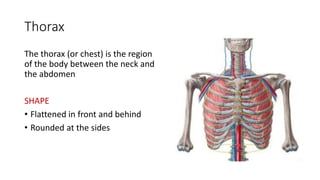
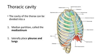
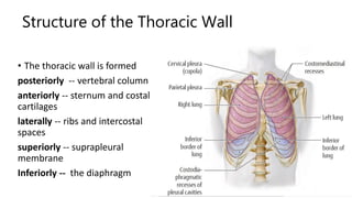
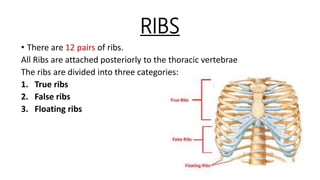
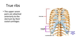
Thorax
Dr. Aliza Moin
Surface Anatomy
Thorax
The thorax (or chest) is the region
of the body between the neck and
the abdomen
SHAPE
• Flattened in front and behind
• Rounded at the sides
Thoracic cage
Boundaries
• Vertebral column behind
• Ribs and intercostal spaces on
either side
• Sternum and costal cartilages
in front
Communications
• Superiorly communicates with
neck
• Inferiorly separated from
viscera by diaphragm
Thoracic cavity
• The cavity of the thorax can be
divided into a
1. Median partition, called the
mediastinum
1. laterally place pleurae and
lungs
Thoracic Wall
Structure of the Thoracic Wall
• The thoracic wall is formed
posteriorly -- vertebral column
anteriorly -- sternum and costal
cartilages
laterally -- ribs and intercostal
spaces
superiorly -- suprapleural
membrane
Inferiorly -- the diaphragm
Bones Of Thorax
STERNUM
• Location -- sternum lies
in the midline of the
anterior chest wall
• Shape – flat bone
• Parts
1. Manubrium
2. Body of sternum
3. Xiphoid process
Manubrium
It makes the upper part of the sternum
• Location : opposite the 3rd and 4th
thoracic vertebrae
• Articulation
Manubrium articulates with
1. body of the sternum at the
manubriosternal
• joint
2. clavicles
3. 1st costal cartilage and the upper part of
the 2nd costal cartilages.
Body of the sternum
It articulates with
1. above with the manubrium at the
manubriosternal joint
2. below with the xiphoid process at
the xiphisternal joint.
3. On each side it articulates with the
2nd to the 7th costal cartilages
Xiphoid process
• It is a thin plate of cartilage making the lower most
part of sternum
• It ossifies at its proximal end during adult life.
• No ribs or costal cartilages are attached to it.
Sternal angle – Angle of Louis
• sternal angle formed by the articulation of the manubrium with the
body of the sternum,
• can be recognized by the presence of a transverse ridge on
• the anterior aspect of the sternum
• This ridge lies at the level of the 2nd costal cartilage
• importance
• all costal cartilages and ribs are counted from this point. The
• sternal angle lies opposite the intervertebral disc between
• the 4th and 5th thoracic vertebrae.
RIBS
• There are 12 pairs of ribs.
All Ribs are attached posteriorly to the thoracic vertebrae
The ribs are divided into three categories:
1. True ribs
2. False ribs
3. Floating ribs
True ribs
• The upper seven
pairs are attached
anteriorly to the
sternum by their
costal cartilages
False ribs
• The 8th, 9th, and 10th
pairs of ribs are
attached anteriorly to
each other and to the
7th rib by means of
their costal cartilages
and small synovial joints
• The 11th and 12th pairs
have no anterior
attachment
Typical Rib
• A typical rib is a long, twisted,
flat bone.
• It has two borders
• superior border -- rounded,
smooth
• inferior border -- sharp, thin.
• The anterior end of each rib is
attached to the corresponding
costal cartilage
Parts of typical Rib
• Head
• Neck
• Tubercle
• Shaft
• angle
Head
• The head has two facets.
• These articulate with
1. the numerically corresponding
vertebral body
2. vertebra immediately above
Tubercle
A prominence on the outer surface
of the rib at the junction of the neck
with the shaft.
It has a facet for articulation with
the transverse process of the
numerically corresponding
vertebrae.
Neck
• a constricted portion situated between the
• head and the tubercle
Shaft
• shaft is thin and flattened and
twisted.
• Its inferior border has the
costal groove.
• This accommodates the
intercostal vessels and nerve
Atypical Rib
• 1st, 2nd, and 10th–12th
• The 1st rib is important clinically
because of its close relationship
• to the lower nerves of the brachial
plexus and the
• This rib is small and flattened
Typical and Atypical Ribs
Costal Cartilages
• Costal cartilages are bars of
cartilage connecting the upper
seven ribs to the lateral edge of the
sternum
• 8th, 9th, and 10th ribs to the
cartilage immediately above
• cartilages of the 11th and 12th ribs
end in the abdominal Musculature
• Function: They contribute
significantly to the elasticity and
mobility of the thoracic walls.
Joints of the Chest Wall
Joints Of The Manubrium
Manubriosternal joint
is a cartilaginous joint
between the manubrium
and the body of the
sternum. A small amount
of angular movement is
possible during respiration.
Xiphisternal joint
• cartilaginous joint between
the xiphoid process
(cartilage) and the body of
the sternum. The xiphoid
process usually fuses with
the body
• of the sternum during
middle age
Joints of the Ribs
The Joints of the Heads of the Ribs
• 1st rib and the three lowest ribs
have a single synovial joint with their
corresponding vertebral body.
• For the 2nd to 9th ribs, the head
articulates by means of a synovial
joint with the corresponding vertebral
body and that of the vertebra above
it .
Joints of the Tubercles of the Ribs
• The tubercle of a rib articulates
by means of a synovial joint
with the transverse process of
the corresponding vertebra
• This joint is absent on the 11th
and 12th ribs
Joints of the Ribs and Costal Cartilages
• These joints are cartilaginous joints.
No movement is possible.
Joints of the Costal Cartilages with the
Sternum
• The 1st costal cartilages articulate with the manubrium, by
cartilaginous joints that permit no movement.
• The 2nd to 7th costal cartilages articulate with the lateral border of
the sternum by synovial joints.
• 6th, 7th, 8th, 9th, and 10th costal cartilages articulate with one
another along their borders by small synovial joints.
• The cartilages of the 11th and 12th ribs are embedded in the
abdominal musculature
Thank you

Thorax.pptx

  • 1.
  • 2.
  • 5.
    Thorax The thorax (orchest) is the region of the body between the neck and the abdomen SHAPE • Flattened in front and behind • Rounded at the sides
  • 6.
    Thoracic cage Boundaries • Vertebralcolumn behind • Ribs and intercostal spaces on either side • Sternum and costal cartilages in front Communications • Superiorly communicates with neck • Inferiorly separated from viscera by diaphragm
  • 7.
    Thoracic cavity • Thecavity of the thorax can be divided into a 1. Median partition, called the mediastinum 1. laterally place pleurae and lungs
  • 8.
  • 9.
    Structure of theThoracic Wall • The thoracic wall is formed posteriorly -- vertebral column anteriorly -- sternum and costal cartilages laterally -- ribs and intercostal spaces superiorly -- suprapleural membrane Inferiorly -- the diaphragm
  • 11.
  • 12.
    STERNUM • Location --sternum lies in the midline of the anterior chest wall • Shape – flat bone • Parts 1. Manubrium 2. Body of sternum 3. Xiphoid process
  • 13.
    Manubrium It makes theupper part of the sternum • Location : opposite the 3rd and 4th thoracic vertebrae • Articulation Manubrium articulates with 1. body of the sternum at the manubriosternal • joint 2. clavicles 3. 1st costal cartilage and the upper part of the 2nd costal cartilages.
  • 14.
    Body of thesternum It articulates with 1. above with the manubrium at the manubriosternal joint 2. below with the xiphoid process at the xiphisternal joint. 3. On each side it articulates with the 2nd to the 7th costal cartilages
  • 15.
    Xiphoid process • Itis a thin plate of cartilage making the lower most part of sternum • It ossifies at its proximal end during adult life. • No ribs or costal cartilages are attached to it.
  • 16.
    Sternal angle –Angle of Louis • sternal angle formed by the articulation of the manubrium with the body of the sternum, • can be recognized by the presence of a transverse ridge on • the anterior aspect of the sternum • This ridge lies at the level of the 2nd costal cartilage • importance • all costal cartilages and ribs are counted from this point. The • sternal angle lies opposite the intervertebral disc between • the 4th and 5th thoracic vertebrae.
  • 18.
    RIBS • There are12 pairs of ribs. All Ribs are attached posteriorly to the thoracic vertebrae The ribs are divided into three categories: 1. True ribs 2. False ribs 3. Floating ribs
  • 19.
    True ribs • Theupper seven pairs are attached anteriorly to the sternum by their costal cartilages
  • 20.
    False ribs • The8th, 9th, and 10th pairs of ribs are attached anteriorly to each other and to the 7th rib by means of their costal cartilages and small synovial joints
  • 21.
    • The 11thand 12th pairs have no anterior attachment
  • 22.
    Typical Rib • Atypical rib is a long, twisted, flat bone. • It has two borders • superior border -- rounded, smooth • inferior border -- sharp, thin. • The anterior end of each rib is attached to the corresponding costal cartilage
  • 23.
    Parts of typicalRib • Head • Neck • Tubercle • Shaft • angle
  • 24.
    Head • The headhas two facets. • These articulate with 1. the numerically corresponding vertebral body 2. vertebra immediately above
  • 25.
    Tubercle A prominence onthe outer surface of the rib at the junction of the neck with the shaft. It has a facet for articulation with the transverse process of the numerically corresponding vertebrae.
  • 26.
    Neck • a constrictedportion situated between the • head and the tubercle
  • 27.
    Shaft • shaft isthin and flattened and twisted. • Its inferior border has the costal groove. • This accommodates the intercostal vessels and nerve
  • 28.
    Atypical Rib • 1st,2nd, and 10th–12th • The 1st rib is important clinically because of its close relationship • to the lower nerves of the brachial plexus and the • This rib is small and flattened
  • 29.
  • 30.
    Costal Cartilages • Costalcartilages are bars of cartilage connecting the upper seven ribs to the lateral edge of the sternum • 8th, 9th, and 10th ribs to the cartilage immediately above • cartilages of the 11th and 12th ribs end in the abdominal Musculature • Function: They contribute significantly to the elasticity and mobility of the thoracic walls.
  • 31.
    Joints of theChest Wall
  • 32.
    Joints Of TheManubrium Manubriosternal joint is a cartilaginous joint between the manubrium and the body of the sternum. A small amount of angular movement is possible during respiration.
  • 33.
    Xiphisternal joint • cartilaginousjoint between the xiphoid process (cartilage) and the body of the sternum. The xiphoid process usually fuses with the body • of the sternum during middle age
  • 34.
    Joints of theRibs The Joints of the Heads of the Ribs • 1st rib and the three lowest ribs have a single synovial joint with their corresponding vertebral body. • For the 2nd to 9th ribs, the head articulates by means of a synovial joint with the corresponding vertebral body and that of the vertebra above it .
  • 35.
    Joints of theTubercles of the Ribs • The tubercle of a rib articulates by means of a synovial joint with the transverse process of the corresponding vertebra • This joint is absent on the 11th and 12th ribs
  • 36.
    Joints of theRibs and Costal Cartilages • These joints are cartilaginous joints. No movement is possible.
  • 37.
    Joints of theCostal Cartilages with the Sternum • The 1st costal cartilages articulate with the manubrium, by cartilaginous joints that permit no movement. • The 2nd to 7th costal cartilages articulate with the lateral border of the sternum by synovial joints. • 6th, 7th, 8th, 9th, and 10th costal cartilages articulate with one another along their borders by small synovial joints. • The cartilages of the 11th and 12th ribs are embedded in the abdominal musculature
  • 39.