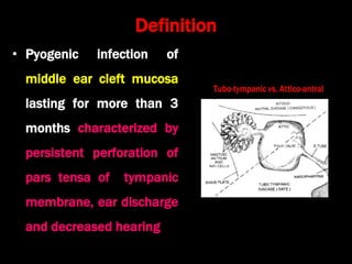
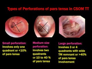
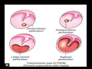
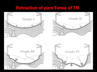
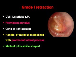
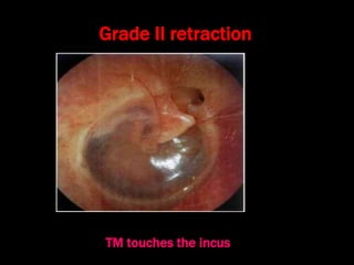
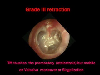
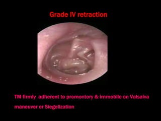
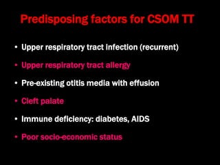
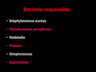
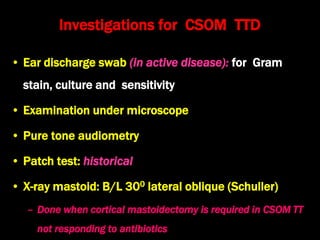
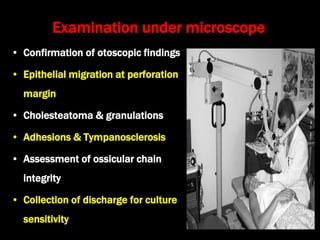
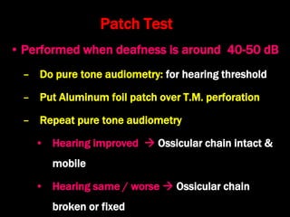
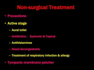
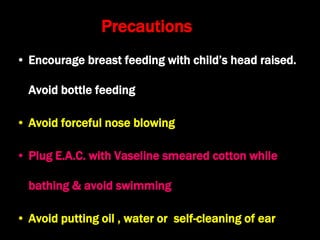
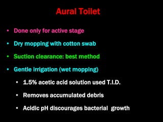
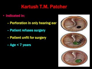
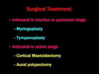
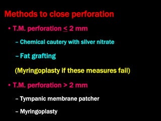
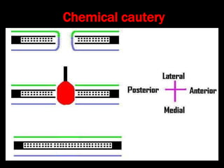
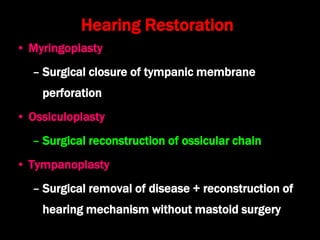
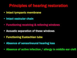
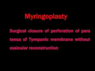
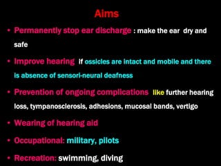
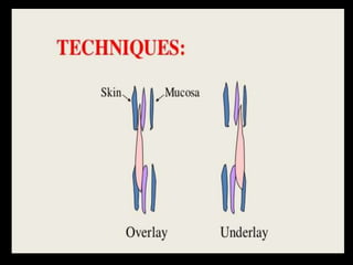
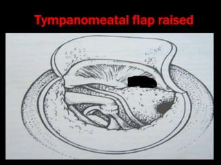
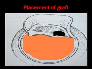
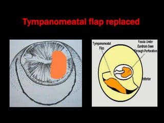
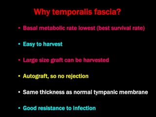
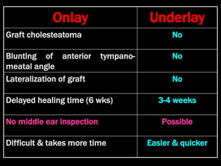
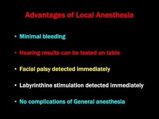
Chronic Suppurative Otitis Media:
Tubotympanic Disease (CSOM TT,
COM Mucosal type)
Dr. Krishna Koirala
2020-05-18
Definition
• Pyogenic infection of
middle ear cleft mucosa
lasting for more than 3
months characterized by
persistent perforation of
pars tensa of tympanic
membrane, ear discharge
and decreased hearing
Small perforation:
Involves only one
quadrant or <10%
of pars tensa
Types of Perforations of pars tensa in CSOM TT
Medium size
perforation:
Involves two
quadrants
or 10 to 40 %
of pars tensa
Large perforation:
Involves 3 or 4
quadrants with wide
TM remnant or >40%
of pars tensa
involvement
Retraction of pars Tensa of TM
Grade I retraction
• Dull, lusterless T.M.
• Prominent annulus
• Cone of light absent
• Handle of malleus medialized
with prominent lateral process
• Malleal folds sickle shaped
Grade II retraction
TM touches the incus
Grade III retraction
TM touches the promontory (atelectasis) but mobile
on Valsalva maneuver or Siegelization
Grade IV retraction
TM firmly adherent to promontory & immobile on Valsalva
maneuver or Siegelization
Predisposing factors for CSOM TT
• Upper respiratory tract infection (recurrent)
• Upper respiratory tract allergy
• Pre-existing otitis media with effusion
• Cleft palate
• Immune deficiency: diabetes, AIDS
• Poor socio-economic status
Bacteria responsible
• Staphylococcus aureus
• Pseudomonas aeruginosa
• Klebsiella
• Proteus
• Streptococcus
• Bacteroides
Routes of infection
1. Via Eustachian tube
– U.R.T.I., nose blowing, regurgitation of milk
2. Via tympanic membrane perforation
– Following A.S.O.M. or post-traumatic
3. Haematogenous (rare): exanthematous fever
Pathological Changes
1. Eardrum
– Central perforation, myringosclerosis
2. Ossicles
– Destruction (hyperemic decalcification)
– Tympanosclerosis, fibrosis and adhesions
3. Middle ear mucosa: edematous, pale, congested
4. Mastoid bone: sclerosis
Clinical Features
• Ear discharge: intermittent, profuse, mucoid to muco-
purulent, whitish, odorless, not blood-stained
• Hearing Loss:
– Usually conductive (25-50 dB) but might be normal
in small, dry perforations
– Round window shielding by ear discharge may lead
to better hearing in acute exacerbations
• Tympanic membrane: central perforation
Stages of tubotympanic disease
Stage Otorrhoea Eardrum
perforation
Last ear
discharge
Active + +
Quiescent _ + < 6 months
Inactive _ + > 6 months
Healed _ _ > 6 months
Investigations for CSOM TTD
• Ear discharge swab (in active disease): for Gram
stain, culture and sensitivity
• Examination under microscope
• Pure tone audiometry
• Patch test: historical
• X-ray mastoid: B/L 300 lateral oblique (Schuller)
– Done when cortical mastoidectomy is required in CSOM TT
not responding to antibiotics
Examination under microscope
• Confirmation of otoscopic findings
• Epithelial migration at perforation
margin
• Cholesteatoma & granulations
• Adhesions & Tympanosclerosis
• Assessment of ossicular chain
integrity
• Collection of discharge for culture
sensitivity
Pure Tone Audiometry
• Uses
– Presence of hearing loss
– Degree of hearing loss
– Type of hearing loss
– Hearing status of other ear
– Record to compare hearing post-operatively
– Medico legal purpose
Patch Test
• Performed when deafness is around 40-50 dB
– Do pure tone audiometry: for hearing threshold
– Put Aluminum foil patch over T.M. perforation
– Repeat pure tone audiometry
• Hearing improved  Ossicular chain intact &
mobile
• Hearing same / worse  Ossicular chain
broken or fixed
Treatment of CSOM Tubo-tympanic
Disease
Non-surgical Treatment
• Precautions
• Active stage
– Aural toilet
– Antibiotics : Systemic & Topical
– Antihistamines
– Nasal decongestants
– Treatment of respiratory infection & allergy
• Tympanic membrane patcher
Precautions
• Encourage breast feeding with child’s head raised.
Avoid bottle feeding
• Avoid forceful nose blowing
• Plug E.A.C. with Vaseline smeared cotton while
bathing & avoid swimming
• Avoid putting oil , water or self-cleaning of ear
• Done only for active stage
• Dry mopping with cotton swab
• Suction clearance: best method
• Gentle irrigation (wet mopping)
• 1.5% acetic acid solution used T.I.D.
• Removes accumulated debris
• Acidic pH discourages bacterial growth
Aural Toilet
Antibiotics
• Topical Antibiotics:
• Ciprofloxacin, Gentamicin, Tobramycin
• Antibiotics + Steroid: for polyps, granulations
• Neosporin + Betamethasone / Hydrocortisone
• Oral Antibiotics: for severe infections
• Cefuroxime, Cefaclor, Cefpodoxime, Cefixime
Antihistamines and Decongestants
• Antihistamines
– Chlorpheniramine
– Cetirizine
– Fexofenadine
– Loratadine
– Levocetrizine
– Azelastine (topical)
• Systemic Decongestants
– Pseudoephedrine
– Phenylephrine
• Topical Decongestants
– Oxymetazoline
– Xylometazoline
– Hypertonic saline
Kartush T.M. Patcher
• Indicated in:
– Perforation in only hearing ear
– Patient refuses surgery
– Patient unfit for surgery
– Age < 7 years
Surgical Treatment
• Indicated in inactive or quiescent stage
–Myringoplasty
–Tympanoplasty
• Indicated in active stage
–Cortical Mastoidectomy
–Aural polypectomy
Methods to close perforation
• T.M. perforation < 2 mm
– Chemical cautery with silver nitrate
–Fat grafting
(Myringoplasty if these measures fail)
• T.M. perforation > 2 mm
– Tympanic membrane patcher
– Myringoplasty
Chemical cautery
Surgical Approaches to the
middle ear
Wilde’s post-aural incision
Lempert’s end-aural incision
Rosen’s permeatal incision
Hearing Restoration
• Myringoplasty
– Surgical closure of tympanic membrane
perforation
• Ossiculoplasty
– Surgical reconstruction of ossicular chain
• Tympanoplasty
– Surgical removal of disease + reconstruction of
hearing mechanism without mastoid surgery
Principles of hearing restoration
• Intact tympanic membrane
• Intact ossicular chain
• Functioning receiving & relieving windows
• Acoustic separation of these windows
• Functioning Eustachian tube
• Absence of sensorineural hearing loss
• Absence of active infection / allergy in middle ear cleft
Myringoplasty
Surgical closure of perforation of pars
tensa of Tympanic membrane without
ossicular reconstruction
Aims
• Permanently stop ear discharge : make the ear dry and
safe
• Improve hearing if ossicles are intact and mobile and there
is absence of sensori-neural deafness
• Prevention of ongoing complications like further hearing
loss, tympanosclerosis, adhesions, mucosal bands, vertigo
• Wearing of hearing aid
• Occupational: military, pilots
• Recreation: swimming, diving
Contraindications
• Purulent ear discharge
• Otitis externa
• Respiratory allergy
• Age < 7 yr (Eustachian tube not fully developed)
• Only hearing ear
• Cholesteatoma
Methods
Techniques
• Underlay: graft placed medial to fibrous annulus
• Overlay: graft placed lateral to fibrous annulus
Grafts used
• Temporalis fascia, Tragal perichondrium, Vein
graft, Fascia lata, Dura mater
Overlay Myringoplasty
Underlay Myringoplasty
Steps of underlay
Myringoplasty
Tympanomeatal flap raised
Placement of graft
Tympanomeatal flap replaced
Why temporalis fascia?
• Basal metabolic rate lowest (best survival rate)
• Easy to harvest
• Large size graft can be harvested
• Autograft, so no rejection
• Same thickness as normal tympanic membrane
• Good resistance to infection
Onlay Underlay
Graft cholesteatoma No
Blunting of anterior tympano-
meatal angle
No
Lateralization of graft No
Delayed healing time (6 wks) 3-4 weeks
No middle ear inspection Possible
Difficult & takes more time Easier & quicker
Advantages of Local Anesthesia
• Minimal bleeding
• Hearing results can be tested on table
• Facial palsy detected immediately
• Labyrinthine stimulation detected immediately
• No complications of General anesthesia
Tympanoplasty
Types
Type Pathology Graft placed on
I Ear drum perforation only Malleus handle
II Malleus handle eroded Incus
III Malleus + Incus eroded Stapes head
IV Only footplate remains: mobile Footplate exposed
V Only stapes remains: fixed Lateral SCC
opening
VI Only footplate remains: mobile Round window
exposed (Sono
inversion )
Ossiculoplasty
• Ossicular graft material
– Autograft
• Ossicles : incus/malleus
• Cartilage : Tragal/ conchal
• Bone : spine of Henle/mastoid
– Homograft: ossicles/cartilage/bone
– Biomaterials: plastic(polyethylene)/ceramic/ teflon/gold
(Biomaterials available as PORP and TORP)

13. csom tt kk

  • 1.
    Chronic Suppurative OtitisMedia: Tubotympanic Disease (CSOM TT, COM Mucosal type) Dr. Krishna Koirala 2020-05-18
  • 2.
    Definition • Pyogenic infectionof middle ear cleft mucosa lasting for more than 3 months characterized by persistent perforation of pars tensa of tympanic membrane, ear discharge and decreased hearing
  • 3.
    Small perforation: Involves onlyone quadrant or <10% of pars tensa Types of Perforations of pars tensa in CSOM TT Medium size perforation: Involves two quadrants or 10 to 40 % of pars tensa Large perforation: Involves 3 or 4 quadrants with wide TM remnant or >40% of pars tensa involvement
  • 5.
    Retraction of parsTensa of TM
  • 6.
    Grade I retraction •Dull, lusterless T.M. • Prominent annulus • Cone of light absent • Handle of malleus medialized with prominent lateral process • Malleal folds sickle shaped
  • 7.
    Grade II retraction TMtouches the incus
  • 8.
    Grade III retraction TMtouches the promontory (atelectasis) but mobile on Valsalva maneuver or Siegelization
  • 9.
    Grade IV retraction TMfirmly adherent to promontory & immobile on Valsalva maneuver or Siegelization
  • 10.
    Predisposing factors forCSOM TT • Upper respiratory tract infection (recurrent) • Upper respiratory tract allergy • Pre-existing otitis media with effusion • Cleft palate • Immune deficiency: diabetes, AIDS • Poor socio-economic status
  • 11.
    Bacteria responsible • Staphylococcusaureus • Pseudomonas aeruginosa • Klebsiella • Proteus • Streptococcus • Bacteroides
  • 12.
    Routes of infection 1.Via Eustachian tube – U.R.T.I., nose blowing, regurgitation of milk 2. Via tympanic membrane perforation – Following A.S.O.M. or post-traumatic 3. Haematogenous (rare): exanthematous fever
  • 13.
    Pathological Changes 1. Eardrum –Central perforation, myringosclerosis 2. Ossicles – Destruction (hyperemic decalcification) – Tympanosclerosis, fibrosis and adhesions 3. Middle ear mucosa: edematous, pale, congested 4. Mastoid bone: sclerosis
  • 14.
    Clinical Features • Eardischarge: intermittent, profuse, mucoid to muco- purulent, whitish, odorless, not blood-stained • Hearing Loss: – Usually conductive (25-50 dB) but might be normal in small, dry perforations – Round window shielding by ear discharge may lead to better hearing in acute exacerbations • Tympanic membrane: central perforation
  • 15.
    Stages of tubotympanicdisease Stage Otorrhoea Eardrum perforation Last ear discharge Active + + Quiescent _ + < 6 months Inactive _ + > 6 months Healed _ _ > 6 months
  • 16.
    Investigations for CSOMTTD • Ear discharge swab (in active disease): for Gram stain, culture and sensitivity • Examination under microscope • Pure tone audiometry • Patch test: historical • X-ray mastoid: B/L 300 lateral oblique (Schuller) – Done when cortical mastoidectomy is required in CSOM TT not responding to antibiotics
  • 17.
    Examination under microscope •Confirmation of otoscopic findings • Epithelial migration at perforation margin • Cholesteatoma & granulations • Adhesions & Tympanosclerosis • Assessment of ossicular chain integrity • Collection of discharge for culture sensitivity
  • 18.
    Pure Tone Audiometry •Uses – Presence of hearing loss – Degree of hearing loss – Type of hearing loss – Hearing status of other ear – Record to compare hearing post-operatively – Medico legal purpose
  • 19.
    Patch Test • Performedwhen deafness is around 40-50 dB – Do pure tone audiometry: for hearing threshold – Put Aluminum foil patch over T.M. perforation – Repeat pure tone audiometry • Hearing improved  Ossicular chain intact & mobile • Hearing same / worse  Ossicular chain broken or fixed
  • 20.
    Treatment of CSOMTubo-tympanic Disease
  • 21.
    Non-surgical Treatment • Precautions •Active stage – Aural toilet – Antibiotics : Systemic & Topical – Antihistamines – Nasal decongestants – Treatment of respiratory infection & allergy • Tympanic membrane patcher
  • 22.
    Precautions • Encourage breastfeeding with child’s head raised. Avoid bottle feeding • Avoid forceful nose blowing • Plug E.A.C. with Vaseline smeared cotton while bathing & avoid swimming • Avoid putting oil , water or self-cleaning of ear
  • 23.
    • Done onlyfor active stage • Dry mopping with cotton swab • Suction clearance: best method • Gentle irrigation (wet mopping) • 1.5% acetic acid solution used T.I.D. • Removes accumulated debris • Acidic pH discourages bacterial growth Aural Toilet
  • 24.
    Antibiotics • Topical Antibiotics: •Ciprofloxacin, Gentamicin, Tobramycin • Antibiotics + Steroid: for polyps, granulations • Neosporin + Betamethasone / Hydrocortisone • Oral Antibiotics: for severe infections • Cefuroxime, Cefaclor, Cefpodoxime, Cefixime
  • 25.
    Antihistamines and Decongestants •Antihistamines – Chlorpheniramine – Cetirizine – Fexofenadine – Loratadine – Levocetrizine – Azelastine (topical) • Systemic Decongestants – Pseudoephedrine – Phenylephrine • Topical Decongestants – Oxymetazoline – Xylometazoline – Hypertonic saline
  • 26.
    Kartush T.M. Patcher •Indicated in: – Perforation in only hearing ear – Patient refuses surgery – Patient unfit for surgery – Age < 7 years
  • 27.
    Surgical Treatment • Indicatedin inactive or quiescent stage –Myringoplasty –Tympanoplasty • Indicated in active stage –Cortical Mastoidectomy –Aural polypectomy
  • 28.
    Methods to closeperforation • T.M. perforation < 2 mm – Chemical cautery with silver nitrate –Fat grafting (Myringoplasty if these measures fail) • T.M. perforation > 2 mm – Tympanic membrane patcher – Myringoplasty
  • 29.
  • 30.
    Surgical Approaches tothe middle ear
  • 31.
  • 32.
  • 33.
  • 34.
    Hearing Restoration • Myringoplasty –Surgical closure of tympanic membrane perforation • Ossiculoplasty – Surgical reconstruction of ossicular chain • Tympanoplasty – Surgical removal of disease + reconstruction of hearing mechanism without mastoid surgery
  • 35.
    Principles of hearingrestoration • Intact tympanic membrane • Intact ossicular chain • Functioning receiving & relieving windows • Acoustic separation of these windows • Functioning Eustachian tube • Absence of sensorineural hearing loss • Absence of active infection / allergy in middle ear cleft
  • 36.
    Myringoplasty Surgical closure ofperforation of pars tensa of Tympanic membrane without ossicular reconstruction
  • 37.
    Aims • Permanently stopear discharge : make the ear dry and safe • Improve hearing if ossicles are intact and mobile and there is absence of sensori-neural deafness • Prevention of ongoing complications like further hearing loss, tympanosclerosis, adhesions, mucosal bands, vertigo • Wearing of hearing aid • Occupational: military, pilots • Recreation: swimming, diving
  • 38.
    Contraindications • Purulent eardischarge • Otitis externa • Respiratory allergy • Age < 7 yr (Eustachian tube not fully developed) • Only hearing ear • Cholesteatoma
  • 39.
    Methods Techniques • Underlay: graftplaced medial to fibrous annulus • Overlay: graft placed lateral to fibrous annulus Grafts used • Temporalis fascia, Tragal perichondrium, Vein graft, Fascia lata, Dura mater
  • 40.
  • 41.
  • 43.
  • 44.
  • 45.
  • 46.
  • 47.
    Why temporalis fascia? •Basal metabolic rate lowest (best survival rate) • Easy to harvest • Large size graft can be harvested • Autograft, so no rejection • Same thickness as normal tympanic membrane • Good resistance to infection
  • 48.
    Onlay Underlay Graft cholesteatomaNo Blunting of anterior tympano- meatal angle No Lateralization of graft No Delayed healing time (6 wks) 3-4 weeks No middle ear inspection Possible Difficult & takes more time Easier & quicker
  • 49.
    Advantages of LocalAnesthesia • Minimal bleeding • Hearing results can be tested on table • Facial palsy detected immediately • Labyrinthine stimulation detected immediately • No complications of General anesthesia
  • 50.
  • 51.
  • 52.
    Type Pathology Graftplaced on I Ear drum perforation only Malleus handle II Malleus handle eroded Incus III Malleus + Incus eroded Stapes head IV Only footplate remains: mobile Footplate exposed V Only stapes remains: fixed Lateral SCC opening VI Only footplate remains: mobile Round window exposed (Sono inversion )
  • 53.
    Ossiculoplasty • Ossicular graftmaterial – Autograft • Ossicles : incus/malleus • Cartilage : Tragal/ conchal • Bone : spine of Henle/mastoid – Homograft: ossicles/cartilage/bone – Biomaterials: plastic(polyethylene)/ceramic/ teflon/gold (Biomaterials available as PORP and TORP)