Downloaded 150 times

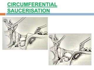
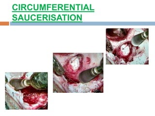
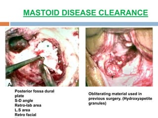
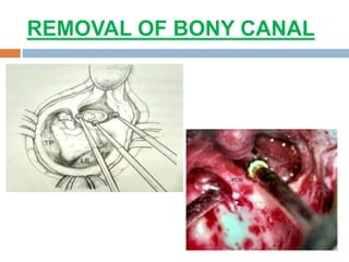
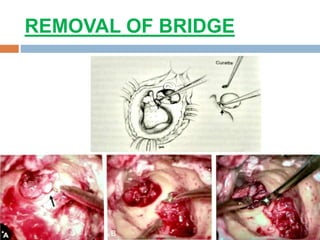
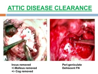
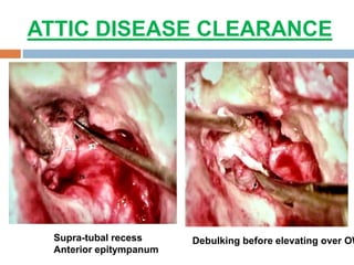
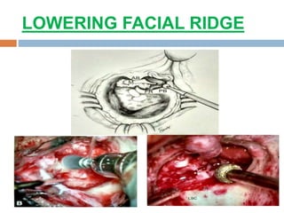
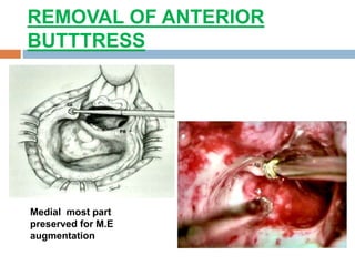
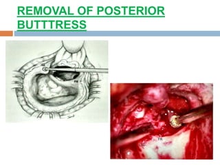
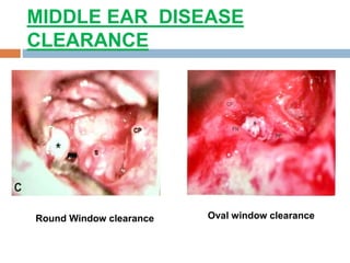
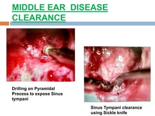
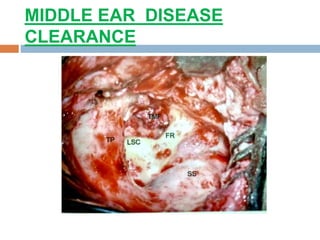
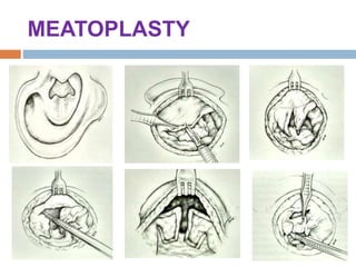
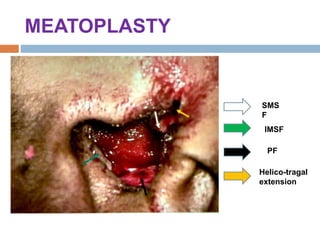

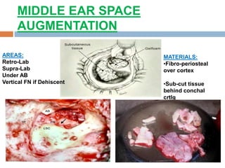
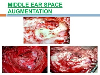
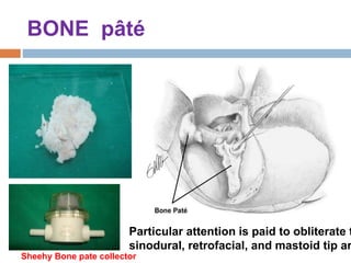
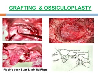
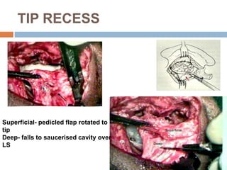
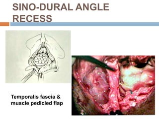
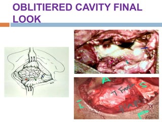
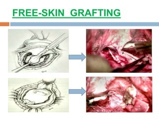
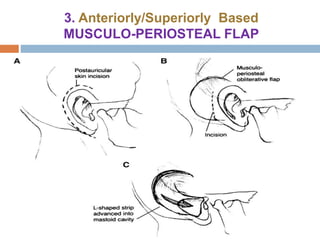
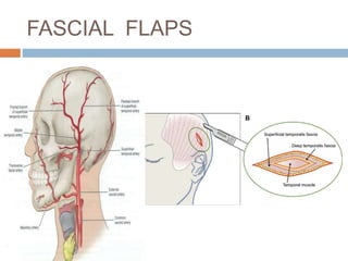
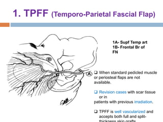
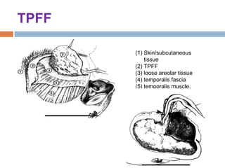
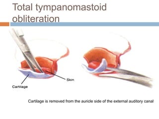
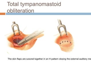
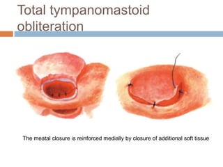
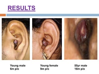


This document discusses the management and surgical techniques for discharging mastoid cavities, outlining causes, pre-operative preparations, and various surgical methods for cavity obliteration. It includes important terminologies related to the anatomy of the mastoid area and highlights the factors leading to discharging cavities, along with indications and contraindications for cavity obliteration. Key techniques mentioned for obliteration include local flaps and free grafts, alongside postoperative considerations.


![Untitled (6) [Autosaved] ear.pptx ear surgeries](https://cdn.slidesharecdn.com/ss_thumbnails/untitled6autosavedear-251128030434-d7ae6d96-thumbnail.jpg?width=640&height=640&fit=bounds)


