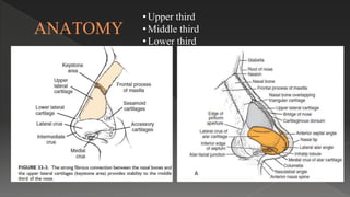
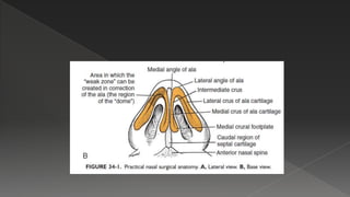
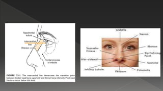
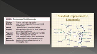
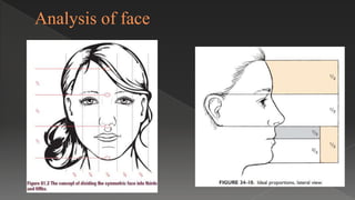
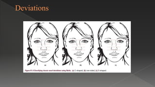
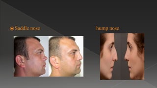
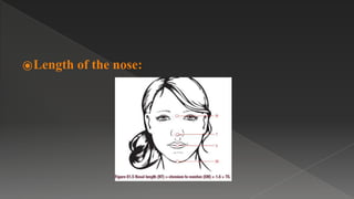
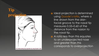
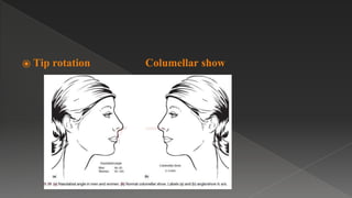
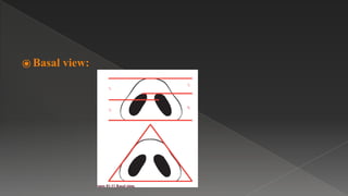
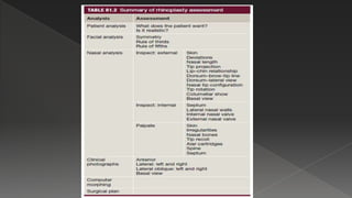
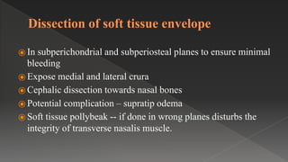
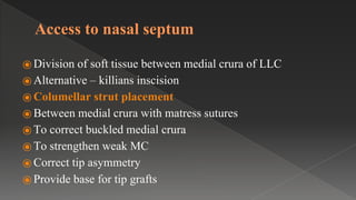
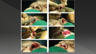
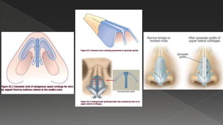
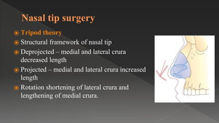
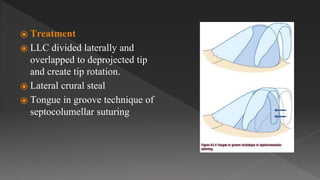
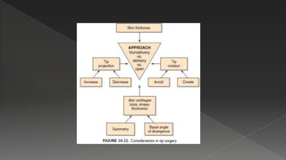
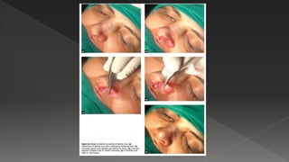
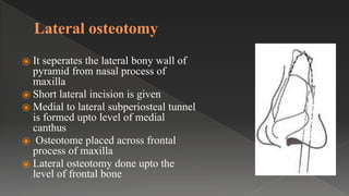
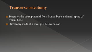
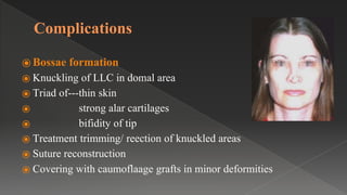
Rhinoplasty
Dr Grace
Postgraduate ent
Nmch Nellore
Introduction
⦿ Challenging procedure in facial plastic surgery
Historical aspects
⦿ First in India 600 b.C external and open approaches
⦿ 1887 - John orlando roe performed first intranasal
rhinoplasty.
⦿ Rethi and sercer in 1934 developed transcolumellar inscisions
ANATOMY
• Upper third
• Middle third
• Lower third
Nasal valve
Preoperative assessment
⦿ Consider facial aesthetics
⦿ nasal function
⦿ Consideration of the patient’s motivations, anxieties
and expectations
⦿ Analysis of the face
⦿ Analysis of the nose
⦿ Examination
⦿ Photography.
Patient
⦿ History
⦿ Expectations
⦿ Body dysmorphic disorder
⦿ High risk patient SIMON
⦿ Ideal patient SLYVIA
⦿ Second opinion from psychiatrist
Analysis of face
• nasofrontal angle 115–135 °
• nasofacial angle 30–40 °
• nasomental angle 120–132 °
• mentocervical angle 80–95 °
Analysis of nose
⦿ Inspection of external nose
⦿ Inspection of internal nose
⦿ Palpation
⦿Skin quality:
⦿ Thick and sebaceous--- refining and narrowing the nasal tip
can be challenging
⦿ Thin skin--- minor irregularities are easily detectable
Deviations
⦿ Saddle nose hump nose
⦿Length of the nose:
⦿ Ideal projection is determined
using Goode’s ratio, where a
line drawn from the alar–
facial groove to the nasal tip
measures 0.55–0.60 of the
distance from the nasion to
the nasal tip.
⦿ A ratio less than this equates
to an underprojected nose
and greater than this
corresponds to overprojection
Tip
projection:
⦿Lip–chin relationship:
Tip configuration
Tip abnormalities
BROAD ,BULLOUS,SQUARE BALL TIP BIFID TIP UPWARDLY ROTATED TIP
ASYMMETRIC TIP
⦿ Dorsum:
⦿ Alar abnormalities
pinching flaring
⦿ Tip rotation Columellar show
⦿ Basal view:
Aetiology
⦿ Nasal fractures – 40% of facial fractures
⦿ Injury – functional
aesthetic problems
⦿ Chilhood/ adult fractures
⦿ Growth in facial skeleton more often asymmetric leading to
appearance of a damaged nose without any prior history of
trauma.
Types of nasal fractures
Stranc and Robertson classification
⦿Lateral blows:
⦿ Simple depressed nasal bone
⦿ Displacement of bony cartilage
⦿ Deviation of nasal axis in lower 2/3rd due to vertical fracture of
septum.
⦿Frontal blows:
⦿ Fracture of tip of nasal bone
⦿ Injury to cartilagenous vault ---widening of bridge
⦿ Horizontal fracture of septum--- loss of height in middle third
leading to saddle nose deformity.
Closed reduction within 2-3 weeks
Types of rhinoplasty
⦿ External --- open approach
⦿ Endonasal--- closed approach
⦿ Advantages of external rhinoplasty
⦿ Extensive exposure
⦿ Binocular vision
⦿ Use of both hands
⦿ Control of bleeding and diathermy
⦿ Precise placement of struts and grafts
⦿ Valve area preserved
⦿ Indications
⦿ Extensive revision surgery
⦿ Severe nasal trauma
⦿ Congenital deformities: cleft lip nose
⦿ Marked tip deformities
⦿ Elaborate reduction and augmentation procedures
⦿ Correction of extreme overprojection
Surgical technique
⦿Inscision:
⦿ Broken line transcolumellar inscision --- less visible scar
⦿ Step, gullwing , inverted v type
⦿ In upper 2/3rd at narrowest point
⦿ In lower1/3rd least support
⦿ Joined to marginal inscions along the lateral crus
Inscisions
Dissection of soft tissue envelope
⦿ In subperichondrial and subperiosteal planes to ensure minimal
bleeding
⦿ Expose medial and lateral crura
⦿ Cephalic dissection towards nasal bones
⦿ Potential complication – supratip odema
⦿ Soft tissue pollybeak -- if done in wrong planes disturbs the
integrity of transverse nasalis muscle.
Access to nasal septum
⦿ Division of soft tissue between medial crura of LLC
⦿ Alternative – killians inscision
⦿ Columellar strut placement
⦿ Between medial crura with matress sutures
⦿ To correct buckled medial crura
⦿ To strengthen weak MC
⦿ Correct tip asymmetry
⦿ Provide base for tip grafts
Closure
⦿ With 6-0 vicryl or with tissue glue.
Specific applications
⦿ Bony pyramid in external rhinoplasty
⦿ Allows use of burr or reduction of the soft tissue envelope at
nasion to deepen the nasofrontal angle
⦿ Application of soft tissue onlay grafts
⦿ Bony dehumping together with lateral, medial, and
intermediate osteotomies
⦿ The middle nasal vault
⦿ Placement of cartilaginous strips or spreader grafts to open up the
nasal valve area and angles
⦿ Auto spreader grafts infolded ULC
⦿ Reconstuction of dorsal nasal roof
⦿ Straightening of high dorsal deviated septum
⦿ Recreation of dorsal aesthetic lines
Nasal tip surgery
⦿ Tripod theory
⦿ Structural framework of nasal tip
⦿ Deprojected – medial and lateral crura
decreased length
⦿ Projected – medial and lateral crura increased
length
⦿ Rotation shortening of lateral crura and
lengthening of medial crura.
⦿ Treatment
⦿ LLC divided laterally and
overlapped to deprojected tip
and create tip rotation.
⦿ Lateral crural steal
⦿ Tongue in groove technique of
septocolumellar suturing
Augumentation rhinoplasty
⦿ Saddle nose --- Pug nose or boxer nose
⦿ Aetiology
1. Trauma
2. Nasal surgery
3. Familial/ethnic
4. Infections
5. Chronic Inflammatory conditions
6. tumour
Daniel and Brenner Classification for
saddle nose
Type 0 pseudosaddle
⦿ Relative depression of cartilaginous dorsum relative to bony
septum
Type 1 minor cosmetic concealment
⦿ Minor decrease in septal support
supratip depression and columella
retraction
⦿ Correction with diced cartilage grafts /
columellar strut
Type 2 moderate cartilage
vault restoration
⦿ Cartilaginous vault collapse,
⦿ Columellar retraction
⦿ Loss of tip support
⦿ Correction combination of
extended vault grafts and
columellar strut.
Type III major–composite
reconstruction
⦿ Total absence of septal support for the
cartilaginous vault, columella, nasal
tip,and external valves.
⦿ Foundation layer extended spreader and
septal strut grafts.
⦿ Esthetic layer columellar strut and diced
cartilage in fascia for dorsal contour.
Type IV severe–structural reconstruction
⦿ End stage of septal collapse.
⦿ Bony vault disruption
⦿ Severe contracture of the nasal lining,
often associated with major septal
perforations
⦿ Composite reconstruction costal
cartilage graft placed in the dorsum
⦿ Crural strut with tip graft
Type V catastrophic–nasal reconstruction
⦿ Esthetic reconstruction of the nose and its
adjacent tissues.
⦿ Extensive bone grafting or plating
Properties of implant materials
⦿ Noncarcinogenic and nonimmunogenic
⦿ Not toxic
⦿ Nondestructive , should not impede healing
⦿ Physical properties match the local tissues
⦿ Nonresorbable
⦿ Easily available
⦿ Cost effective easy to sterilize
Graft Materials
⦿ Autografts - cartilage, bone,
dermis and fascia
⦿ Homografts - Irradiated rib,
pooled acellular dermis
⦿ Xenografts - Leather, duck's
sternum, bovine cartilage
⦿ Precious metals - Titanium, gold,
silver, metal alloys
⦿ Synthetic biomaterials
• Silicone
• Teflon
• Proplast
• Gore-tex
Type of autografts
⦿ Septal cartilage
⦿ Auricular cartilage
⦿ Costal cartilage
⦿ Bone graft
REDUCTION RHINOPLASTY
⦿ Indications
⦿ Patients with ideal height and position of the nasion associated
with excess dorsal convexity
⦿ Oversized alar cartilages producing increase tip and lobule volume
Dorsal hump
Osteotomies
⦿ Medial osteotomy
⦿ Lateral osteotomy
• Low to high
• Low to low
⦿ Transverse osteotomy
⦿ Intermediate osteotomy
Medial osteotomy
⦿ It seperates the nasal bone from the septum
⦿ Made on both side
⦿ Nasal bone seperated at intranasal suture
⦿ Outer peritosteun is pushed to the side
⦿ Osteotome is placed at about 2mm
paramedially • Osteotome is worked through
the bone slightly below the level of frontal
bone
Lateral osteotomy
⦿ It seperates the lateral bony wall of
pyramid from nasal process of
maxilla
⦿ Short lateral incision is given
⦿ Medial to lateral subperiosteal tunnel
is formed upto level of medial
canthus
⦿ Osteotome placed across frontal
process of maxilla
⦿ Lateral osteotomy done upto the
level of frontal bone
Tranverse osteotomy
⦿ Seperates the bony pyramid from frontal bone and nasal spine of
frontal bone
⦿ Osteotomy made at a level just below nasion
Complications
⦿ Bossae formation
⦿ Knuckling of LLC in domal area
⦿ Triad of---thin skin
⦿ strong alar cartilages
⦿ bifidity of tip
⦿ Treatment trimming/ reection of knuckled areas
⦿ Suture reconstruction
⦿ Covering with caumoflaage grafts in minor deformities
⦿ Alar retraction
⦿ Over zealous cephalic strip
resection
⦿ Absolute minimum of 6 mm
cartilage should be preserved.
⦿ Hanging columella
⦿ Over resection of caudal septum
⦿ Tongue in groove suturing / strut
placement
Revision rhinoplasty
⦿ 5 – 10% require revision
⦿ Unpredictable healing beyond surgeons control
⦿ Inadequate primary procedure
⦿ Safian 3 basic rules to prevent revision
⦿ Never change normal basic anatomy/relationships and never totally
remove a component
⦿ Never allow skin and lining membrane to meet or left in contact
⦿ Never destroy nasal cartilage
Pollybeak deformity
⦿ Dropping of nasal tip and loss of
projection
Overshortened nose
⦿ Medicolegal
References
⦿ Daniel, R. K., & Brenner, K. A. (2006). Saddle Nose Deformity: A
New Classification and Treatment. Facial Plastic Surgery Clinics of
North America, 14(4), 301–312. doi:10.1016/j.fsc.2006.06.008
⦿ Scott brown 8th edition
⦿ Cummings 6th edition
Rhinoplasty

Rhinoplasty

  • 1.
  • 2.
    Introduction ⦿ Challenging procedurein facial plastic surgery
  • 3.
    Historical aspects ⦿ Firstin India 600 b.C external and open approaches ⦿ 1887 - John orlando roe performed first intranasal rhinoplasty. ⦿ Rethi and sercer in 1934 developed transcolumellar inscisions
  • 4.
    ANATOMY • Upper third •Middle third • Lower third
  • 8.
  • 9.
    Preoperative assessment ⦿ Considerfacial aesthetics ⦿ nasal function ⦿ Consideration of the patient’s motivations, anxieties and expectations ⦿ Analysis of the face ⦿ Analysis of the nose ⦿ Examination ⦿ Photography.
  • 10.
    Patient ⦿ History ⦿ Expectations ⦿Body dysmorphic disorder ⦿ High risk patient SIMON ⦿ Ideal patient SLYVIA ⦿ Second opinion from psychiatrist
  • 11.
  • 12.
    • nasofrontal angle115–135 ° • nasofacial angle 30–40 ° • nasomental angle 120–132 ° • mentocervical angle 80–95 °
  • 13.
    Analysis of nose ⦿Inspection of external nose ⦿ Inspection of internal nose ⦿ Palpation
  • 14.
    ⦿Skin quality: ⦿ Thickand sebaceous--- refining and narrowing the nasal tip can be challenging ⦿ Thin skin--- minor irregularities are easily detectable
  • 15.
  • 16.
    ⦿ Saddle nosehump nose
  • 17.
  • 18.
    ⦿ Ideal projectionis determined using Goode’s ratio, where a line drawn from the alar– facial groove to the nasal tip measures 0.55–0.60 of the distance from the nasion to the nasal tip. ⦿ A ratio less than this equates to an underprojected nose and greater than this corresponds to overprojection Tip projection:
  • 19.
  • 20.
  • 21.
    Tip abnormalities BROAD ,BULLOUS,SQUAREBALL TIP BIFID TIP UPWARDLY ROTATED TIP ASYMMETRIC TIP
  • 22.
  • 23.
  • 24.
    ⦿ Tip rotationColumellar show
  • 25.
  • 27.
    Aetiology ⦿ Nasal fractures– 40% of facial fractures ⦿ Injury – functional aesthetic problems ⦿ Chilhood/ adult fractures ⦿ Growth in facial skeleton more often asymmetric leading to appearance of a damaged nose without any prior history of trauma.
  • 28.
    Types of nasalfractures
  • 29.
    Stranc and Robertsonclassification
  • 30.
    ⦿Lateral blows: ⦿ Simpledepressed nasal bone ⦿ Displacement of bony cartilage ⦿ Deviation of nasal axis in lower 2/3rd due to vertical fracture of septum. ⦿Frontal blows: ⦿ Fracture of tip of nasal bone ⦿ Injury to cartilagenous vault ---widening of bridge ⦿ Horizontal fracture of septum--- loss of height in middle third leading to saddle nose deformity.
  • 32.
  • 33.
    Types of rhinoplasty ⦿External --- open approach ⦿ Endonasal--- closed approach ⦿ Advantages of external rhinoplasty ⦿ Extensive exposure ⦿ Binocular vision ⦿ Use of both hands ⦿ Control of bleeding and diathermy ⦿ Precise placement of struts and grafts ⦿ Valve area preserved
  • 34.
    ⦿ Indications ⦿ Extensiverevision surgery ⦿ Severe nasal trauma ⦿ Congenital deformities: cleft lip nose ⦿ Marked tip deformities ⦿ Elaborate reduction and augmentation procedures ⦿ Correction of extreme overprojection
  • 36.
    Surgical technique ⦿Inscision: ⦿ Brokenline transcolumellar inscision --- less visible scar ⦿ Step, gullwing , inverted v type ⦿ In upper 2/3rd at narrowest point ⦿ In lower1/3rd least support ⦿ Joined to marginal inscions along the lateral crus
  • 37.
  • 38.
    Dissection of softtissue envelope ⦿ In subperichondrial and subperiosteal planes to ensure minimal bleeding ⦿ Expose medial and lateral crura ⦿ Cephalic dissection towards nasal bones ⦿ Potential complication – supratip odema ⦿ Soft tissue pollybeak -- if done in wrong planes disturbs the integrity of transverse nasalis muscle.
  • 39.
    Access to nasalseptum ⦿ Division of soft tissue between medial crura of LLC ⦿ Alternative – killians inscision ⦿ Columellar strut placement ⦿ Between medial crura with matress sutures ⦿ To correct buckled medial crura ⦿ To strengthen weak MC ⦿ Correct tip asymmetry ⦿ Provide base for tip grafts
  • 40.
    Closure ⦿ With 6-0vicryl or with tissue glue.
  • 42.
    Specific applications ⦿ Bonypyramid in external rhinoplasty ⦿ Allows use of burr or reduction of the soft tissue envelope at nasion to deepen the nasofrontal angle ⦿ Application of soft tissue onlay grafts ⦿ Bony dehumping together with lateral, medial, and intermediate osteotomies
  • 43.
    ⦿ The middlenasal vault ⦿ Placement of cartilaginous strips or spreader grafts to open up the nasal valve area and angles ⦿ Auto spreader grafts infolded ULC ⦿ Reconstuction of dorsal nasal roof ⦿ Straightening of high dorsal deviated septum ⦿ Recreation of dorsal aesthetic lines
  • 45.
    Nasal tip surgery ⦿Tripod theory ⦿ Structural framework of nasal tip ⦿ Deprojected – medial and lateral crura decreased length ⦿ Projected – medial and lateral crura increased length ⦿ Rotation shortening of lateral crura and lengthening of medial crura.
  • 46.
    ⦿ Treatment ⦿ LLCdivided laterally and overlapped to deprojected tip and create tip rotation. ⦿ Lateral crural steal ⦿ Tongue in groove technique of septocolumellar suturing
  • 50.
    Augumentation rhinoplasty ⦿ Saddlenose --- Pug nose or boxer nose ⦿ Aetiology 1. Trauma 2. Nasal surgery 3. Familial/ethnic 4. Infections 5. Chronic Inflammatory conditions 6. tumour
  • 51.
    Daniel and BrennerClassification for saddle nose Type 0 pseudosaddle ⦿ Relative depression of cartilaginous dorsum relative to bony septum
  • 52.
    Type 1 minorcosmetic concealment ⦿ Minor decrease in septal support supratip depression and columella retraction ⦿ Correction with diced cartilage grafts / columellar strut
  • 53.
    Type 2 moderatecartilage vault restoration ⦿ Cartilaginous vault collapse, ⦿ Columellar retraction ⦿ Loss of tip support ⦿ Correction combination of extended vault grafts and columellar strut.
  • 54.
    Type III major–composite reconstruction ⦿Total absence of septal support for the cartilaginous vault, columella, nasal tip,and external valves. ⦿ Foundation layer extended spreader and septal strut grafts. ⦿ Esthetic layer columellar strut and diced cartilage in fascia for dorsal contour.
  • 55.
    Type IV severe–structuralreconstruction ⦿ End stage of septal collapse. ⦿ Bony vault disruption ⦿ Severe contracture of the nasal lining, often associated with major septal perforations ⦿ Composite reconstruction costal cartilage graft placed in the dorsum ⦿ Crural strut with tip graft
  • 56.
    Type V catastrophic–nasalreconstruction ⦿ Esthetic reconstruction of the nose and its adjacent tissues. ⦿ Extensive bone grafting or plating
  • 57.
    Properties of implantmaterials ⦿ Noncarcinogenic and nonimmunogenic ⦿ Not toxic ⦿ Nondestructive , should not impede healing ⦿ Physical properties match the local tissues ⦿ Nonresorbable ⦿ Easily available ⦿ Cost effective easy to sterilize
  • 58.
    Graft Materials ⦿ Autografts- cartilage, bone, dermis and fascia ⦿ Homografts - Irradiated rib, pooled acellular dermis ⦿ Xenografts - Leather, duck's sternum, bovine cartilage ⦿ Precious metals - Titanium, gold, silver, metal alloys ⦿ Synthetic biomaterials • Silicone • Teflon • Proplast • Gore-tex
  • 59.
    Type of autografts ⦿Septal cartilage ⦿ Auricular cartilage ⦿ Costal cartilage ⦿ Bone graft
  • 60.
    REDUCTION RHINOPLASTY ⦿ Indications ⦿Patients with ideal height and position of the nasion associated with excess dorsal convexity ⦿ Oversized alar cartilages producing increase tip and lobule volume Dorsal hump
  • 61.
    Osteotomies ⦿ Medial osteotomy ⦿Lateral osteotomy • Low to high • Low to low ⦿ Transverse osteotomy ⦿ Intermediate osteotomy
  • 62.
    Medial osteotomy ⦿ Itseperates the nasal bone from the septum ⦿ Made on both side ⦿ Nasal bone seperated at intranasal suture ⦿ Outer peritosteun is pushed to the side ⦿ Osteotome is placed at about 2mm paramedially • Osteotome is worked through the bone slightly below the level of frontal bone
  • 63.
    Lateral osteotomy ⦿ Itseperates the lateral bony wall of pyramid from nasal process of maxilla ⦿ Short lateral incision is given ⦿ Medial to lateral subperiosteal tunnel is formed upto level of medial canthus ⦿ Osteotome placed across frontal process of maxilla ⦿ Lateral osteotomy done upto the level of frontal bone
  • 64.
    Tranverse osteotomy ⦿ Seperatesthe bony pyramid from frontal bone and nasal spine of frontal bone ⦿ Osteotomy made at a level just below nasion
  • 66.
    Complications ⦿ Bossae formation ⦿Knuckling of LLC in domal area ⦿ Triad of---thin skin ⦿ strong alar cartilages ⦿ bifidity of tip ⦿ Treatment trimming/ reection of knuckled areas ⦿ Suture reconstruction ⦿ Covering with caumoflaage grafts in minor deformities
  • 67.
    ⦿ Alar retraction ⦿Over zealous cephalic strip resection ⦿ Absolute minimum of 6 mm cartilage should be preserved. ⦿ Hanging columella ⦿ Over resection of caudal septum ⦿ Tongue in groove suturing / strut placement
  • 68.
    Revision rhinoplasty ⦿ 5– 10% require revision ⦿ Unpredictable healing beyond surgeons control ⦿ Inadequate primary procedure ⦿ Safian 3 basic rules to prevent revision ⦿ Never change normal basic anatomy/relationships and never totally remove a component ⦿ Never allow skin and lining membrane to meet or left in contact ⦿ Never destroy nasal cartilage
  • 69.
    Pollybeak deformity ⦿ Droppingof nasal tip and loss of projection
  • 70.
  • 71.
    References ⦿ Daniel, R.K., & Brenner, K. A. (2006). Saddle Nose Deformity: A New Classification and Treatment. Facial Plastic Surgery Clinics of North America, 14(4), 301–312. doi:10.1016/j.fsc.2006.06.008 ⦿ Scott brown 8th edition ⦿ Cummings 6th edition