1. Mastoidectomy is a surgical procedure that opens the mastoid cavity to clean infected air cells and improve ventilation. It has evolved from simple trephination for acute infections to modern techniques that preserve normal anatomy.
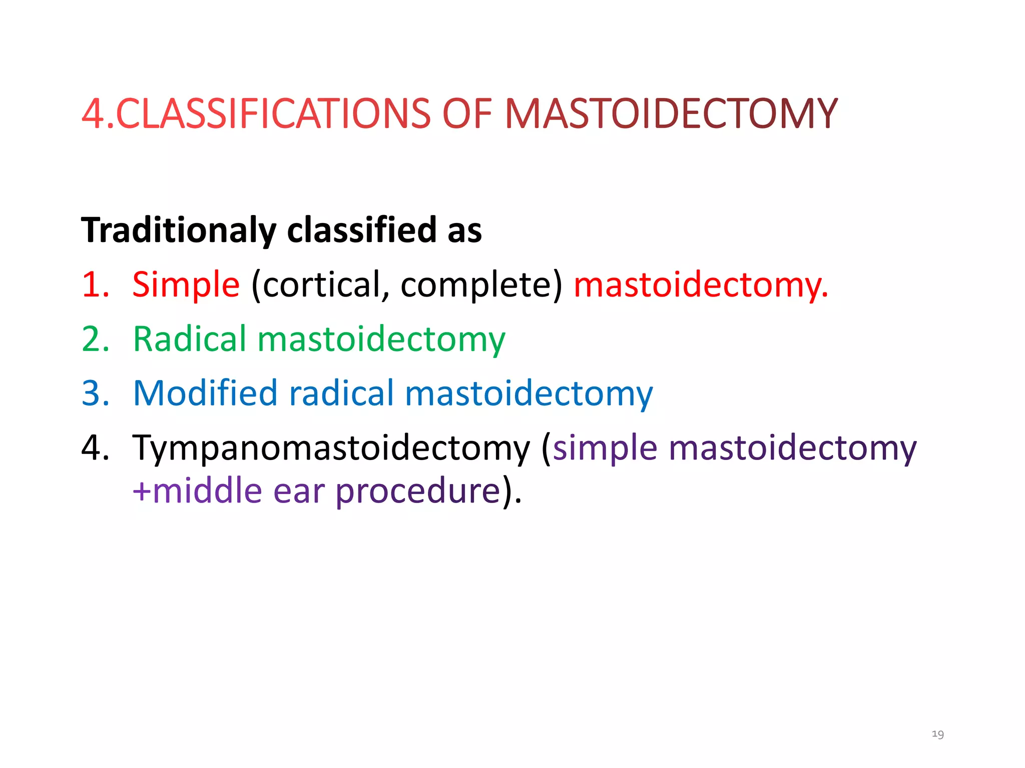
2. The document outlines the history, surgical anatomy, types (cortical, radical, modified radical), indications, and techniques of mastoidectomy. Types are classified as open (canal wall down) or closed (canal wall up) approaches.
3. Potential complications are discussed briefly. Controversies remain regarding the best surgical techniques and approaches to different pathologies.


![Untitled (6) [Autosaved] ear.pptx ear surgeries](https://cdn.slidesharecdn.com/ss_thumbnails/untitled6autosavedear-251128030434-d7ae6d96-thumbnail.jpg?width=640&height=640&fit=bounds)


