CHOLESTEATOMA
AND COMPLICATIONS OF CSOM
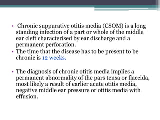
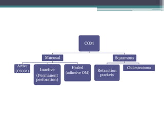
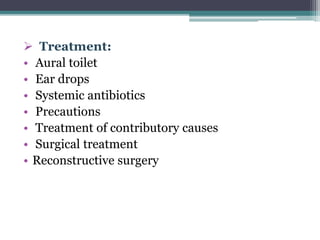
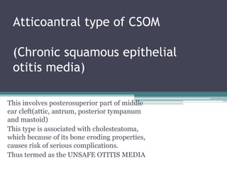
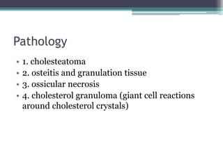
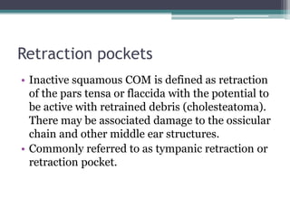
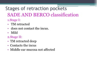
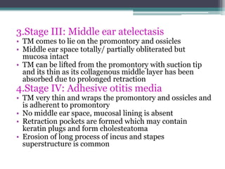
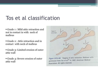
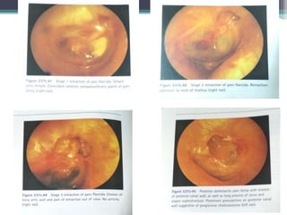
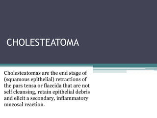
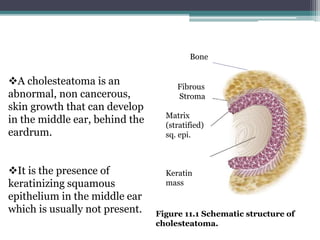
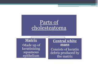
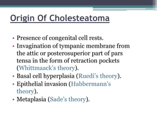
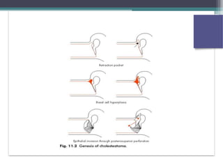
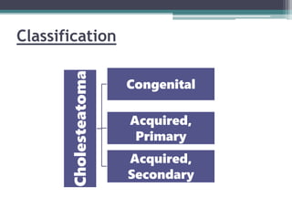
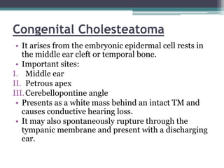
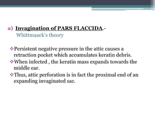
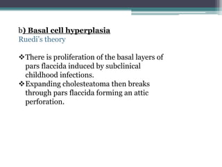
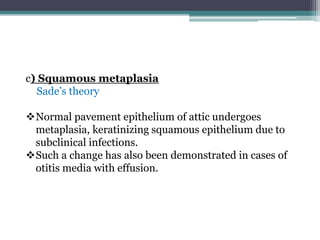
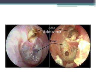
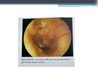
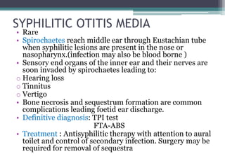
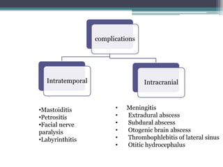
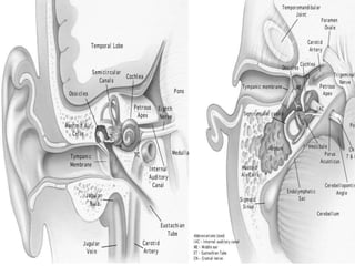
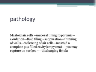
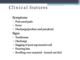
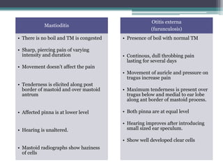
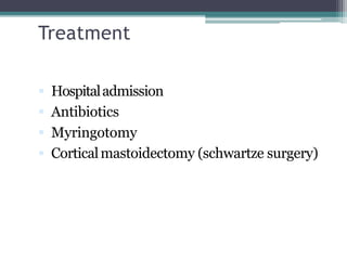
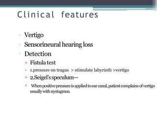
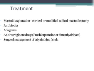
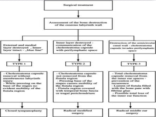
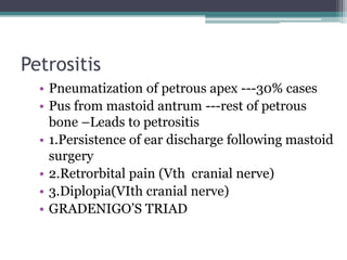
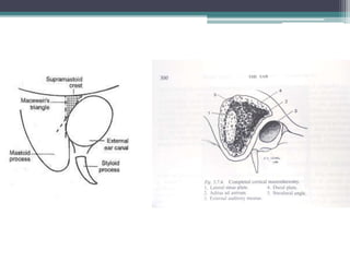
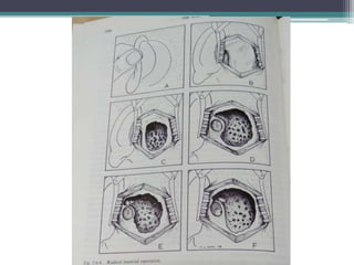
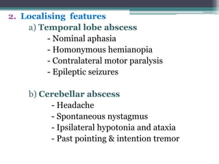
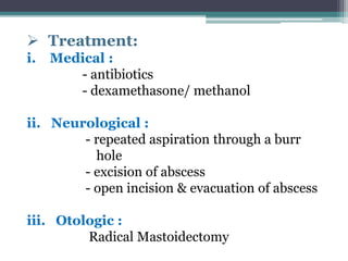
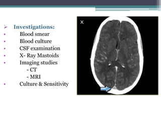
• Chronic suppurative otitis media (CSOM) is a long
standing infection of a part or whole of the middle
ear cleft characterised by ear discharge and a
permanent perforation.
• The time that the disease has to be present to be
chronic is 12 weeks.
• The diagnosis of chronic otitis media implies a
permanent abnormality of the pars tensa or flaccida,
most likely a result of earlier acute otitis media,
negative middle ear pressure or otitis media with
effusion.
COM
Mucosal
Active
(CSOM) Inactive
(Permanent
perforation)
Healed
(adhesive OM)
Squamous
Retraction
pockets
Cholesteatoma
Perforation
of TM
Pars tensa
Central
perforation
Marginal
perforation
Pars
flaccida
Attic
perforation
Unsafe otitis media
Safe otitis media
Tubotympanic type
 Etiology
• sequela of acute otitis media
• ascending infections via Eustachian tube
• persistent mucoid otorrhea
 Pathology
• Perforation of Pars tensa
• Middle ear mucosa (red and velvety)
• Polyp (pale-smooth mass of edematous mucosa
protruding through perforation)
• Ossicular chain (may show necrosis)
• Tympanosclerosis (hyalinization & calcification
of subepithelial cells)
• Fibrosis and adhesions
 Clinical features:
• Ear discharge (profuse, mucoid, odourless)
• Hearing loss (conductive upto 50dB)
• Perforation (central)
• Middle ear mucosa (red, edematous, swollen)
• Inactive mucosal COM(dry perforation)
Perforation in the pars tensa, but middle ear
mucosa and mastoid mucosa not inflamed.
Squamous epithelium can migrate medially into
the middle ear. At time of tympanoplasty, it is
important for an otologic surgeon to excise such
ingrown squamous epithelium, which can be
recognized by its velvety appearance under the
operating microscope.
Active mucosal COM
• Chronic inflammation within the mucosa of the
middle ear and mastoid, with varying degrees of
edema, submucosal fibrosis, hypervascularity
and infiltration with lymphocytes, plasma cells
and histiocytes.
• Mucopurulent discharge which drains via TM
perforation.
• Mucosal changes Aural polyps
coalesce
 Assessment:
• Examination under microscope
• Audiogram
• Culture and sensitivity
• Mastoid X- Rays/ CT Scan temporal bone
 Treatment:
• Aural toilet
• Ear drops
• Systemic antibiotics
• Precautions
• Treatment of contributory causes
• Surgical treatment
• Reconstructive surgery
Atticoantral type of CSOM
(Chronic squamous epithelial
otitis media)
This involves posterosuperior part of middle
ear cleft(attic, antrum, posterior tympanum
and mastoid)
This type is associated with cholesteatoma,
which because of its bone eroding properties,
causes risk of serious complications.
Thus termed as the UNSAFE OTITIS MEDIA
Pathology
• 1. cholesteatoma
• 2. osteitis and granulation tissue
• 3. ossicular necrosis
• 4. cholesterol granuloma (giant cell reactions
around cholesterol crystals)
Symptoms
• 1. Ear discharge
• (scanty, purulent,
odourless)
• 2. hearing loss
(conductive/ mixed
deafness)
• 3. bleeding
1. Perforation
(attic/posterosuperior
marginal type)
2. Retraction pocket
(Invagination of tympanic
membrane)
3. Cholesteatoma
Signs
Retraction pockets
• Inactive squamous COM is defined as retraction
of the pars tensa or flaccida with the potential to
be active with retrained debris (cholesteatoma).
There may be associated damage to the ossicular
chain and other middle ear structures.
• Commonly referred to as tympanic retraction or
retraction pocket.
Stages of retraction pockets
SADE AND BERCO classification
1.Stage I:
• TM retracted
• does not contact the incus.
• Mild
2.Stage II:
• TM retracted deep
• Contacts the incus
• Middle ear mucosa not affected
3.Stage III: Middle ear atelectasis
• TM comes to lie on the promontory and ossicles
• Middle ear space totally/ partially obliterated but
mucosa intact
• TM can be lifted from the promontory with suction tip
and its thin as its collagenous middle layer has been
absorbed due to prolonged retraction
4.Stage IV: Adhesive otitis media
• TM very thin and wraps the promontory and ossicles and
is adherent to promontory
• No middle ear space, mucosal lining is absent
• Retraction pockets are formed which may contain
keratin plugs and form cholesteatoma
• Erosion of long process of incus and stapes
superstructure is common
Tos et al classification
 Grade 1 : Mild attic retraction and
not in contact in with neck of
malleus
 Grade 2 : Attic retraction and in
contact with neck of malleus
 Grade 3: Limited erosion of outer
attic wall
 Grade 4: Severe erosion of outer
attic wall
CHOLESTEATOMA
Cholesteatomas are the end stage of
(squamous epithelial) retractions of
the pars tensa or flaccida that are not
self cleansing, retain epithelial debris
and elicit a secondary, inflammatory
mucosal reaction.
Fibrous
Stroma
Matrix
(stratified)
sq. epi.
Keratin
mass
Figure 11.1 Schematic structure of
cholesteatoma.
Bone
A cholesteatoma is an
abnormal, non cancerous,
skin growth that can develop
in the middle ear, behind the
eardrum.
It is the presence of
keratinizing squamous
epithelium in the middle ear
which is usually not present.
Parts of
cholesteatoma
Matrix
•Made up of
keratinizing
squamous
epithelium
Central white
mass
Consists of keratin
debris produced by
the matrix
Origin Of Cholesteatoma
• Presence of congenital cell rests.
• Invagination of tympanic membrane from
the attic or posterosuperior part of pars
tensa in the form of retraction pockets
(Whittmaack’s theory).
• Basal cell hyperplasia (Ruedi’s theory).
• Epithelial invasion (Habbermann’s
theory).
• Metaplasia (Sade’s theory).
Cholesteatoma Congenital
Acquired,
Primary
Acquired,
Secondary
Classification
Congenital Cholesteatoma
• It arises from the embryonic epidermal cell rests in
the middle ear cleft or temporal bone.
• Important sites:
I. Middle ear
II. Petrous apex
III.Cerebellopontine angle
• Presents as a white mass behind an intact TM and
causes conductive hearing loss.
• It may also spontaneously rupture through the
tympanic membrane and present with a discharging
ear.
Primary Acquired Cholesteatoma
• It is called primary as there is no history of
previous otitis media or a pre-existing
perforation.
• Theories on its genesis are:-
a) Invagination of PARS FLACCIDA.-
Whittmaack’s theory
Persistent negative pressure in the attic causes a
retraction pocket which accumulates keratin debris.
When infected , the keratin mass expands towards the
middle ear.
Thus, attic perforation is in fact the proximal end of an
expanding invaginated sac.
b) Basal cell hyperplasia
Ruedi’s theory
There is proliferation of the basal layers of
pars flaccida induced by subclinical
childhood infections.
Expanding cholesteatoma then breaks
through pars flaccida forming an attic
perforation.
c) Squamous metaplasia
Sade’s theory
Normal pavement epithelium of attic undergoes
metaplasia, keratinizing squamous epithelium due to
subclinical infections.
Such a change has also been demonstrated in cases of
otitis media with effusion.
Secondary Acquired Cholesteatoma
• Pre-existing perforation in pars tensa.
• Associated with posterosuperior marginal
perforation or sometimes large central
perforation.
• Theories of genesis include:
• Squamous metaplasia
• Misplaced epithelium
Most likely to arise from retraction pocket in the pars
flaccida or posterosuperior part of pars tensa.(initiating
factor is probably dysfunction of the Eustachian tube
resulting in negative middle ear pressure)
(best evidence suggests that cholesteatoma arises from skin cells of tympanic
membrane.
The middle ear cholesteatoma has cytokeratin pattern typical of skin and
closely resembling skin of the external auditory meatus. )
• Because of greater blood supply of compared
with the rest of the tympanic membrane, pars
flaccida and the posterosuperior quadrant are
more affected by inflammatory cell infiltration in
otitis media.
• This may leave fibrous layer in these areas
thinner than the remainder of TM and the
overcrowding of ossicles further adds to it.
Expansion of Cholesteatoma and
Destruction of Bone
• An attic cholesteatoma may extend backwards into
the mesotympanum; medially, it may surround the
incus and/or head of malleus.
• It may cause destruction of ear ossicles, erosion of
bony labyrinth, canal of facial nerve, sinus plate or
tegmen tympani and thus cause several
complications.
• Resorption of bone is a feature of both active
mucosal and squamous epithelial COM.
• Number of triggers such as infection, inflammation,
pressure and keratin can lead to elaboration of a
variety of molecular factors including cytokines such
as interleukins- IL-1, IL-6 and TNF, other protein
mediators such as GF, and non protein mediators
PGs, NTs and NO. These molecular factors are
believed to provide the initiating signals that lead to
the recruitment , development and activation of
osteoclasts.
• Enzymes implicated include collagenase, acid
phosphatase and proteolytic enzymes, liberated
by osteoclasts and mononuclear inflammatory
cells, seen in association with cholesteatoma.
Symptoms
• Presentation with foul smelling otorrhoea and
hearing impairment.
• Sometimes the discharge is very scanty
Assessment
• Examination under microscope
• Tuning fork tests and audiogram
• X-ray mastoids/ CT scan temporal bone
• Culture and sensitivity of ear discharge
Treatment
1. SURGICAL:
(a) Canal wall down procedures
(b) Canal wall up procedures
2.RECONSTRUCTIVE SURGERY
3. CONSERVATIVE TREATMENT
• CANAL WALL UP PROCEDURE
• It’s a combined approach through the meatus and
mastoid to remove disease but retaining the
posterior bony meatal wall intact, thereby avoiding
an open mastoid cavity.
• Gives dry ear and permits easy reconstruction of
hearing mechanism.
• However, there is danger of leaving some
cholesteatoma behind.
• Incidence of residual or recurrent cholesteatoma is
very high and therefore long term follow up is
required.
• Advised only in selected cases.
• CANAL WALL DOWN PROCEDURE
• (1)Traditional method for removal- modified radical
mastoidectomy using posterior to anterior approach.
• Disadvantages: larger cavity(much larger than required
to control the disease) (mean volume 2.4cm³)
• These lager cavities even if they do epithelialize, often
don’t self clean.
• Small cavity mastoidectomy(attico-antrostomy).
• Newer approach- Anterior to posterior
The cholesteatoma is identified in the epitympanum
or the posterior mesotympanum and followed
backwards.
Much smaller cavity is resulted (mean volume 1.4cm³)
Canal wall down surgery has lower rates of recurrence
of cholesteatoma(5-15%) and recurrences are easily
identified in OPD.
A significant proportion of patients(20-25%) continue
to have otorrhoea, either intermittent or continous,
after mastoid surgery.
• Intact canal wall mastoidectomy
• Disease is removed both premeatally, and through
cortical mastoidectomy and posterior tympanotomy
approach, in which a window is created between the
mastoid and middle ear, through the facial recess, to
reach sinus tympani.
FEATURES
• Meatus
• Dependence
• Recurrence or residual
disease
• Second look surgery
• Patients limitations
• Auditory rehabilitation
CANAL WALL UP
• Normal appearance
• Does not require routine
cleaning
• High rate of recurrent or
residual cholesteatoma
• Requires second look
surgery after 6 months or
so to rule out
cholesteatoma
• No limitation. Patient
allowed swimming
• Easy to wear a hearing
aid if needed
CANAL WALL DOWN
• Widely open meatus
communicating with
mastoid
• Dependence on doctor for
cleaning mastoid cavity
once or twice a year
• Low rate of recurrence or
residual disease and thus
a safe procedure
• Not required
• Swimming can lead to
infection of mastoid
cavity and it is thus
curtailed
• Problems in fitting a
hearing aid due to large
meatus and mastoid
cavity which sometimes
gets infected
Features indicating complications in
CSOM
1. Pain
2. Vertigo
3. Persistent headache
4. Facial weakness
5. A listless child refusing to take feeds
6. Fever, nausea and vomitting
7. Irritability and neck rigidity
8. Diplopia
9. Ataxia
10. Abscess round the ear
TUBERCULAR OTITIS MEDIA
Aetiology:
 In most cases, infection is
secondary to pulmonary tuberculosis.
 Infection reaches the middle ear
through Eustachian tube.
Pathology
 Slow and insidious
 Tubercles appear in the submucosal layers of
middle ear cleft and caseate.
painless necrosis of TM
multiple perforations may form which coalesce to
form a single large perforation.
Caries of bone and ossicles may occur leading to
complications.
Mastoiditis, facial paralysis, postauricular fistula
osteomyelitis with formation of bony sequestra and
profound hearing loss.
Clinical features
1. Painless ear discharge
2. Perforation
3. Hearing loss
4. Facial paralysis
Diagnosis
• Culture of ear discharge for tubercle bacilli
• Histopathological examination of granulations
• X-ray chest
• Other evidence of tuberculosis in the body
• DNA probe and PCR from the ear discharge can
give early diagnosis in 3-7 days.
Treatment
1. Systemic antitubercular therapy
2. Local treatment
(aural toilet and control of secondary pyogenic
infection)
3. Mastoid surgery
(indicated for complications)
SYPHILITIC OTITIS MEDIA
• Rare
• Spirochaetes reach middle ear through Eustachian tube
when syphilitic lesions are present in the nose or
nasopharynx.(infection may also be blood borne )
• Sensory end organs of the inner ear and their nerves are
soon invaded by spirochaetes leading to:
o Hearing loss
o Tinnitus
o Vertigo
• Bone necrosis and sequestrum formation are common
complications leading foetid ear discharge.
• Definitive diagnosis: TPI test
FTA-ABS
• Treatment : Antisyphilitic therapy with attention to aural
toilet and control of secondary infection. Surgery may be
required for removal of sequestra
COMPLICATIONS OF CSOM
complications
Intratemporal Intracranial
•Mastoiditis
•Petrositis
•Facial nerve
paralysis
•Labyrinthitis
• Meningitis
• Extradural abscess
• Subdural abscess
• Otogenic brain abscess
• Thrombophlebitis of lateral sinus
• Otitic hydrocephalus
FACTORS influencing of
complications
 Age
 Poor socioeconomic group
 Virulence of organisms
 Preformed pathways
 cholesteatoma
 immunocompromised host
Intratemporal
complications
 Mastoiditis
 Facialnerveparalysis
 Labyrinthitis
 Petrositis
Mastoid a i r c e l l system
 Mastoiditis
 mastoidabscess
 Mastoidfistula
Mastoiditis
 Mostcommonextracranialcomplication
 Pus production undertension
 Decalcification & osteoclastic resorption of
bony walls
pathology
Mastoid air cells –mucosal lining hyperemic—
exudation—fluid filing –suppuration—thinning
of walls—coalescing of air cells—mastoid a
complete pus filled cavity(empyema)---pus may
rupture on surface ----discharging fistula
Clinical features
 Symptoms
 Postauralpain
 Fever
 Discharge(profuse and purulent)
 Signs
 Tenderness
 Discharge
 Saggingof postsupmeatalwall
 Hearingloss
 Swelling over mastoid –ironed out feel
Mastioditis
• There is no boil and TM is congested
• Sharp, piercing pain of varying
intensity and duration
• Movement doesn’t affect the pain
• Tenderness is elicited along post
border of mastoid and over mastoid
antrum
• Affected pinna is at lower level
• Hearing is unaltered.
• Mastoid radiographs show haziness
of cells
Otitis externa
(furunculosis)
• Presence of boil with normal TM
• Continous, dull throbbing pain
lasting for several days
• Movement of auricle and pressure on
tragus increase pain
• Maximum tenderness is present over
tragus below and medial to ear lobe
along ant border of mastoid process.
• Both pinna are at equal level
• Hearing improves after introducing
small sized ear speculum.
• Show well developed clear cells
Treatment
 Hospitaladmission
 Antibiotics
 Myringotomy
 Corticalmastoidectomy (schwartze surgery)
Facial nerve paralysis
Treatment
 1.Urgentexploration of middle ear and
mastoid
 2.Facial nerve decompression
Labyrinthitis
 Thinningorerosionof bony capsule
 Twotypes
 Circumscribed(fistula of labyrinth)
 There is thinning or erosion of bony capsule of
labyrinth, usually of the horizontal semicircular
canal
 Diffuse-
 Serous-Itisdiffuseintralabyrinthineinflammationwithoutpus
formationandisareversibleconditioniftreatedearly-
suppurative-This is diffuse pyogenic infection of the labyrinth
with permanent loss of vestibular and cochlear functions.
LABYRINTHINE FISTULA
• A labyrinthine fistula is an abnormal opening in
the inner ear. This can result in leakage of the
perilymph into the middle ear. This includes
specifically a perilymph fistula (PLF), an abnormal
connection between the fluid of the inner ear and the
air-filled middle ear. This is caused by a rupture of the
round window or oval window ligaments separating
the inner and middle ear.
• SIGNS AND SYMPTOMS
• dizziness, imbalance, and hearing loss
• CAUSES
• Cholesteatoma, glomus tumor, during surgery or
trauma
• thinning of the otic capsule by the persistent
pulsations of the intracranial pressures
Clinical features
 Vertigo
 Sensorineuralhearingloss
 Detection
 Fistulatest
 1.pressure on tragus > stimulate labyrinth >vertigo
 2.Seigel’sspeculum—
 Whenpositivepressureisappliedtoearcanal,patientcomplainsofvertigo
usuallywithnystagmus.
Treatment
 Mastoidexploration--cortical or modified radical mastoidectomy
 Antibiotics
 Analgesics
 Anti vertiginousdrugs(Prochlorperazineordimenhydrinate)
 Surgicalmanagementoflabyrinthinefistula
Petrositis
• Pneumatization of petrous apex ---30% cases
• Pus from mastoid antrum ---rest of petrous
bone –Leads to petrositis
• 1.Persistence of ear discharge following mastoid
surgery
• 2.Retrorbital pain (Vth cranial nerve)
• 3.Diplopia(VIth cranial nerve)
• GRADENIGO’S TRIAD
 Osteitisof the petrousbone
 Headache
 DIAGNOSIS: HRCTscan
 Exploration
 Treatment :Antibiotics
INTRACRANIAL
COMPLICATIONS
• Meningitis
• Extradural abscess
• Subdural abscess
• Otogenic brain abscess
• Thrombophlebitis of lateral sinus
• Otitic hydrocephalus
1. MENINGITIS
 usually presents as acute illness with:
- pyrexia (102- 104º F)
- severe headache
- neck rigidity
- nausea & vomiting
- cranial nerve palsies
- drowsiness and coma (in later stages)
 Signs:
• Kernig’s sign positive
• Brudzinski’s sign positive
• Diagnosis :
- CT
- MRI
- Lumbar puncture and CSF examination
• Treatment:
i. medical:
- broad spectrum antibiotics
- corticosteroids may be added
ii. surgical:
Mastoidectomy
2. EXTRADURAL ABSCESS
Clinical features:
• persistent headache
• severe earache
• low grade fever
• pulsatile purulent ear discharge
Treatment:
• Cortical or modified radical Mastoidectomy
• An antibiotic cover
3. SUBDURAL ABSCESS
Clinical features:
• Meningeal irritation
• Cortical venous thrombophlebitis
• Raised intracranial tension
Treatment:
• series of burr holes or craniotomy to drain the
abscess
• intravenous antibiotics
• lumbar puncture should not be done
4. OTOGENIC BRAIN ABSCESS
Pathology:
Stage of invasion
Stage of
localisation
Stage of
enlargement
Stage of
termination
Clinical
features
Localising
features
Temporal
lobe abscess
Cerebellar
abscess
Due to
raised ICT
 Clinical features:
1. Due to increased intracranial tension
• headache
• nausea & vomiting
• level of unconsciousness
• papilloedema
• slow pulse and subnormal temperature
2. Localising features
a) Temporal lobe abscess
- Nominal aphasia
- Homonymous hemianopia
- Contralateral motor paralysis
- Epileptic seizures
b) Cerebellar abscess
- Headache
- Spontaneous nystagmus
- Ipsilateral hypotonia and ataxia
- Past pointing & intention tremor
 Investigations:
• X- Rays
• CT Scan
• Lumbar puncture
 Treatment:
i. Medical :
- antibiotics
- dexamethasone/ methanol
ii. Neurological :
- repeated aspiration through a burr
hole
- excision of abscess
- open incision & evacuation of abscess
iii. Otologic :
Radical Mastoidectomy
5. LATERAL SINUS
THROMBOPHLEBITIS
PATHOLOGY:
Formation of
perisinus
abscess
Endophthalmitis
and mural
thrombus
formation
Obliteration
of sinus
lumen and
intrasinus
abscess
Extension
of thrombus
 Clinical features:
• Hectic Picket-Fence type of fever with
rigors
• headache
• progressive anaemia
• Gresienger’s sign
• papilloedema
• Tobey Ayer test
• Crowe –Beck test
• Tenderness along Jugular vein
 Investigations:
• Blood smear
• Blood culture
• CSF examination
• X- Ray Mastoids
• Imaging studies
- CT
- MRI
• Culture & Sensitivity
 Complications:
• Septicaemia and pyaemic abscess
• Meningitis & subdural abscess
• Cerebellar abscess
• Thrombosis of jugular bulb
• Cavernous sinus thrombosis
• Otitic hydrocephalus
 Treatment:
• Intravenous antibacterial therapy
• Mastoidectomy & exposure of sinus
• Ligation of internal jugular vein
• Anticoagulant therapy
• Supportive treatment
6. OTITIC HYDROCEPHALUS
• Etiology: results from sigmoid sinus
thrombosis with obstruction of other
intracranial venous channels
• Symptoms:
- severe headache
- diplopia
- blurring of vision
• Signs:
- Papilloedema
- Nystagmus
- Lumbar
puncture ( CSF >
300 mm water )
• Investigation:
CT scan
• Treatment:
- Acetazolamide
- Corticosteroids
- lumbar drain
- lumboperitoneal shunt
REFERENCES
• PL Dhingra diseases of ear, nose throat - 7th
edition
• Scott Brown’s otorhinolaryngology, Head and
neck surgery volume 3 -7th edition
• Logan Turner’s diseases of the nose, throat and
ear -10th edition
THANK YOU

Chronic suppurative otitis media

  • 1.
  • 2.
    • Chronic suppurativeotitis media (CSOM) is a long standing infection of a part or whole of the middle ear cleft characterised by ear discharge and a permanent perforation. • The time that the disease has to be present to be chronic is 12 weeks. • The diagnosis of chronic otitis media implies a permanent abnormality of the pars tensa or flaccida, most likely a result of earlier acute otitis media, negative middle ear pressure or otitis media with effusion.
  • 3.
  • 4.
  • 5.
    Tubotympanic type  Etiology •sequela of acute otitis media • ascending infections via Eustachian tube • persistent mucoid otorrhea
  • 6.
     Pathology • Perforationof Pars tensa • Middle ear mucosa (red and velvety) • Polyp (pale-smooth mass of edematous mucosa protruding through perforation) • Ossicular chain (may show necrosis) • Tympanosclerosis (hyalinization & calcification of subepithelial cells) • Fibrosis and adhesions
  • 7.
     Clinical features: •Ear discharge (profuse, mucoid, odourless) • Hearing loss (conductive upto 50dB) • Perforation (central) • Middle ear mucosa (red, edematous, swollen)
  • 8.
    • Inactive mucosalCOM(dry perforation) Perforation in the pars tensa, but middle ear mucosa and mastoid mucosa not inflamed. Squamous epithelium can migrate medially into the middle ear. At time of tympanoplasty, it is important for an otologic surgeon to excise such ingrown squamous epithelium, which can be recognized by its velvety appearance under the operating microscope.
  • 9.
    Active mucosal COM •Chronic inflammation within the mucosa of the middle ear and mastoid, with varying degrees of edema, submucosal fibrosis, hypervascularity and infiltration with lymphocytes, plasma cells and histiocytes. • Mucopurulent discharge which drains via TM perforation. • Mucosal changes Aural polyps coalesce
  • 11.
     Assessment: • Examinationunder microscope • Audiogram • Culture and sensitivity • Mastoid X- Rays/ CT Scan temporal bone
  • 12.
     Treatment: • Auraltoilet • Ear drops • Systemic antibiotics • Precautions • Treatment of contributory causes • Surgical treatment • Reconstructive surgery
  • 13.
    Atticoantral type ofCSOM (Chronic squamous epithelial otitis media) This involves posterosuperior part of middle ear cleft(attic, antrum, posterior tympanum and mastoid) This type is associated with cholesteatoma, which because of its bone eroding properties, causes risk of serious complications. Thus termed as the UNSAFE OTITIS MEDIA
  • 14.
    Pathology • 1. cholesteatoma •2. osteitis and granulation tissue • 3. ossicular necrosis • 4. cholesterol granuloma (giant cell reactions around cholesterol crystals)
  • 15.
    Symptoms • 1. Eardischarge • (scanty, purulent, odourless) • 2. hearing loss (conductive/ mixed deafness) • 3. bleeding 1. Perforation (attic/posterosuperior marginal type) 2. Retraction pocket (Invagination of tympanic membrane) 3. Cholesteatoma Signs
  • 16.
    Retraction pockets • Inactivesquamous COM is defined as retraction of the pars tensa or flaccida with the potential to be active with retrained debris (cholesteatoma). There may be associated damage to the ossicular chain and other middle ear structures. • Commonly referred to as tympanic retraction or retraction pocket.
  • 17.
    Stages of retractionpockets SADE AND BERCO classification 1.Stage I: • TM retracted • does not contact the incus. • Mild 2.Stage II: • TM retracted deep • Contacts the incus • Middle ear mucosa not affected
  • 18.
    3.Stage III: Middleear atelectasis • TM comes to lie on the promontory and ossicles • Middle ear space totally/ partially obliterated but mucosa intact • TM can be lifted from the promontory with suction tip and its thin as its collagenous middle layer has been absorbed due to prolonged retraction 4.Stage IV: Adhesive otitis media • TM very thin and wraps the promontory and ossicles and is adherent to promontory • No middle ear space, mucosal lining is absent • Retraction pockets are formed which may contain keratin plugs and form cholesteatoma • Erosion of long process of incus and stapes superstructure is common
  • 19.
    Tos et alclassification  Grade 1 : Mild attic retraction and not in contact in with neck of malleus  Grade 2 : Attic retraction and in contact with neck of malleus  Grade 3: Limited erosion of outer attic wall  Grade 4: Severe erosion of outer attic wall
  • 21.
    CHOLESTEATOMA Cholesteatomas are theend stage of (squamous epithelial) retractions of the pars tensa or flaccida that are not self cleansing, retain epithelial debris and elicit a secondary, inflammatory mucosal reaction.
  • 22.
    Fibrous Stroma Matrix (stratified) sq. epi. Keratin mass Figure 11.1Schematic structure of cholesteatoma. Bone A cholesteatoma is an abnormal, non cancerous, skin growth that can develop in the middle ear, behind the eardrum. It is the presence of keratinizing squamous epithelium in the middle ear which is usually not present.
  • 23.
    Parts of cholesteatoma Matrix •Made upof keratinizing squamous epithelium Central white mass Consists of keratin debris produced by the matrix
  • 24.
    Origin Of Cholesteatoma •Presence of congenital cell rests. • Invagination of tympanic membrane from the attic or posterosuperior part of pars tensa in the form of retraction pockets (Whittmaack’s theory). • Basal cell hyperplasia (Ruedi’s theory). • Epithelial invasion (Habbermann’s theory). • Metaplasia (Sade’s theory).
  • 26.
  • 27.
    Congenital Cholesteatoma • Itarises from the embryonic epidermal cell rests in the middle ear cleft or temporal bone. • Important sites: I. Middle ear II. Petrous apex III.Cerebellopontine angle • Presents as a white mass behind an intact TM and causes conductive hearing loss. • It may also spontaneously rupture through the tympanic membrane and present with a discharging ear.
  • 28.
    Primary Acquired Cholesteatoma •It is called primary as there is no history of previous otitis media or a pre-existing perforation. • Theories on its genesis are:-
  • 29.
    a) Invagination ofPARS FLACCIDA.- Whittmaack’s theory Persistent negative pressure in the attic causes a retraction pocket which accumulates keratin debris. When infected , the keratin mass expands towards the middle ear. Thus, attic perforation is in fact the proximal end of an expanding invaginated sac.
  • 30.
    b) Basal cellhyperplasia Ruedi’s theory There is proliferation of the basal layers of pars flaccida induced by subclinical childhood infections. Expanding cholesteatoma then breaks through pars flaccida forming an attic perforation.
  • 31.
    c) Squamous metaplasia Sade’stheory Normal pavement epithelium of attic undergoes metaplasia, keratinizing squamous epithelium due to subclinical infections. Such a change has also been demonstrated in cases of otitis media with effusion.
  • 32.
    Secondary Acquired Cholesteatoma •Pre-existing perforation in pars tensa. • Associated with posterosuperior marginal perforation or sometimes large central perforation. • Theories of genesis include:
  • 33.
    • Squamous metaplasia •Misplaced epithelium Most likely to arise from retraction pocket in the pars flaccida or posterosuperior part of pars tensa.(initiating factor is probably dysfunction of the Eustachian tube resulting in negative middle ear pressure) (best evidence suggests that cholesteatoma arises from skin cells of tympanic membrane. The middle ear cholesteatoma has cytokeratin pattern typical of skin and closely resembling skin of the external auditory meatus. )
  • 34.
    • Because ofgreater blood supply of compared with the rest of the tympanic membrane, pars flaccida and the posterosuperior quadrant are more affected by inflammatory cell infiltration in otitis media. • This may leave fibrous layer in these areas thinner than the remainder of TM and the overcrowding of ossicles further adds to it.
  • 36.
    Expansion of Cholesteatomaand Destruction of Bone • An attic cholesteatoma may extend backwards into the mesotympanum; medially, it may surround the incus and/or head of malleus. • It may cause destruction of ear ossicles, erosion of bony labyrinth, canal of facial nerve, sinus plate or tegmen tympani and thus cause several complications.
  • 37.
    • Resorption ofbone is a feature of both active mucosal and squamous epithelial COM. • Number of triggers such as infection, inflammation, pressure and keratin can lead to elaboration of a variety of molecular factors including cytokines such as interleukins- IL-1, IL-6 and TNF, other protein mediators such as GF, and non protein mediators PGs, NTs and NO. These molecular factors are believed to provide the initiating signals that lead to the recruitment , development and activation of osteoclasts.
  • 38.
    • Enzymes implicatedinclude collagenase, acid phosphatase and proteolytic enzymes, liberated by osteoclasts and mononuclear inflammatory cells, seen in association with cholesteatoma.
  • 40.
    Symptoms • Presentation withfoul smelling otorrhoea and hearing impairment. • Sometimes the discharge is very scanty
  • 41.
    Assessment • Examination undermicroscope • Tuning fork tests and audiogram • X-ray mastoids/ CT scan temporal bone • Culture and sensitivity of ear discharge
  • 43.
    Treatment 1. SURGICAL: (a) Canalwall down procedures (b) Canal wall up procedures 2.RECONSTRUCTIVE SURGERY 3. CONSERVATIVE TREATMENT
  • 44.
    • CANAL WALLUP PROCEDURE • It’s a combined approach through the meatus and mastoid to remove disease but retaining the posterior bony meatal wall intact, thereby avoiding an open mastoid cavity. • Gives dry ear and permits easy reconstruction of hearing mechanism. • However, there is danger of leaving some cholesteatoma behind. • Incidence of residual or recurrent cholesteatoma is very high and therefore long term follow up is required. • Advised only in selected cases.
  • 45.
    • CANAL WALLDOWN PROCEDURE • (1)Traditional method for removal- modified radical mastoidectomy using posterior to anterior approach. • Disadvantages: larger cavity(much larger than required to control the disease) (mean volume 2.4cm³) • These lager cavities even if they do epithelialize, often don’t self clean.
  • 46.
    • Small cavitymastoidectomy(attico-antrostomy). • Newer approach- Anterior to posterior The cholesteatoma is identified in the epitympanum or the posterior mesotympanum and followed backwards. Much smaller cavity is resulted (mean volume 1.4cm³) Canal wall down surgery has lower rates of recurrence of cholesteatoma(5-15%) and recurrences are easily identified in OPD. A significant proportion of patients(20-25%) continue to have otorrhoea, either intermittent or continous, after mastoid surgery.
  • 47.
    • Intact canalwall mastoidectomy • Disease is removed both premeatally, and through cortical mastoidectomy and posterior tympanotomy approach, in which a window is created between the mastoid and middle ear, through the facial recess, to reach sinus tympani.
  • 48.
    FEATURES • Meatus • Dependence •Recurrence or residual disease • Second look surgery • Patients limitations • Auditory rehabilitation CANAL WALL UP • Normal appearance • Does not require routine cleaning • High rate of recurrent or residual cholesteatoma • Requires second look surgery after 6 months or so to rule out cholesteatoma • No limitation. Patient allowed swimming • Easy to wear a hearing aid if needed CANAL WALL DOWN • Widely open meatus communicating with mastoid • Dependence on doctor for cleaning mastoid cavity once or twice a year • Low rate of recurrence or residual disease and thus a safe procedure • Not required • Swimming can lead to infection of mastoid cavity and it is thus curtailed • Problems in fitting a hearing aid due to large meatus and mastoid cavity which sometimes gets infected
  • 49.
    Features indicating complicationsin CSOM 1. Pain 2. Vertigo 3. Persistent headache 4. Facial weakness 5. A listless child refusing to take feeds 6. Fever, nausea and vomitting 7. Irritability and neck rigidity 8. Diplopia 9. Ataxia 10. Abscess round the ear
  • 50.
    TUBERCULAR OTITIS MEDIA Aetiology: In most cases, infection is secondary to pulmonary tuberculosis.  Infection reaches the middle ear through Eustachian tube.
  • 51.
    Pathology  Slow andinsidious  Tubercles appear in the submucosal layers of middle ear cleft and caseate. painless necrosis of TM multiple perforations may form which coalesce to form a single large perforation. Caries of bone and ossicles may occur leading to complications. Mastoiditis, facial paralysis, postauricular fistula osteomyelitis with formation of bony sequestra and profound hearing loss.
  • 52.
    Clinical features 1. Painlessear discharge 2. Perforation 3. Hearing loss 4. Facial paralysis
  • 53.
    Diagnosis • Culture ofear discharge for tubercle bacilli • Histopathological examination of granulations • X-ray chest • Other evidence of tuberculosis in the body • DNA probe and PCR from the ear discharge can give early diagnosis in 3-7 days.
  • 54.
    Treatment 1. Systemic antituberculartherapy 2. Local treatment (aural toilet and control of secondary pyogenic infection) 3. Mastoid surgery (indicated for complications)
  • 55.
    SYPHILITIC OTITIS MEDIA •Rare • Spirochaetes reach middle ear through Eustachian tube when syphilitic lesions are present in the nose or nasopharynx.(infection may also be blood borne ) • Sensory end organs of the inner ear and their nerves are soon invaded by spirochaetes leading to: o Hearing loss o Tinnitus o Vertigo • Bone necrosis and sequestrum formation are common complications leading foetid ear discharge. • Definitive diagnosis: TPI test FTA-ABS • Treatment : Antisyphilitic therapy with attention to aural toilet and control of secondary infection. Surgery may be required for removal of sequestra
  • 56.
  • 57.
    complications Intratemporal Intracranial •Mastoiditis •Petrositis •Facial nerve paralysis •Labyrinthitis •Meningitis • Extradural abscess • Subdural abscess • Otogenic brain abscess • Thrombophlebitis of lateral sinus • Otitic hydrocephalus
  • 58.
    FACTORS influencing of complications Age  Poor socioeconomic group  Virulence of organisms  Preformed pathways  cholesteatoma  immunocompromised host
  • 61.
  • 62.
    Mastoid a ir c e l l system  Mastoiditis  mastoidabscess  Mastoidfistula
  • 63.
    Mastoiditis  Mostcommonextracranialcomplication  Pusproduction undertension  Decalcification & osteoclastic resorption of bony walls
  • 64.
    pathology Mastoid air cells–mucosal lining hyperemic— exudation—fluid filing –suppuration—thinning of walls—coalescing of air cells—mastoid a complete pus filled cavity(empyema)---pus may rupture on surface ----discharging fistula
  • 65.
    Clinical features  Symptoms Postauralpain  Fever  Discharge(profuse and purulent)  Signs  Tenderness  Discharge  Saggingof postsupmeatalwall  Hearingloss  Swelling over mastoid –ironed out feel
  • 66.
    Mastioditis • There isno boil and TM is congested • Sharp, piercing pain of varying intensity and duration • Movement doesn’t affect the pain • Tenderness is elicited along post border of mastoid and over mastoid antrum • Affected pinna is at lower level • Hearing is unaltered. • Mastoid radiographs show haziness of cells Otitis externa (furunculosis) • Presence of boil with normal TM • Continous, dull throbbing pain lasting for several days • Movement of auricle and pressure on tragus increase pain • Maximum tenderness is present over tragus below and medial to ear lobe along ant border of mastoid process. • Both pinna are at equal level • Hearing improves after introducing small sized ear speculum. • Show well developed clear cells
  • 67.
    Treatment  Hospitaladmission  Antibiotics Myringotomy  Corticalmastoidectomy (schwartze surgery)
  • 68.
  • 69.
    Treatment  1.Urgentexploration ofmiddle ear and mastoid  2.Facial nerve decompression
  • 71.
    Labyrinthitis  Thinningorerosionof bonycapsule  Twotypes  Circumscribed(fistula of labyrinth)  There is thinning or erosion of bony capsule of labyrinth, usually of the horizontal semicircular canal  Diffuse-  Serous-Itisdiffuseintralabyrinthineinflammationwithoutpus formationandisareversibleconditioniftreatedearly- suppurative-This is diffuse pyogenic infection of the labyrinth with permanent loss of vestibular and cochlear functions.
  • 72.
    LABYRINTHINE FISTULA • Alabyrinthine fistula is an abnormal opening in the inner ear. This can result in leakage of the perilymph into the middle ear. This includes specifically a perilymph fistula (PLF), an abnormal connection between the fluid of the inner ear and the air-filled middle ear. This is caused by a rupture of the round window or oval window ligaments separating the inner and middle ear. • SIGNS AND SYMPTOMS • dizziness, imbalance, and hearing loss • CAUSES • Cholesteatoma, glomus tumor, during surgery or trauma • thinning of the otic capsule by the persistent pulsations of the intracranial pressures
  • 73.
    Clinical features  Vertigo Sensorineuralhearingloss  Detection  Fistulatest  1.pressure on tragus > stimulate labyrinth >vertigo  2.Seigel’sspeculum—  Whenpositivepressureisappliedtoearcanal,patientcomplainsofvertigo usuallywithnystagmus.
  • 74.
    Treatment  Mastoidexploration--cortical ormodified radical mastoidectomy  Antibiotics  Analgesics  Anti vertiginousdrugs(Prochlorperazineordimenhydrinate)  Surgicalmanagementoflabyrinthinefistula
  • 76.
    Petrositis • Pneumatization ofpetrous apex ---30% cases • Pus from mastoid antrum ---rest of petrous bone –Leads to petrositis • 1.Persistence of ear discharge following mastoid surgery • 2.Retrorbital pain (Vth cranial nerve) • 3.Diplopia(VIth cranial nerve) • GRADENIGO’S TRIAD
  • 77.
     Osteitisof thepetrousbone  Headache  DIAGNOSIS: HRCTscan  Exploration  Treatment :Antibiotics
  • 78.
    INTRACRANIAL COMPLICATIONS • Meningitis • Extraduralabscess • Subdural abscess • Otogenic brain abscess • Thrombophlebitis of lateral sinus • Otitic hydrocephalus
  • 80.
    1. MENINGITIS  usuallypresents as acute illness with: - pyrexia (102- 104º F) - severe headache - neck rigidity - nausea & vomiting - cranial nerve palsies - drowsiness and coma (in later stages)  Signs: • Kernig’s sign positive • Brudzinski’s sign positive
  • 81.
    • Diagnosis : -CT - MRI - Lumbar puncture and CSF examination • Treatment: i. medical: - broad spectrum antibiotics - corticosteroids may be added ii. surgical: Mastoidectomy
  • 84.
    2. EXTRADURAL ABSCESS Clinicalfeatures: • persistent headache • severe earache • low grade fever • pulsatile purulent ear discharge Treatment: • Cortical or modified radical Mastoidectomy • An antibiotic cover
  • 85.
    3. SUBDURAL ABSCESS Clinicalfeatures: • Meningeal irritation • Cortical venous thrombophlebitis • Raised intracranial tension Treatment: • series of burr holes or craniotomy to drain the abscess • intravenous antibiotics • lumbar puncture should not be done
  • 86.
    4. OTOGENIC BRAINABSCESS Pathology: Stage of invasion Stage of localisation Stage of enlargement Stage of termination
  • 88.
  • 89.
     Clinical features: 1.Due to increased intracranial tension • headache • nausea & vomiting • level of unconsciousness • papilloedema • slow pulse and subnormal temperature
  • 90.
    2. Localising features a)Temporal lobe abscess - Nominal aphasia - Homonymous hemianopia - Contralateral motor paralysis - Epileptic seizures b) Cerebellar abscess - Headache - Spontaneous nystagmus - Ipsilateral hypotonia and ataxia - Past pointing & intention tremor
  • 91.
     Investigations: • X-Rays • CT Scan • Lumbar puncture
  • 92.
     Treatment: i. Medical: - antibiotics - dexamethasone/ methanol ii. Neurological : - repeated aspiration through a burr hole - excision of abscess - open incision & evacuation of abscess iii. Otologic : Radical Mastoidectomy
  • 93.
    5. LATERAL SINUS THROMBOPHLEBITIS PATHOLOGY: Formationof perisinus abscess Endophthalmitis and mural thrombus formation Obliteration of sinus lumen and intrasinus abscess Extension of thrombus
  • 94.
     Clinical features: •Hectic Picket-Fence type of fever with rigors • headache • progressive anaemia • Gresienger’s sign • papilloedema • Tobey Ayer test • Crowe –Beck test • Tenderness along Jugular vein
  • 96.
     Investigations: • Bloodsmear • Blood culture • CSF examination • X- Ray Mastoids • Imaging studies - CT - MRI • Culture & Sensitivity
  • 97.
     Complications: • Septicaemiaand pyaemic abscess • Meningitis & subdural abscess • Cerebellar abscess • Thrombosis of jugular bulb • Cavernous sinus thrombosis • Otitic hydrocephalus
  • 98.
     Treatment: • Intravenousantibacterial therapy • Mastoidectomy & exposure of sinus • Ligation of internal jugular vein • Anticoagulant therapy • Supportive treatment
  • 99.
    6. OTITIC HYDROCEPHALUS •Etiology: results from sigmoid sinus thrombosis with obstruction of other intracranial venous channels • Symptoms: - severe headache - diplopia - blurring of vision • Signs: - Papilloedema - Nystagmus - Lumbar puncture ( CSF > 300 mm water )
  • 100.
    • Investigation: CT scan •Treatment: - Acetazolamide - Corticosteroids - lumbar drain - lumboperitoneal shunt
  • 101.
    REFERENCES • PL Dhingradiseases of ear, nose throat - 7th edition • Scott Brown’s otorhinolaryngology, Head and neck surgery volume 3 -7th edition • Logan Turner’s diseases of the nose, throat and ear -10th edition
  • 102.