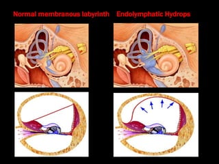
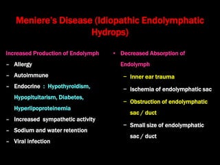
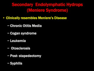
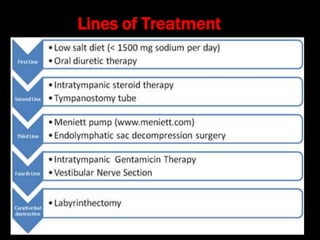
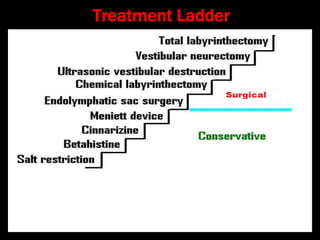
Meniere’s Disease
Dr. Krishna Koirala
8/6/2020
Define Meniere’s disease
• Symptom triad of recurrent episodes of vertigo
of sudden onset, tinnitus, hearing loss and
aural fullness with endolymphatic hydrops as
the principal pathological feature
Normal membranous labyrinth Endolymphatic Hydrops
What is the etiology of Meniere's disease?
What is Meniere’s syndrome? List the
causes of Meniere’s syndrome
Meniere’s Disease (Idiopathic Endolymphatic
Hydrops)
Increased Production of Endolymph
– Allergy
– Autoimmune
– Endocrine : Hypothyroidism,
Hypopituitarism, Diabetes,
Hyperlipoproteinemia
– Increased sympathetic activity
– Sodium and water retention
– Viral infection
• Decreased Absorption of
Endolymph
− Inner ear trauma
− Ischemia of endolymphatic sac
− Obstruction of endolymphatic
sac / duct
− Small size of endolymphatic
sac / duct
Secondary Endolymphatic Hydrops
(Meniere Syndrome)
• Clinically resembles Meniere’s Disease
− Chronic Otitis Media
− Cogan syndrome
− Leukemia
− Otosclerosis
− Post- stapedectomy
− Syphilis
Discuss the pathology of Meniere’s
disease.
• Membrane Rupture Theory
– Endolymphatic hydrops  Rupture of membranous
labyrinth  Potassium rich endolymph (toxic to
neural tissues) gets mixed with perilymph 
Sustained depolarization and inactivation of the
hair cells and neurons of the VIII nerve bathed in
perilymph
• Increased Sympathetic activity
– Ischemia of cochlear & vestibular end organs 
deafness + vertigo
Write down the clinical features of Meniere’s
disease
• Age: 30 - 60 years , F >M, Unilateral > Bilateral
• Vertigo
– Sudden onset, episodic, rotatory, lasting for 24 minutes to
24 hrs , associated with nausea, vomiting & diaphoresis
with normal level of consciousness and orientation
– Vertigo caused by loud , low frequency sound  Tulio
phenomenon
• Deafness:
– Accompanies vertigo and improves after vertigo
attack (fluctuant), sensorineural, progressive
– Intolerance to loud sound (due to recruitment)
– Distortion of sound frequency (Diplacusis
Binauralis Dysharmonica)
• Tinnitus
– Low-pitched, roaring, non-pulsatile, intermittent or
continuous
– Increases during vertigo attacks
• Aural fullness
– Fluctuating , not relieved by swallowing
• Emotional upset and anxiety /depression
AAO-HNS Diagnostic Criteria (1995)
A. Vertigo: Spontaneous, > 2 episodes lasting > 20 min
B. Audiogram documented sensorineural deafness
C. Tinnitus or Aural fullness in diseased ear
D. Other cases excluded
Meniere’s Disease variants
• Lermoyez reverse Meniere syndrome
– Deafness and tinnitus  vertigo hearing improves
• Tumarkin's otolitic Catastrophe (drop attack Meniere's)
– Patient falls without vertigo / loss of consciousness
• Cochlear hydrops
– Deafness & tinnitus only
• Vestibular hydrops: vertigo only ( mimics BPPV)
Examination
• Otoscopy : normal tympanic membrane
• Rinne test : Positive (A.C. > B.C.)
• Weber test : lateralizes towards better ear
• A.B.C. test : reduced in diseased ear
• Nystagmus : beats towards affected ear initially
(Irritative), then to the healthy ear (Paralytic), and
again beating towards the affected ear (Recovery)
Rising Curve
(7% ): Low
frequency
sensorineural
deafness
Discuss the investigations of Meniere’s disease
Flat (42%) :
Uniform
sensorineural
deafness
Downsloping
curve (19%) :
Sensorineural
deafness more in
high frequency
• Pure Tone Audiometry
• Speech Audiometry
– Score  50 - 80 %
• A.B.L.B.
– Presence of Recruitment
(Rapid increase in loudness
above the Threshold level)
• S.I.S.I. : Positive (> 70 % score)
– Presentation of a tone 20 dB above threshold,
followed by a series of 200 millisecond tones 1 dB
louder
– Percentage of small increments detected by the
subject is the small increment sensitivity index
– Perception of these increments indicates cochlear
damage
• Tone Decay Test : Negative (decay < 20 dB)
AP represents the summed response of the synchronous firing of
the nerve fibers
SP is the direct current (DC) response of the hair cells as they move
in conjunction with the basilar membrane
• Transtympanic Electro- cochleography
– Summation Potential : Compound Action Potential
ratio > 30 %
– Widened waveform of SP/ AP complex (>2 mS)
– Distorted cochlear microphonics
• Bithermal Caloric Test
• Ipsilateral canal paresis in 75 % cases
• Contralateral directional preponderance
• Glycerol Test
• Do P.T.A. & speech audiogram
• Glycerol (1 .5 ml / Kg), mixed in lemon juice given orally
and audiometry repeated after 2 hrs
• Test is positive if
• Pure Tone threshold improves > 10 dB
• Speech Discrimination Score increases > 15 %
• S.P. / A.P. ratio in ECOG > 15 %
Treatment
Discuss the treatment of acute attack of
Meniere’s disease
• Reassurance ,bed rest , head support
• Vestibular sedatives
– Inj. Prochlorperazine (Stemetil) : 12.5 mg I.V.,
T.I.D.
– Inj. Promethazine (Phenergan) : 25 mg I.V., T.I.D.
– Inj. Diazepam (Calmpose) : 5 mg I.V. stat
• Intravenous Fluids
Non-surgical treatment
• Discussion
• Reassurance
• Avoid tea, coffee, chocolate, allergens, stress,
smoking, alcohol
• Diet
• Low salt (1.5 g/day), less fluids, Exercise
• Vestibular Depressants
• Cinnarizine, Prochlorperazine
• Cochlear Vasodilators
• Betahistine, Xanthinol nicotinate, Carbogen (5 % CO2
+ 95
% O2
), L.M.W. Dextran, Histamine drip
• Diuretics
• Thiazide + Triamterene
• Dexamethasone / Ig G
• Decreases auto-immunity
• Dehydration by hyperosmolar fluids
• Hormone replacement therapy
Meniett Device
• Low pressure pulse generator
• Pressure pulses transmitted to
round window via grommet 
displaces endolymph and thus
relieves endolymphatic hydrops
• Used for 5 min, TID
Chemical Labyrinthectomy
• Transtympanic injection of ototoxic drugs
• Intra-tympanic drug instillation via grommet or
Silverstein micro wick
• Trans-tympanic drug perfusion
• Drugs used are
− Gentamicin (vestibulotoxic)
− Dexamethasone ( anti-inflammatory)
Trans-tympanic injection Intra-tympanic instillation
Silverstein microwick
Trans-tympanic Drug perfusion
Surgical Treatment:
• Endolymphatic sac decompression
• Endolymphatic sac shunting into subarachnoid space or
mastoid cavity
• Sacculotomy
– Fick’s needle puncture of footplate
– Cody’s tack puncture of footplate
• Cochlear duct piercing via round window
Decompression Surgery
Vestibular Surgery
• Denervation of vestibule by vestibular neurectomy
via middle cranial fossa
• Destruction of vestibule (via round window or lateral
semicircular canal) by cryo -probe or ultrasound
probe
Vestibular Destruction
Ultrasound Probe
Vestibular Neurectomy
Total Destructive Surgery
• Destroys both cochlear & vestibular functions
• Done in patients with severe deafness
• Types of surgery are
– Section of vestibular + cochlear nerves
– Total Labyrinthectomy
Total Destructive Surgery
Lines of Treatment
Treatment Ladder

Meniere's disease

  • 1.
  • 2.
    Define Meniere’s disease •Symptom triad of recurrent episodes of vertigo of sudden onset, tinnitus, hearing loss and aural fullness with endolymphatic hydrops as the principal pathological feature
  • 3.
    Normal membranous labyrinthEndolymphatic Hydrops
  • 4.
    What is theetiology of Meniere's disease? What is Meniere’s syndrome? List the causes of Meniere’s syndrome
  • 5.
    Meniere’s Disease (IdiopathicEndolymphatic Hydrops) Increased Production of Endolymph – Allergy – Autoimmune – Endocrine : Hypothyroidism, Hypopituitarism, Diabetes, Hyperlipoproteinemia – Increased sympathetic activity – Sodium and water retention – Viral infection • Decreased Absorption of Endolymph − Inner ear trauma − Ischemia of endolymphatic sac − Obstruction of endolymphatic sac / duct − Small size of endolymphatic sac / duct
  • 6.
    Secondary Endolymphatic Hydrops (MeniereSyndrome) • Clinically resembles Meniere’s Disease − Chronic Otitis Media − Cogan syndrome − Leukemia − Otosclerosis − Post- stapedectomy − Syphilis
  • 7.
    Discuss the pathologyof Meniere’s disease.
  • 8.
    • Membrane RuptureTheory – Endolymphatic hydrops  Rupture of membranous labyrinth  Potassium rich endolymph (toxic to neural tissues) gets mixed with perilymph  Sustained depolarization and inactivation of the hair cells and neurons of the VIII nerve bathed in perilymph • Increased Sympathetic activity – Ischemia of cochlear & vestibular end organs  deafness + vertigo
  • 9.
    Write down theclinical features of Meniere’s disease • Age: 30 - 60 years , F >M, Unilateral > Bilateral • Vertigo – Sudden onset, episodic, rotatory, lasting for 24 minutes to 24 hrs , associated with nausea, vomiting & diaphoresis with normal level of consciousness and orientation – Vertigo caused by loud , low frequency sound  Tulio phenomenon
  • 10.
    • Deafness: – Accompaniesvertigo and improves after vertigo attack (fluctuant), sensorineural, progressive – Intolerance to loud sound (due to recruitment) – Distortion of sound frequency (Diplacusis Binauralis Dysharmonica)
  • 11.
    • Tinnitus – Low-pitched,roaring, non-pulsatile, intermittent or continuous – Increases during vertigo attacks • Aural fullness – Fluctuating , not relieved by swallowing • Emotional upset and anxiety /depression
  • 12.
    AAO-HNS Diagnostic Criteria(1995) A. Vertigo: Spontaneous, > 2 episodes lasting > 20 min B. Audiogram documented sensorineural deafness C. Tinnitus or Aural fullness in diseased ear D. Other cases excluded
  • 13.
    Meniere’s Disease variants •Lermoyez reverse Meniere syndrome – Deafness and tinnitus  vertigo hearing improves • Tumarkin's otolitic Catastrophe (drop attack Meniere's) – Patient falls without vertigo / loss of consciousness • Cochlear hydrops – Deafness & tinnitus only • Vestibular hydrops: vertigo only ( mimics BPPV)
  • 14.
    Examination • Otoscopy :normal tympanic membrane • Rinne test : Positive (A.C. > B.C.) • Weber test : lateralizes towards better ear • A.B.C. test : reduced in diseased ear • Nystagmus : beats towards affected ear initially (Irritative), then to the healthy ear (Paralytic), and again beating towards the affected ear (Recovery)
  • 15.
    Rising Curve (7% ):Low frequency sensorineural deafness Discuss the investigations of Meniere’s disease Flat (42%) : Uniform sensorineural deafness Downsloping curve (19%) : Sensorineural deafness more in high frequency • Pure Tone Audiometry
  • 16.
    • Speech Audiometry –Score  50 - 80 % • A.B.L.B. – Presence of Recruitment (Rapid increase in loudness above the Threshold level)
  • 17.
    • S.I.S.I. :Positive (> 70 % score) – Presentation of a tone 20 dB above threshold, followed by a series of 200 millisecond tones 1 dB louder – Percentage of small increments detected by the subject is the small increment sensitivity index – Perception of these increments indicates cochlear damage • Tone Decay Test : Negative (decay < 20 dB)
  • 18.
    AP represents thesummed response of the synchronous firing of the nerve fibers SP is the direct current (DC) response of the hair cells as they move in conjunction with the basilar membrane
  • 19.
    • Transtympanic Electro-cochleography – Summation Potential : Compound Action Potential ratio > 30 % – Widened waveform of SP/ AP complex (>2 mS) – Distorted cochlear microphonics
  • 21.
    • Bithermal CaloricTest • Ipsilateral canal paresis in 75 % cases • Contralateral directional preponderance • Glycerol Test • Do P.T.A. & speech audiogram • Glycerol (1 .5 ml / Kg), mixed in lemon juice given orally and audiometry repeated after 2 hrs • Test is positive if • Pure Tone threshold improves > 10 dB • Speech Discrimination Score increases > 15 % • S.P. / A.P. ratio in ECOG > 15 %
  • 22.
  • 23.
    Discuss the treatmentof acute attack of Meniere’s disease • Reassurance ,bed rest , head support • Vestibular sedatives – Inj. Prochlorperazine (Stemetil) : 12.5 mg I.V., T.I.D. – Inj. Promethazine (Phenergan) : 25 mg I.V., T.I.D. – Inj. Diazepam (Calmpose) : 5 mg I.V. stat • Intravenous Fluids
  • 24.
    Non-surgical treatment • Discussion •Reassurance • Avoid tea, coffee, chocolate, allergens, stress, smoking, alcohol • Diet • Low salt (1.5 g/day), less fluids, Exercise • Vestibular Depressants • Cinnarizine, Prochlorperazine
  • 25.
    • Cochlear Vasodilators •Betahistine, Xanthinol nicotinate, Carbogen (5 % CO2 + 95 % O2 ), L.M.W. Dextran, Histamine drip • Diuretics • Thiazide + Triamterene • Dexamethasone / Ig G • Decreases auto-immunity • Dehydration by hyperosmolar fluids • Hormone replacement therapy
  • 26.
    Meniett Device • Lowpressure pulse generator • Pressure pulses transmitted to round window via grommet  displaces endolymph and thus relieves endolymphatic hydrops • Used for 5 min, TID
  • 27.
    Chemical Labyrinthectomy • Transtympanicinjection of ototoxic drugs • Intra-tympanic drug instillation via grommet or Silverstein micro wick • Trans-tympanic drug perfusion • Drugs used are − Gentamicin (vestibulotoxic) − Dexamethasone ( anti-inflammatory)
  • 28.
  • 29.
  • 30.
  • 31.
    Surgical Treatment: • Endolymphaticsac decompression • Endolymphatic sac shunting into subarachnoid space or mastoid cavity • Sacculotomy – Fick’s needle puncture of footplate – Cody’s tack puncture of footplate • Cochlear duct piercing via round window
  • 32.
  • 33.
    Vestibular Surgery • Denervationof vestibule by vestibular neurectomy via middle cranial fossa • Destruction of vestibule (via round window or lateral semicircular canal) by cryo -probe or ultrasound probe
  • 34.
  • 35.
  • 36.
    Total Destructive Surgery •Destroys both cochlear & vestibular functions • Done in patients with severe deafness • Types of surgery are – Section of vestibular + cochlear nerves – Total Labyrinthectomy
  • 37.
  • 38.
  • 39.